
1: One of my supervisors in psychiatry residency would explain concepts to patients by using the analogy of the brain as a computer.
Much has been written about this idea over the years. A group of neuroscientists has now written a paper that takes it more literally than most. Specifically, they propose to build an “ultrastructure-to-dynamics compiler” for the brain.
In computer science, a compiler is what translates a program written in a human-understandable language into something that a machine can actually execute. The “neural compiler” proposed in this paper would take detailed images of brain structure, possibly including some degree of molecular annotation, and then translate that into the parameters needed to simulate how the circuit behaves.
Their reasoning is that on one hand, anatomical imaging is getting cheaper at a relatively fast pace (although I certainly wish it were faster and cheaper and better!).
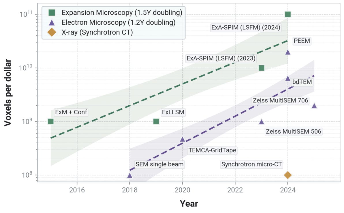
But on the other hand, physiological recording is improving relatively more slowly. The compiler idea is meant to address this problem by using paired structure-function data sets to learn how to predict dynamic parameters (e.g. the synaptic conductance) from morphomolecular data alone.
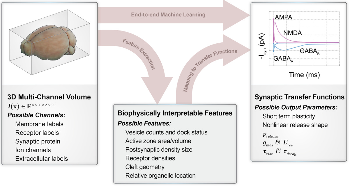
They propose to test the idea in the retina, which is highly characterized and has well defined inputs (photons) and outputs (neuronal spikes).
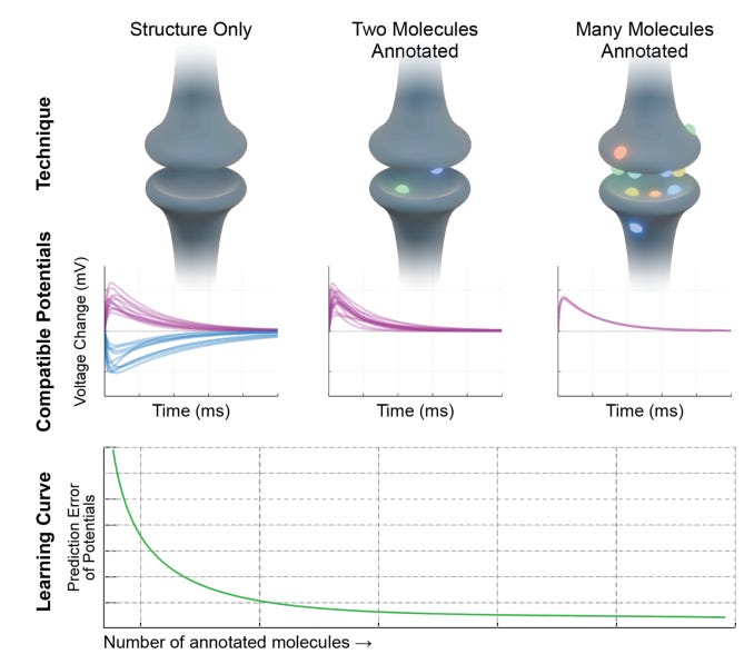
They note that it would not necessarily be possible with current technology to predict the dynamics with arbitrarily high accuracy, at least if there is little or no molecular annotation. Instead, they propose that the compiler should constrain the space of plausible models, with the degree of constraint possible depending on the degree of molecular annotation. The output would be a probability distribution over possible parameters, which could then be further narrowed once it is modeled as a part of a larger network with its own constraints.
The article reads sort of like a grant proposal. But it’s a grant that I would like to fund if I had the capacity to do so! It’s clearly an important problem where more systematic data and analysis would be helpful.
2: One of the long-standing questions people have raised with aldehyde fixation is the extent to which it affects molecular properties of the tissue.
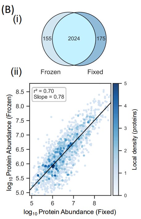
A new study helps to address this question. First, they compared the proteome (i.e. all of the measurable proteins) from fresh-frozen compared to formaldehyde fixed mouse tissue. They found a high correlation, with no evidence of extraction bias affecting any particular cellular compartment.
Next, they developed a method for laser capture microdissection to capture the proteome of single neurons from fixed mouse brain tissue.
They found that they could recover around 1,500 protein groups per single neuron on average, and were able to recover more proteins when they sampled larger areas. The proteins that were missed in single cell samples were mainly low abundance ones. This is consistent with the limits of mass spectrometry in general, as opposed to the loss of a particular protein type due to fixation.
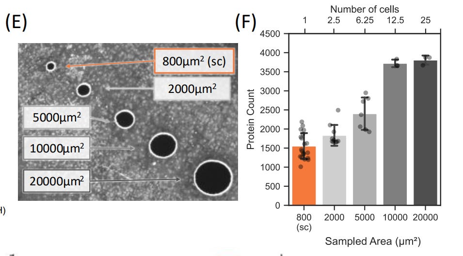
Overall, I consider this to be another solid data point that high-quality fixation preserves the molecular constituents of the brain in their original locations.
3: A new study characterizes a human brain that was immersion fixed 7 hours after death with multiple modalities, including MRI, synchrotron imaging, micro-CT, and electron microscopy.
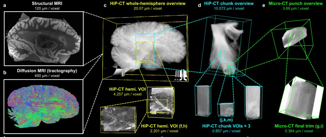
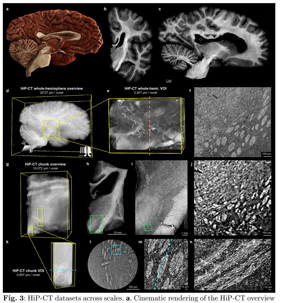
The goal of it is to be a prototype study to show that they can map white matter architecture in human brains across scales (a buzz word, but accurate here). They showed that it is possible to see single myelinated axons using non-invasive synchrotron imaging as well as micro-CT.
Here is some of the synchrotron imaging data they collected (i.e. Hierarchical Phase-Contrast Tomography, or HiP-CT), which shows individual axon fascicles:
In their electron microscopy data, they are able to see individual myelinated axons. However, they note that there are some regions with de-compacted myelin (red arrows).
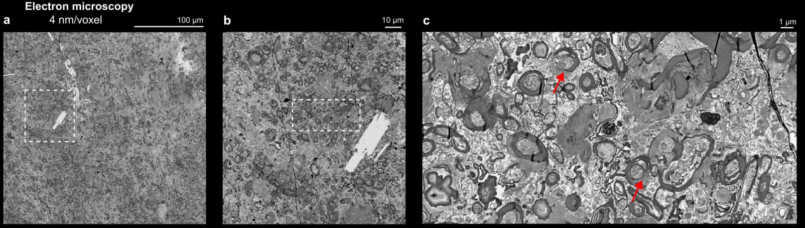
In the discussion, they note that specimens with shorter postmortem intervals would provide better ultrastructural preservation. It’s interesting but not particularly surprising to me that they were able to achieve this level of preservation quality via immersion fixation 7 hours after death.
4: Another methods paper for studying white matter connections in postmortem human brain tissue, this time using 3x hydrogel expansion, immunolabeling for neurofilament H, and light-sheet microscopy imaging. See also the companion methods paper.
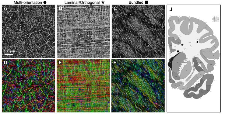
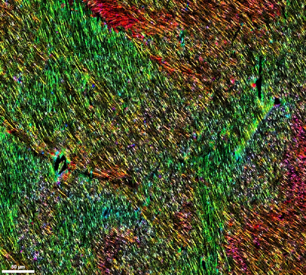
The brain tissue was from a 61 year old male donor obtained from the San Diego Medical Examiner’s Office. The brain was removed and bisected at the midline, and coronal slabs approximately 0.5 cm thick were cut. These were embedded in alginate, which provided mechanical support, then rapidly frozen in a dry ice-isopentane slurry, vacuum sealed, and stored at -80°C. When retrieved from the -80°C freezer for processing, the frozen slab was immersion fixed in ice-cold 4% paraformaldehyde with 10% acrylamide in PBS for 24 hours at 4°C. After all of this, they were still able to see individual axons stained with neurofilament H:
They also provide Neuroglancer links in which you can view the raw data yourself:
A key open question to me is regarding the size of axons they are able to visualize (and thus demonstrate the preservation of).
The abstract notes that they can see trajectories of densely stained large projection axons greater than ~1 μm in diameter. In the results, they note that there was generally a good amount of spacing between axons and that those with a larger caliber, above ~1.5 μm, could be easily traced manually with few ambiguities.
Intriguingly, they wrote in the methods: “NFH was selected based on its established use for myelinated long-range projection axons. Control experiments with viral labeling of excitatory neurons in macaques demonstrated that all virally labeled cortical axons were also labeled with NF antibodies (data not shown [can be included in revision])”.
One thing I am interested in is what fraction of axons are actually being visualized. Their ~1 μm and ~1.5 μm size thresholds are for what can be clearly resolved and easily manually traced, but it seems that they cannot address how many axons fall below those thresholds.
The main reason I care is that I want to use their results as a proxy for what kind of axon preservation quality we can expect in human brains subjected to the relatively harsh conditions reported here, including non-cryoprotected (but rapid) freezing, immersion fixation after thawing, and presumably a decently long postmortem interval (although they do not report it).
It’s confusing to me that all of the large axons look so good here, and yet axons in general often look damaged in other data sets (like electron microscopy) that have undergone similar types of preservation steps. Why is that? Is it just that the smaller axons that are seen more commonly on electron microscopy are more vulnerable to damage? But why are they so much more vulnerable? And if thin axons really are just more fragile, does the good preservation of the large axons suggest that the way smaller axons break down is constrained enough to be likely to be inferable from the breakdown products? Or is there something else going on?
I realize this is probably inside baseball to 99% of my readers, and I apologize about that, but it’s something I’m thinking a lot about right now, so perhaps you might find it interesting.
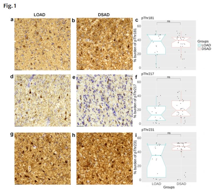
5: Study finds a similar distribution of phosphorylated tau in people with Down syndrome with Alzheimer’s disease neuropathology (DSAD) as people with late-onset Alzheimer’s disease (LOAD). This is despite people with DSAD being substantially younger on average.
6: Study finds that dementia in people with schizophrenia seems to be a distinct entity, insofar as its pattern of demographic, clinical, and genetic correlates cannot be easily explained by comorbid Alzheimer’s disease or cardiovascular risk factors.
7: A randomized trial finds that more hydration doesn’t actually help prevent kidney stones, but does cause more nocturia. Another L for Big Water.
8: Two randomized trials (n = 3808 participants total) find that oral semaglutide is not effective at slowing the progression of disease in early Alzheimer’s. So another drug enters the Alzheimer’s disease randomized trial graveyard.
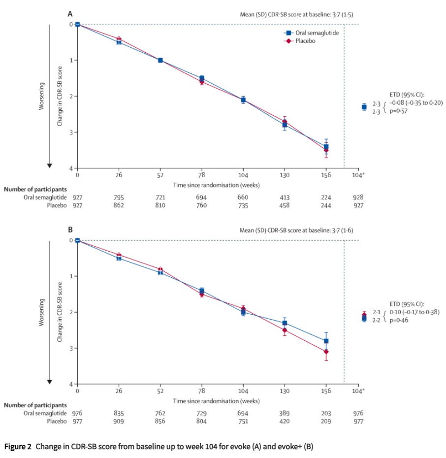
Hindsight is 20/20, but this is not super surprising to me. People with Alzheimer’s disease often have lower BMIs to begin with, for reasons unclear to me.
One interesting note is that there were 5 fatalities attributed by the (blinded) investigators to the study drug in the trial, but 4/5 of them were in the placebo group, which was half of the participants! This is a nice example of how it is hard to figure out what caused someone’s death. And also (weak) evidence of how relatively safe semaglutide is from a mortality perspective.
9: Is social media time actually bad for youth mental health? A meta-analysis of 46 studies finds that the “current pool of research is unable to support claims of harmful effects for social media use on youth internalizing disorders.”
It’s an interesting abstract and may be correct. However, I take issue with their use of the term “there is no evidence that” in the title. People should stop using that phrase once and for all. There is nearly always evidence of things on both sides of any active debate. The question is nearly always about what the preponderance of evidence best supports. I know this point has been made many times before, but apparently it has not yet reached everyone’s cortex.
10: Kevin Kennedy has an article with some nice criticism of the SAINT trial of TMS for depression.
11: A case of HPPD reported in a clinical trial of 25 mg psilocybin for treatment resistant depression. “One participant with a history of anxiety and dissociative symptoms acutely experienced severe panic, which subsided first, but reoccurred in form of anxiety and panic attacks, dissociation/depersonalization, flashback phenomena and symptoms of hallucinogen persisting perception disorder (HPPD) requiring hospitalization.” The overall trial did not show a significant effect on the primary outcome.
12: An article on the HHMI approach to journal publications. They are trying to move away from journal publications and towards evaluating preprints. In order to do this, journal names are now stripped away from the citations in the HHMI review submission materials. Also, at HHMI’s Janelia campus, scientists are not allowed to write the journal name on their presentations.
This is such. A. Good. Idea.
Chasing journal prestige may be good for individual scientist careers (actually, I’d bet that on average, it is), but it is a collective action problem that ends up enriching publishing companies at the cost of precious taxpayer and philanthropic dollars for research. Incentive realignment to prevent researchers from chasing prestige over actually advancing science is exactly the way to solve this problem.
This is why, at NN, I try to avoid mentioning the journal name of articles, and I try to ignore this when I choose which articles to write about.
13: The Amaranth Foundation on why they are funding neuroscience research in order to get safer AI, as a form of defensive accelerationism.
14: Interview with the founder of Bexorg, a new neuroscience company that is using perfusion of postmortem human brains as a platform to investigate new drugs. I am thrilled that they were able to raise 42.5 million dollars for this, because maybe the market for this type of human neuropharmacologic research is larger than I had realized.
15: Mark Woodward’s custom liquid handler at his cryobiology company Wake Bio.
16: There is a literature on using focused-beam microwave irradiation to rapidly heat rodent brains to 75-90°C. Here is a recent paper I just saw on this, which reminded me of it.
Microwave irradiation can denature enzymes and halts post-mortem metabolism within a second. The argument is that most of postmortem decomposition is enzyme-mediated. So if you could rapidly inactivate enzymes, you would dramatically slow autolysis, and so you would have much more time to preserve the structure of the brain.
I’ve looked into applying this to human brains, but the main problem is penetration depth. Microwave energy at these frequencies only penetrates a few centimeters into the brain tissue, which works for a mouse brain but wouldn’t work for a human brain.
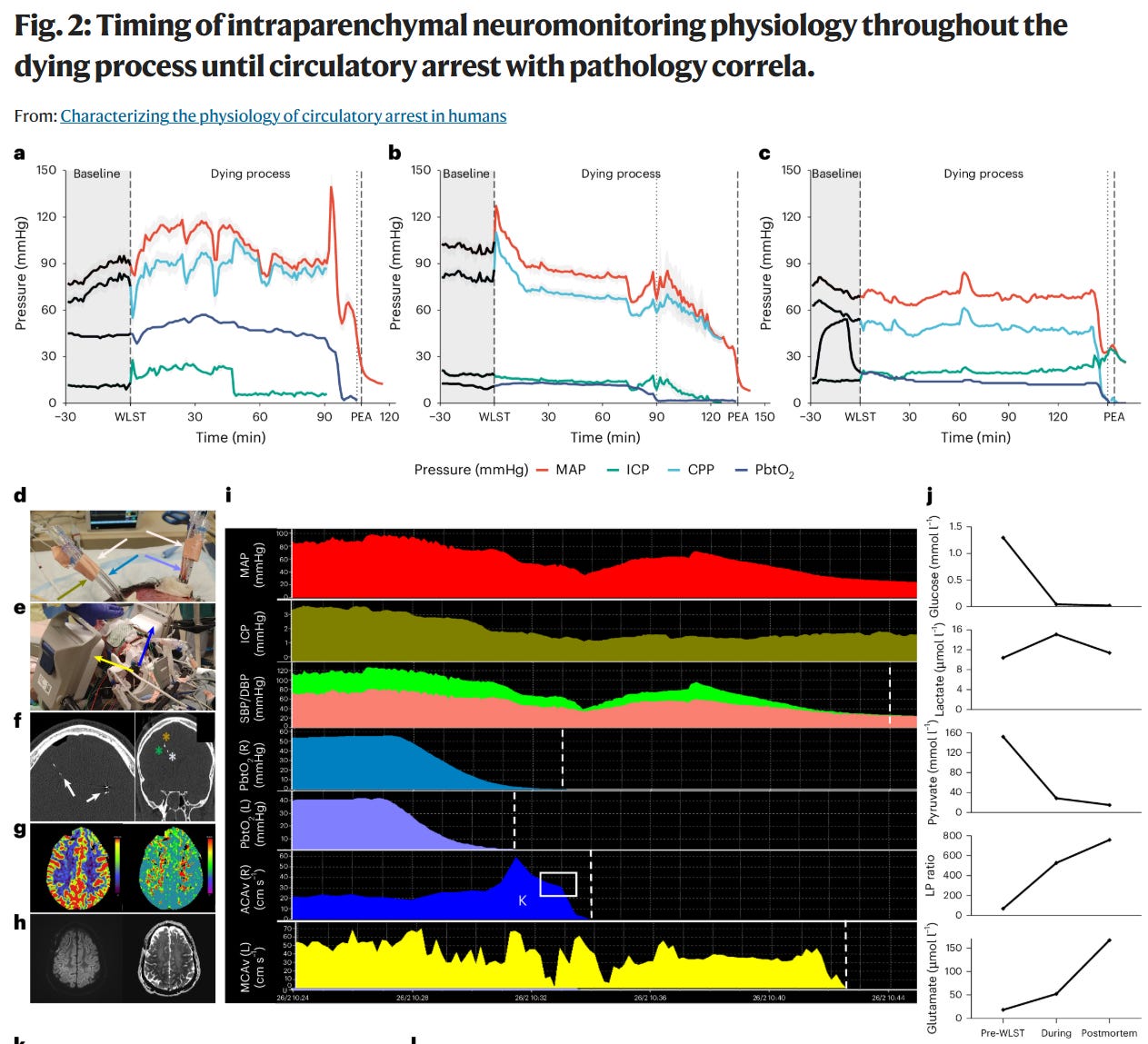
17: A paper from last year on the pathophysiology of the agonal phase, which is a highly understudied topic very relevant to brain preservation (HT: Mike Darwin).
Specifically, they studied 39 critically ill patients undergoing withdrawal of life-sustaining treatment while monitoring their cerebrovascular physiology until circulatory arrest occurred.
They found that in general, cerebral blood velocity declined linearly with hypotension. However, brain blood flow velocities and brain tissue oxygen tension stopped before systemic circulatory arrest. The median time from the cessation of brain perfusion to pulseless electrical activity was around 1.5 minutes, and the time to electrical asystole (ECG flatline) was around 6.5 minutes.
Because legal death is often declared a couple of minutes after pulseless electrical activity, the “warm ischemia” clock for the brain probably starts several minutes earlier than the time of declaration suggests.
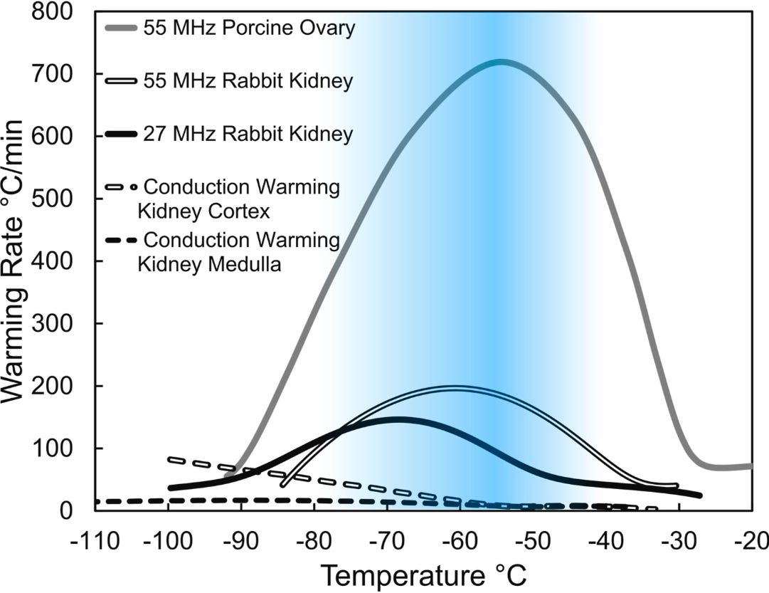
18: Review by Brian Wowk on rewarming cryopreserved tissue with electric fields.
19: Study on the attitudes to cryopreservation among people living in Switzerland. In general the public is highly supportive of the practice being legal, with 83.6% of respondents expressing no support for legal restrictions nor making a change from its current status. However, that is a bit of a low bar IMO. Surprisingly, 20.1% expressed interest or intent to sign up for preservation services.
20: Very interesting interview of the superstar brain preservation researcher Alex German by the journalist Kristen French.
Q: Would you personally want to be cryopreserved?
A: If the alternative were facing certain death, I currently would. It’s important to note that you expose the person to a lot of uncertainty in the future. They become more vulnerable to long-term variance in the developmental trajectory of humanity. There might be risks that we’re not aware of today. In a representative survey of 1,000 Germans, about 22 percent of them said they want to undergo these procedures, the majority of these being male.
My personal preferences could change in the future. But my professional opinion, as a physician, is that patients with bleak prognosis, and the public in general, should be informed about these possibilities. And these should be discussed in a measured, rational way. And this is what we try to contribute to with our research.
No posts
Comments
Nothing yet. Say the first thing.
Sign in to join the conversation.