
1: Now that we are getting increasingly good at generating connectomes, if we only got better at neuron typing, we could theoretically test Sebastian Seung’s vision of simulating neuronal information flow via connectomes and cell types.
The two major neuron types are excitatory and inhibitory. Most of the neurons (around 80-90%) in the cerebral cortex are excitatory (meaning that their synaptic transmission makes downstream neurons more likely to fire an action potential), while around 10-20% of them are inhibitory.
It turns out that despite being a minority, inhibitory neurons are much more diverse than excitatory neurons (in terms of anatomy, gene expression, and electrophysiology), which naturally makes them more interesting.
A new study examines inhibitory neuron typing in the whisker barrel cortex of the rat:
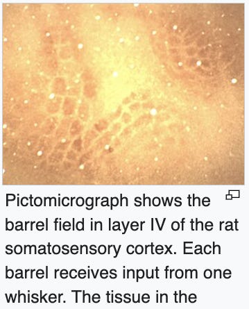
The authors note in the introduction that there is no consensus on how many inhibitory neuron “types” there are. However, this is utterly unsurprising because various aspects of neuronal features (including electrophysiology) are on a multidimensional set of hierarchical spectrums, so there’s generally no objective answer to how many types of a neuron there are.
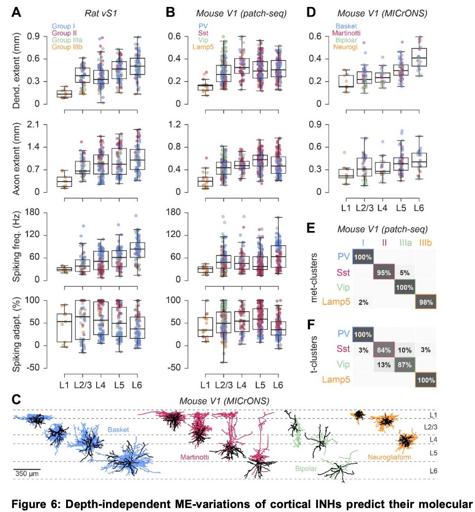
The authors recorded electrophysiology from individual cells, preserved their brains via perfusion fixation, and then labeled and imaged the cells via light microscopy:
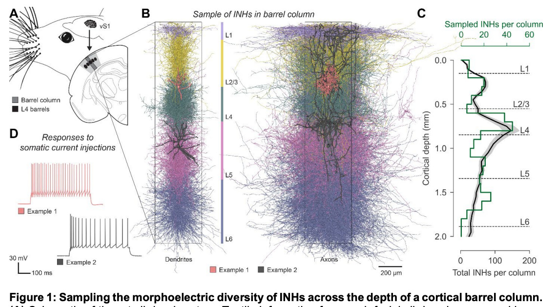
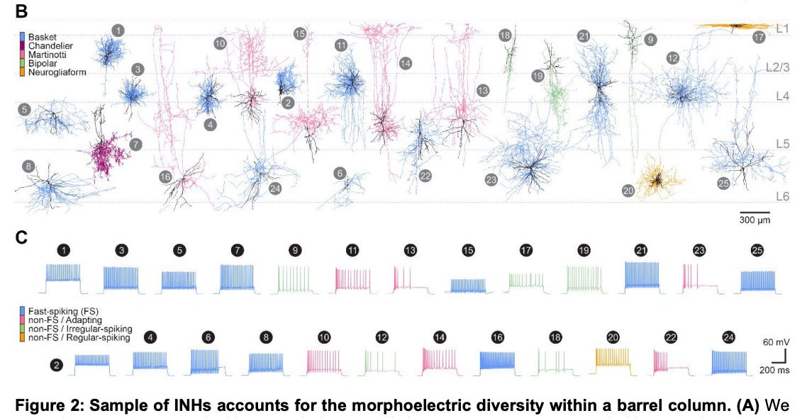
They first clustered the neurons that they profiled based on both their morphology (B) and electrophysiology (C):
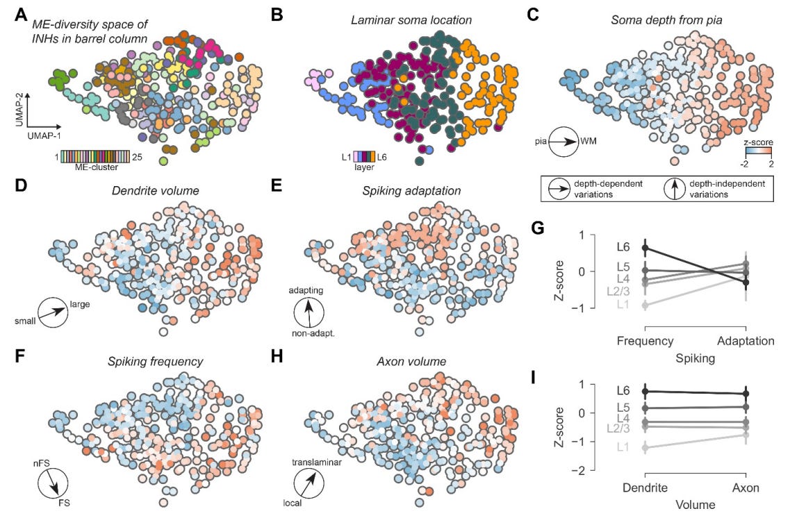
They then clustered neurons into a space that combines both morphology and electrophysiology, which they call “morphoelectric”. They noticed that these clusters are highly correlated with the depth of the cell body in the cortex (from layers 1 to 6):
They found that the morphoelectric properties of a neuron could predict which of the classic four molecular markers of inhibitory neurons it expresses (PV, Sst, Vip or Lamp5 ). The accuracy was pretty high:
Orthogonal to a given cell’s molecular expression, they also found that cortical depth was predictive of the morphoelectric properties of cells, in different data sets:
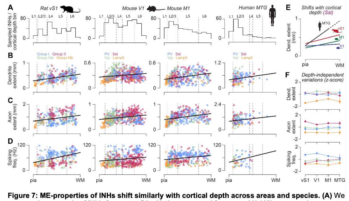
The authors speculate that the cortical depth has such a large effect independently of the molecular cell type because it is associated with local environmental effects on the morphology and electrophysiology of cells.
Extrinsic environmental effects on neuronal function are a potential problem for the connectome + cell type = simulation idea. Some extrinsic environmental effects, like the ones described in this paper, might be predictable based on the depth of the soma in the cortex, in which case you could extend the equation to connectome + cell type/cell location = simulation.
But other extrinsic environmental effects on neuronal function are not necessarily going to be easily predictable based on the location of the soma. They might require inferring the functioning of local glial cells based on morphological images, or doing molecular profiling. The extent to which this will matter for simulating any particular type of neural function is very much an open question.
2: Speaking of brain simulation, the nascent brain emulation company Eon Systems went a bit viral this month with an announcement from their founder Michael Andregg that “We’ve uploaded a fruit fly.” More detail was eventually described in the Eon Systems blog post.
Ken Hayworth expressed skepticism. So did Michał Januszewski, who noted “extraordinary claims require extraordinary evidence.” Alexander Bates had some good questions:

Alexander Bates@as_bates
.@michaelandregg @Philip_Shiu I’ve worked on the fly connectome 10 yrs. Good-faith questions: Do you have known internal dynamics: head-direction ring attractor bump (working memory)? "Spinal cord" analogue not modelled, so behaviour is "descending neuron X on, play animation"?

Michael Andregg @michaelandregg
We've uploaded a fruit fly. We took the @FlyWireNews connectome of the fruit fly brain, applied a simple neuron model (@Philip_Shiu Nature 2024) and used it to control a MuJoCo physics-simulated body, closing the loop from neural activation to action. A few things I want to
5:47 PM · Mar 9, 2026 · 10.2K Views
3 Replies · 7 Reposts · 87 Likes
I actually think Bates’s grandmother may have the best point of all: “But - I thought flies didn’t have brains?” (from Bates’s personal website)
3: Speaking of the head direction ring attractor bump, what exactly is that, anyway? And what kind of research are people doing on it right now?
First, a bunch of background. The term “manifold” has been one of the hottest in neuroscience over the past decade:
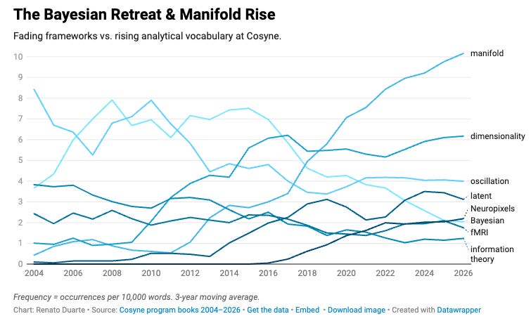
When neuroscientists use the term “manifold”, they use it differently from the formal mathematical definition.
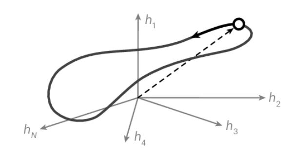
Instead, they are generally referring to the common phenomenon that neural activity, which technically exists in a high-dimensional space (i.e. one dimension per neuron), usually actually “lives in” a lower-dimensional surface, called a manifold. A classic example is a ring, which is a one dimensional manifold embedded in a two dimensional space. Here’s a toy example of a ring-like manifold of neural activity in a higher dimension space:
Another important concept here is an attractor. In a dynamical system, an attractor is a state that the system tends to settle into over time.
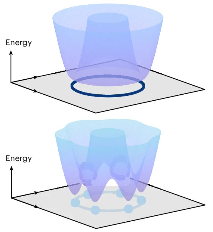
A ring attractor, then, is a neural activity space that is constrained to a ring manifold with attractors at different discrete points. An energy landscape is one way to visualize this. Low points (valleys) are the states the network naturally settles into, while high points are the states it tends to move away from.
Neural state spaces made of large numbers of neurons tend to have smooth energy landscapes (top), while spaces made of small number of neurons (bottom, n = 6 here) tend to have “bumpy” energy landscapes:
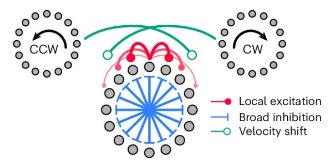
Computational models of neurons that have local excitation and diffuse inhibition can create a ring-like attractor system. And as it turns out, this is a very good way to model how flies maintain their head direction when moving through space:
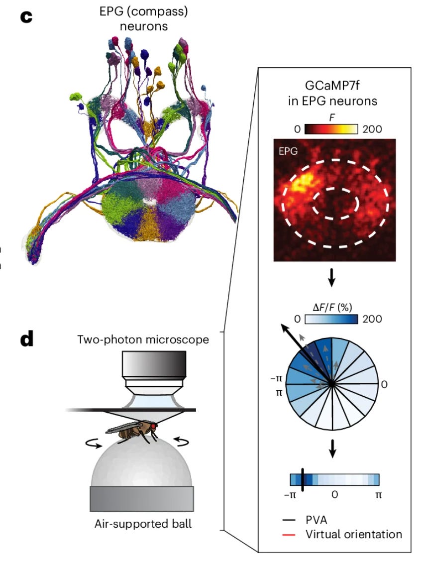
With the recent mapping of the fly connectome, we now know how the neurons responsible for maintaining head direction connect to one another. These are called “compass” neurons. You can see the local connectome and also an example of a calcium imaging activity map:
Bates’s point — which is a good one — is that anybody who claims to have “uploaded” a fly should be able to tell that these internal dynamics are working as expected. Which is challenging, because we don’t even know exactly how they work yet!
Finally I can get to a recent paper which can be used to show some of the uncertainty in this field.
A key structure that helps to maintain head direction is the ellipsoid body, which is a donut-shaped structure made entirely of axons and dendrites. The cell bodies of the compass neurons whose neurites project into it are found elsewhere in the fly brain.
The ellipsoid body can be divided into 8 wedges. Each wedge has processes from a different group of compass neurons. Here is a reconstruction of the ellipsoid body connectome, color-coded by wedge:
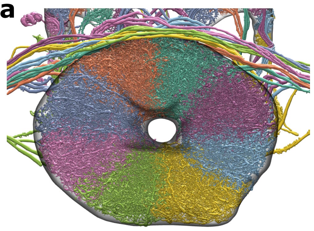
At any given moment, the compass neurons in a few adjacent wedges are more active than the rest, forming a localized “bump” of activity. As the fly turns, this currently-active bump travels around the ring, so that its position at any given moment encodes the fly’s current head direction.
This study also uses calcium imaging (electrophysiology) data from the ellipsoid body, where darker regions indicate higher neural activity, an example of which is here:
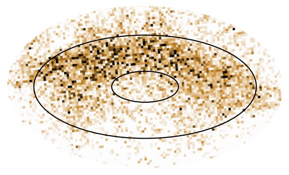
What this article found was that the fly connectome is well positioned in the space of possible connectomes for supporting robust ring attractor dynamics. Even when synapse counts were varied by nearly 90%, the scaling parameters could nearly always be adjusted to restore ring attractor functionality.
In particular, even though all four of the available fly connectomes (male CNS, hemibrain, FAFB, and BANC) have different connectivity in this area, they all fall well within the region of connectome space where ring attractor dynamics are achievable. This means that evolution seems to have found a connectivity pattern that is tolerant of natural synaptic variability between flies:
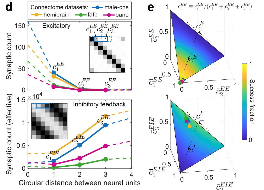
However, we do not know what the scaling factors that the authors used to tune the network actually correspond to biologically. The authors suggest they could reflect neuromodulation, which can up- or down-regulate synaptic strengths across local groups of cell types all at once.
Could these scaling factors be inferred purely from electron microscopy data, given enough “side experiments” to figure out how they work? Hard to say, but perhaps. Either way, we definitely don’t have this kind of information yet, which is another reminder of how far we are from being able to simulate even relatively simple neural circuits at a very detailed level.
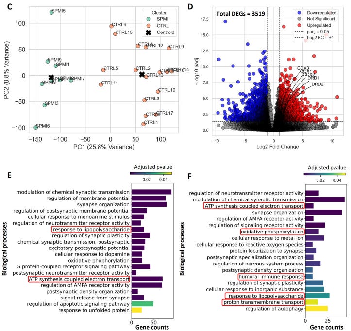
4: A new study on the human brain thanatotranscriptome, i.e. gene expression changes occurring after death. They find that postmortem time induces artifactual upregulation of genes involved in oxidative phosphorylation, ATP synthesis, and inflammatory responses. Notably, 6 hours of storage of the brain at 4°C (sort of mimicking rapid morgue refrigeration) produced zero genes that were differentially expressed by their significance thresholds, which suggests that the changes in gene expression after death are not necessarily immediate and huge.
5: A big retrospective study of n = 606,434 US veterans with type 2 diabetes finds that, compared to SGLT-2 inhibitors, GLP-1 receptor agonists were associated with an around 13-25% relative risk reduction of developing new substance use disorders (i.e. alcohol, cannabis, cocaine, nicotine, and opioid use disorders) over three years of treatment.
6: Nils Wendel on the new one year open-label study of the non-dopaminergic antipsychotic xanomeline. It continues to have a low side effect burden, including no significant EPS or metabolic effects, which is obviously just amazing.
7: A nice essay about schizophrenia trajectories by Michael Halassa. He covers a GWAS-by-subtraction study which suggests that there may be two components within the schizophrenia GWAS signal: (a) one component associated with lower educational attainment, with mechanisms more related to early neurodevelopmental genes expressed across the brain, and (b) one component associated with higher educational attainment, with mechanisms more related to synaptic function later in life that tend to be found more in the frontal cortex. It’s a preliminary hypothesis that requires more study, but I find it interesting.
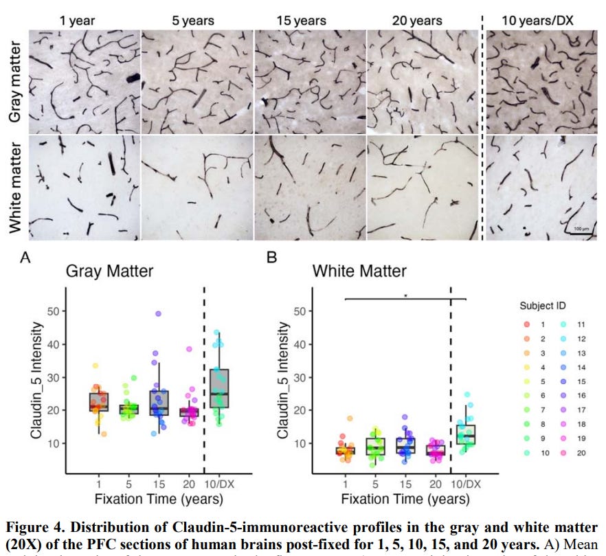
8: A study on the effect of long formalin fixation periods on the immunohistochemistry of different vascular markers in the postmortem human brain. Claudin-5 is one antigen that was found to not really change, even after 20 years of storage:
9: Study of archival brain tissue stored in formalin fixative at room temperature for decades. They reported: “In general, good staining quality was achieved across all decades, ranging from 1946 to 1980, and across both types of tissue preservation, i.e. original blocks stored in cardboard boxes for up to 78 years and newly sampled blocks with fixation times up to 78 years.”
10: A new paper from Adam Higgins’s lab describes a robotic platform to identify the minimum concentration of cryoprotectant needed to vitrify to find lower toxicity cryoprotectant formulations:
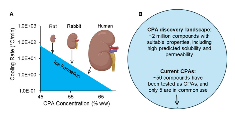
11: Alexander German et al’s article describing the use of interleaved equilibration to maintain electrophysiological properties of brain tissue after cryopreservation is now published in a peer-reviewed journal. This has been widely covered in the scientific press (for good reason), but loyal NN readers already knew about it last February when the preprint was published.
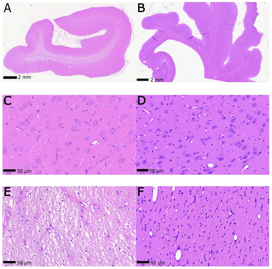
12: A new paper from Aurelia Song et al on the use of aldehyde-stabilized cryopreservation to preserve the ultrastructure of whole pig brains. They report unsuccessful preservation in one case where the cannulation of the aorta was not successful (A, C, E) and successful preservation in one case where perfusion started 14 minutes after cardiac arrest (B, D, F):
In the successful preservation case, they also report electron microscopy data, one image of which can be seen here:
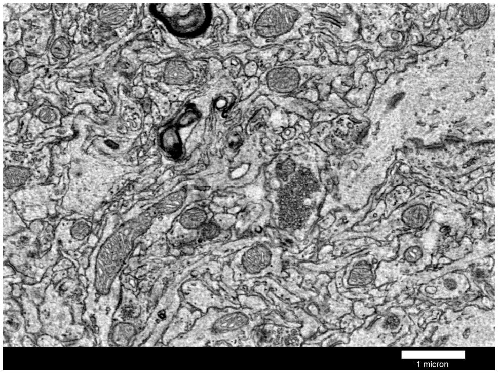
While these are amazing and encouraging results, I’m not sure if they will necessarily translate to humans in the context of medical aid in dying (MAID), which the authors suggest in the paper. There are many potential differences between these situations, including humans having a 10x larger brain, generally being much older and likely having some degree of vascular disease (e.g. atherosclerosis can begin as early as age 2), and having a longer agonal period (even with MAID). And plus, biological variability means that perfusion might work well in some cases and less well in others, even at the same time point.
13: On a related note, Aurelia Song and I discuss some of our differences in opinion about brain preservation in this Less Wrong comment thread.
No posts
Comments
Nothing yet. Say the first thing.
Sign in to join the conversation.