
1: Synapses are clearly important aspects of the brain, but they obviously aren’t the only thing. A new paper re-analyzes publicly available volume electron microscopy (“connectomics”) data to describe what they call the “contactome,” which is defined as a description of the places where the cell membranes from two cells touch one another. (This term has been used in this context a few times before, e.g. see here for C. elegans.)
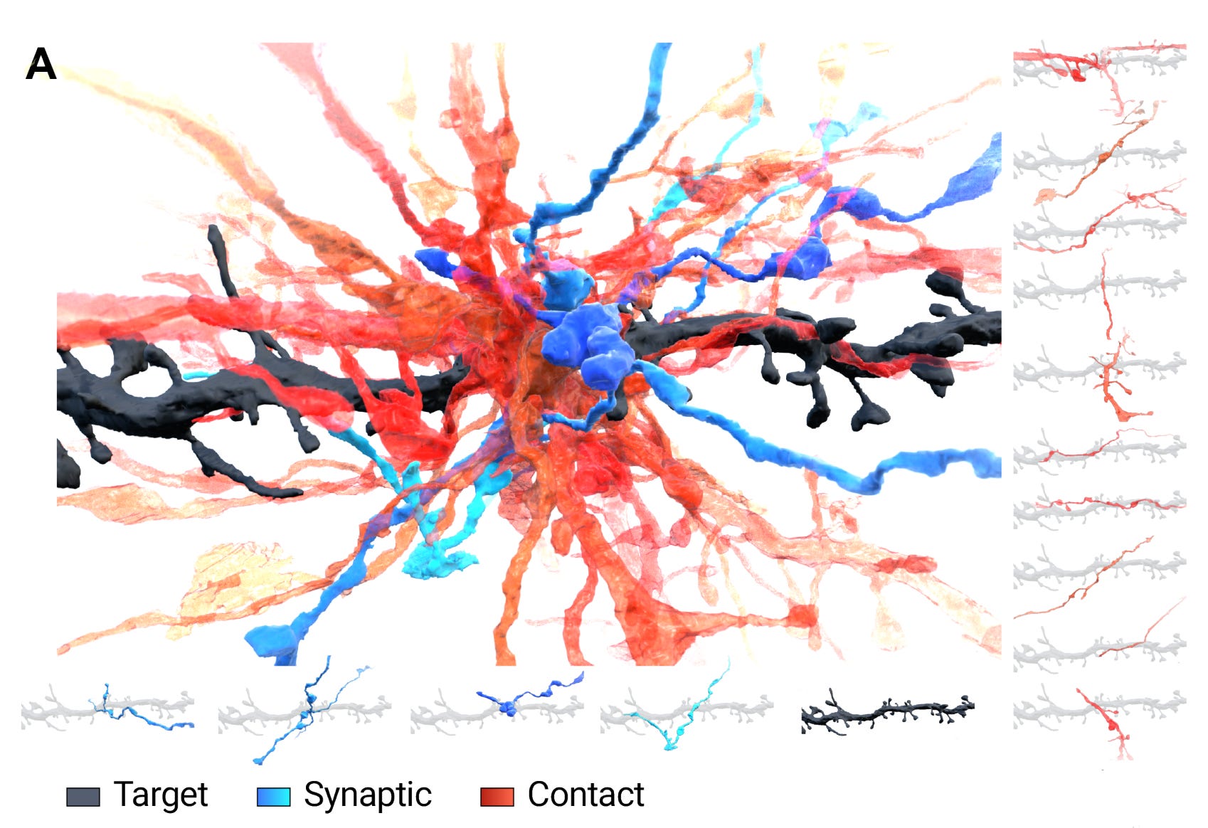
Here’s an example of what they mean by the contactome. A particular part of a single dendritic spine has four other neurons that it shares synapses with (colored blue), while it has fifteen other neurons that its membrane contacts without a synapse (colored red, and seen individually on the right):
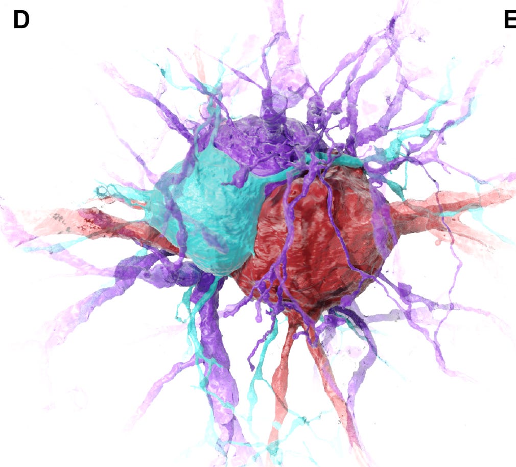
And here is an example of three neuronal somas (teal, purple, and red) whose membranes make a lot of contact with one another but don’t share any synapses — possibly they have some sort of other interaction:
They also found that astrocytes form a large syncytium-like network that spreads across the brain. This is well known from smaller scale studies — there’s a reason for the concept of a tripartite synapse — but this seems to be (one of) the first to describe it on such a large scale, with a single network containing 6146 of the 7562 total astrocytes in the volume.
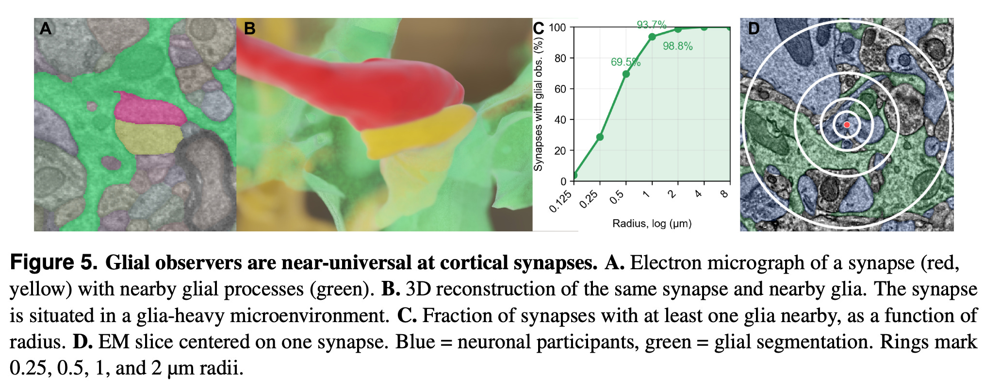
They also find that the majority of synapses are <500 nm from a glial cell:
All in all it’s a nice study with some good visualizations. I do have some areas where it might be improved. First, it’s not always clear to me when they’re talking about glia broadly versus astrocytes specifically. This matters because astrocytes, oligodendrocytes, OPCs, and microglia all have very different functional roles in the brain in general, and viz a viz synapses in particular. I actually think that people should try to avoid using the term “glia”.
Second, what I would really like to know is how these cell-cell contacts might be functioning. They suggest a few possibilities in the intro: (a) gap junctions, (b) hormones, (c) neuropeptides, and (d) ephaptic coupling. It’s not entirely clear to me that these connectomes are at a high enough resolution to even visualize gap junctions, so I would have like to see that discussed. Ephaptic coupling is the most interesting possibility to me. It would be nice in a future study to model if that might be playing a role or not.
Finally, I’m not sure it’s fair to today’s field of connectomics to say it just maps synapses. I would say that’s more like “synaptomics.” Modern connectomics is usually more than that, including cell shapes and inferred cell types. Although I’m certainly not an expert in the field, I have previously defined a few different uses/levels of the term “connectome.” What I have called the “cell membrane connectome” is the 3D map of all cell membrane locations, which would include the contactome. So at least based on how I think about it, the contactome isn’t really a separate thing from the connectome, so much as a particular aspect of it that the field hasn’t emphasized much. And in fact, this paper uses the segmentations that the original “connectomics” studies already generated to extract its contactome.
That said, I do agree that these cell-cell contacts have not been studied or considered as much as they should be (probably because the field is just not very big), and maybe branding them with an -ome is a way to move the needle in that direction.
2: One of the world’s leading connectomics experts, Moritz Helmstaedter, has written an article about accelerating human brain connectomics. He argues that a human connectome could be completed within 10 years if $10-20 billion were invested.
He proposes that this be done in the context of a rapid body donation program. His threshold for high-quality ultrastructure is a 4 hour post-mortem interval (PMI), although he notes that even within that time window, connectome reconstruction becomes increasingly more difficult.
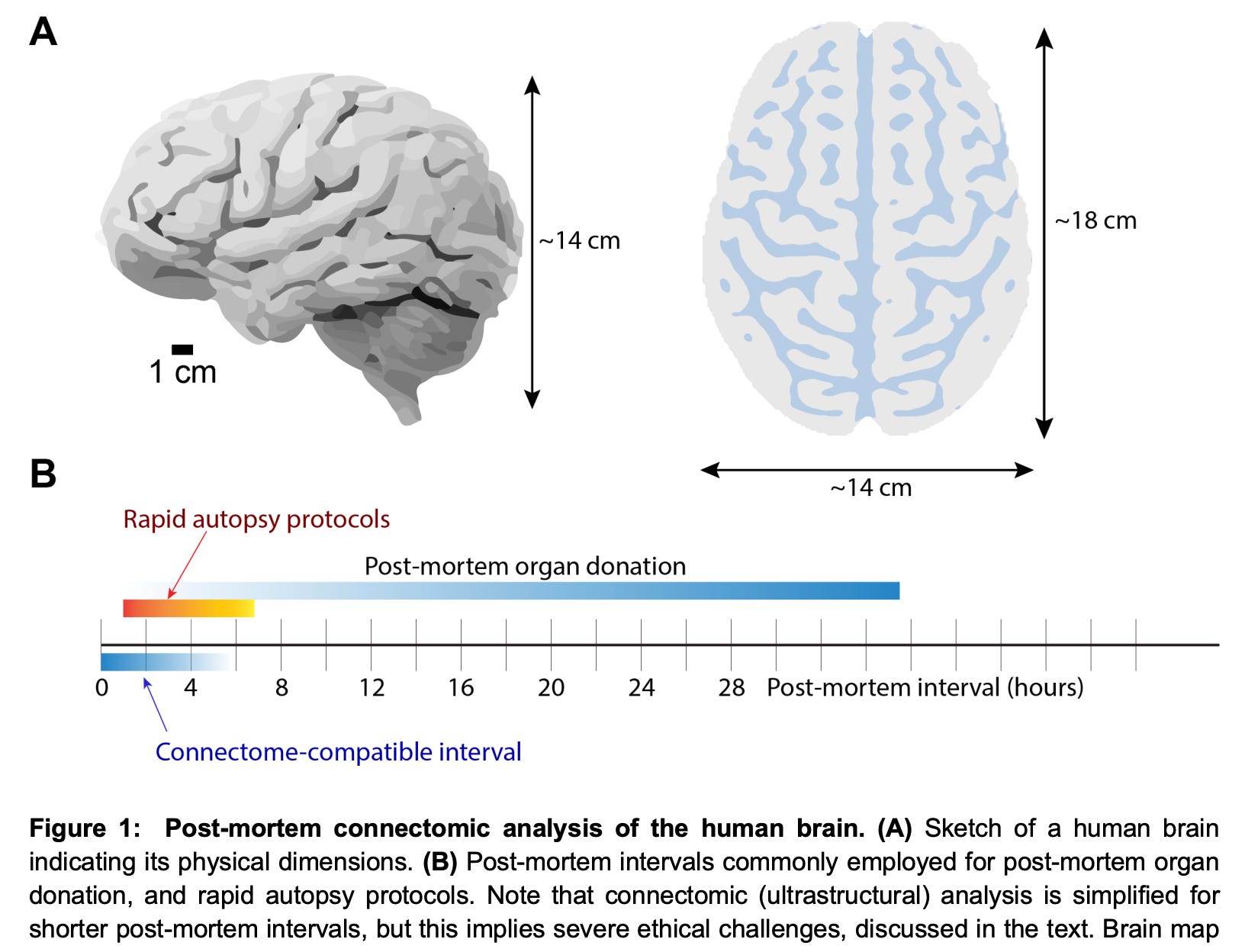
He notes that due to the PMI, two main problems develop: (a) synaptic vesicles are depleted, making synapses more difficult to detect, and (b) the very thin parts of axons (which are frequent in the grey matter) get “severed,” leaving only “spotty axonal traces,” and making them not possible to trace. Dendrite tracing can also be a problem at their thin spine necks, but generally dendrites are not as hard to trace because they are 5-10 fold wider than thin axons.
He also points out that it is usually possible even in highly damaged tissue to find some areas with relatively good ultrastructure for study, but that for a whole brain connectome project, the problem is much harder because it is the worst-preserved areas that are the bottleneck for success.
He notes that this need to lower the PMI raises “serious ethical concerns,” because for short PMIs, the exact time point and definition of death matters, and for this reason he calls for a discussion of the relevant ethics.
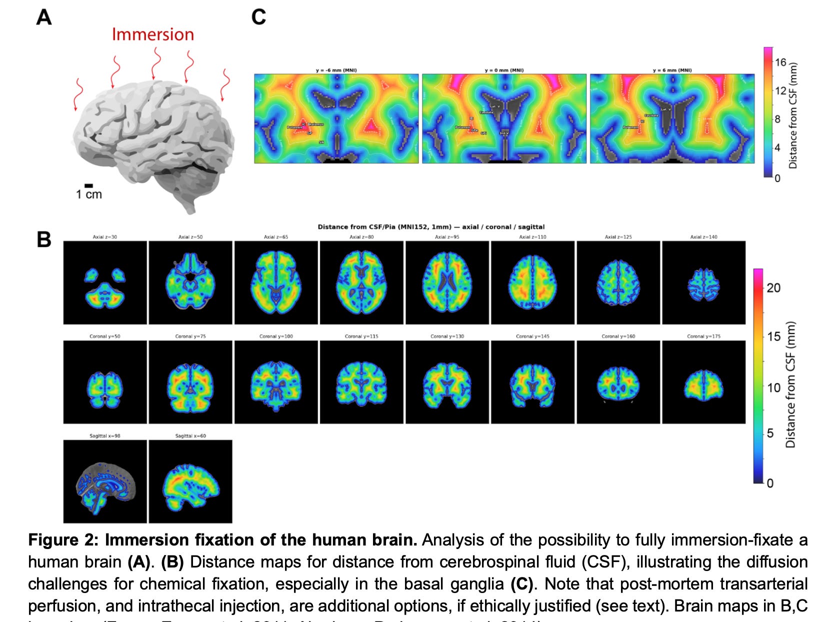
He considers immersion fixation as one potential approach for brain-wide ultrastructural preservation. In this analysis, he seems to be assuming that 3 mm is a short enough distance of immersion for fixative to penetrate the brain tissue fast enough. He notes that the surface of the cerebral cortex of the brain is exposed to CSF within 3-4 mm at maximum, which is encouraging. However, he notes that other areas, such as the basal ganglia, are much further away. Therefore, he rejects immersion fixation as sufficient and claims that perfusion fixation of the brain will be necessary.
The rest of the paper assumes that the brain has somehow been fixed rapidly enough so that the entire brain-wide nanometer-scale tissue is well enough preserved, and describes how it could be imaged at the ultrastructural level.
The main challenge is in finding a parcellation strategy that still allows neurite tracing. His proposal is that instead of intentionally cutting the brain into small pieces, it is actually best for the tissue to become brittle and then crack haphazardly into small pieces. These pieces can then be computationally reconstructed back into the whole brain, even if their order is lost, because they will each be so distinctive. Sort of like a giant jigsaw puzzle. Finally, the main cost is actually imaging them. He notes that 2,000 multibeam scanning electron microscopes working in parallel would allow the tissue to be imaged in 5 years, at a cost of $10-20 billion, which is the primary cost of the project.
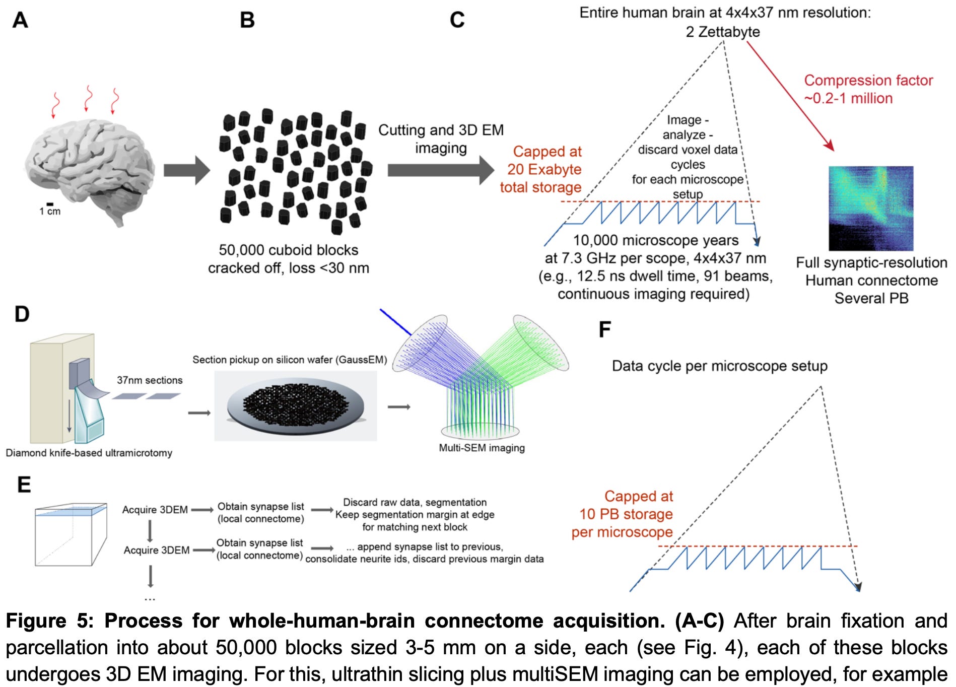
Probably my main concern with the proposal is the idea to only store the synaptome (aka adjacency connectome). As I mentioned in the discussion of the “contactome” paper above, I just don’t think that the synaptome alone is likely to be that useful, and with all of the effort to image this brain, it seems like we should be able to do better at the data storage problem. It seems to me like some sort of mesh representation would not necessarily require storing the raw images but would allow much more information storage. There is precedent here, for example CERN can store an exabyte of data already.
It’s certainly a provocative proposal and I would love it if some government or other large funder got interested in it. I also completely agree with him that human connectomics would significantly advance the study of human brain disorders, indeed being very useful for understanding the cellular-level mechanisms of certain types of psychiatric disorders.
See also: Pelagia Martin’s article “The Connectome is Too Big for One Lab.”
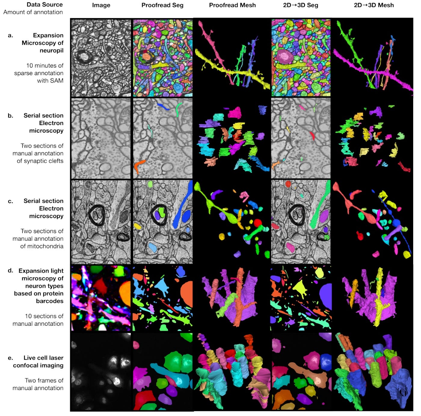
3: A new computational approach for bootstrapping 2d annotations to 3d reconstructions of ultrastructural data. Includes this nice figure showing how the method can work with different input data types:
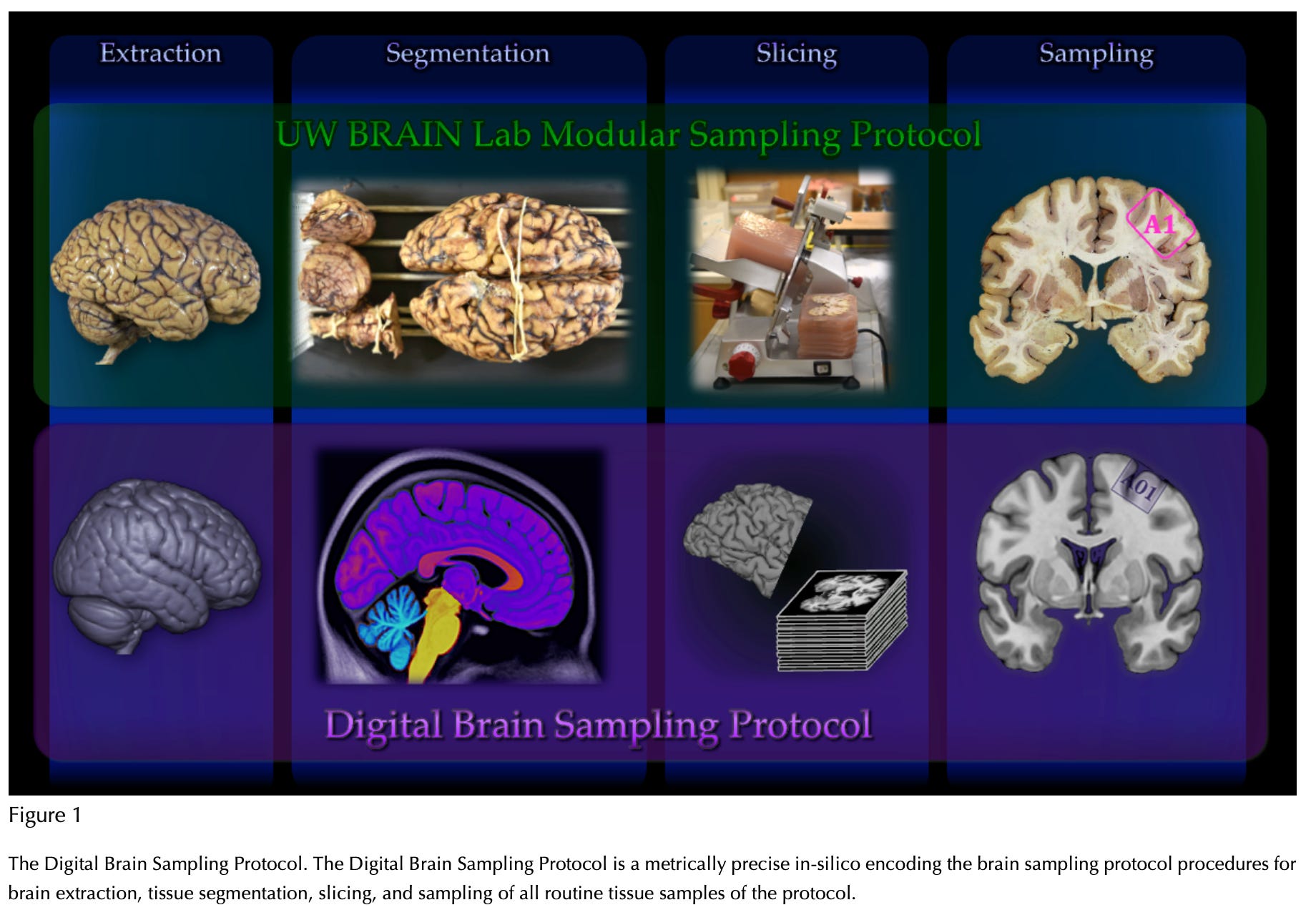
4: A software method for figuring out how to sample tissue banked brain tissue:
5: How does the precise organization of neuron wiring come about? A new study corroborates that in Drosophila, particular axons express unique RNA and protein molecules. This includes the cell surface receptor encoded by the Dscam gene, which undergoes alternative splicing and allows each neuron to express a particular combination of isoforms from tens of thousands of possibilities. Vertebrates have a functional analog of this that also allows for neurite self/non-self recognition (protocadherins).
Here are the axons they focused on (those of the neurons named “pSc” and “Gr59d”):
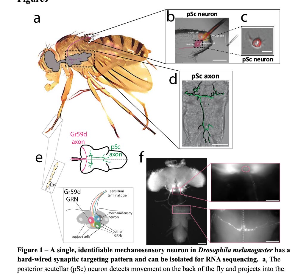
In addition to Dscam, this study also used RNA sequencing and found that Drosophila have a bunch of other transcripts for cell surface and secreted proteins that are differentially expressed between these two axon types (with multiple biological replicates of each):
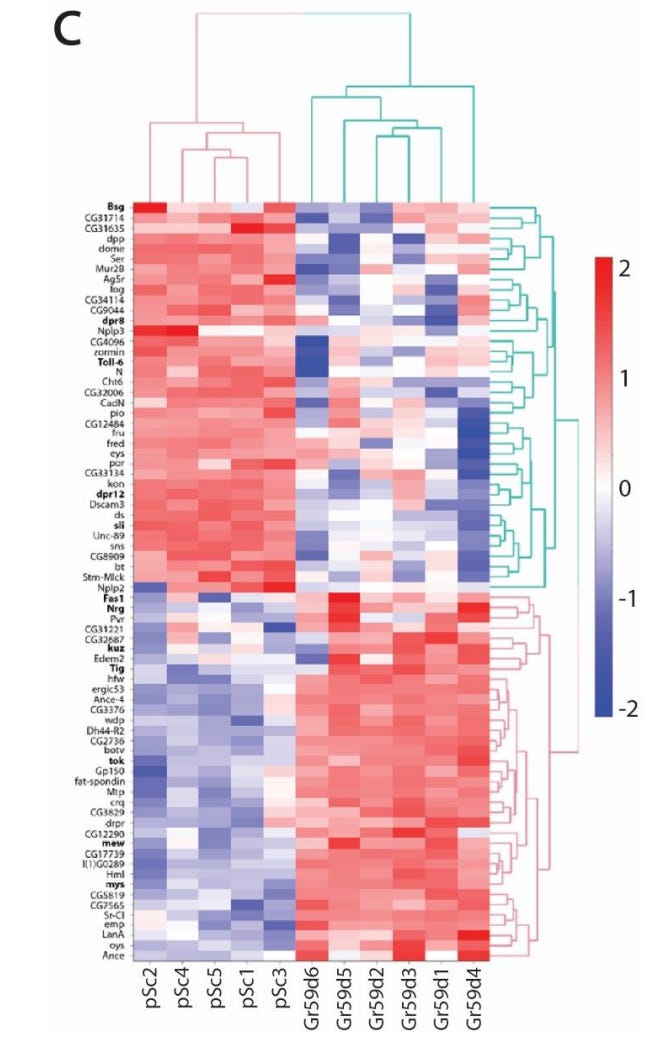
The main reason I am interested in this is because it shows how molecular mapping could potentially be used by future technology to help decode the information in the original wiring diagram in the presence of damage.
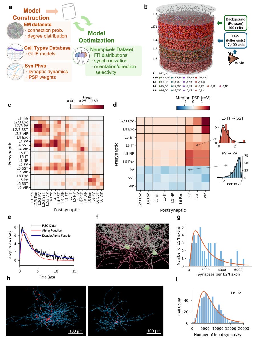
6: Simulation of a cortical circuit based on connectomics data, cell types, and known synaptic physiology:
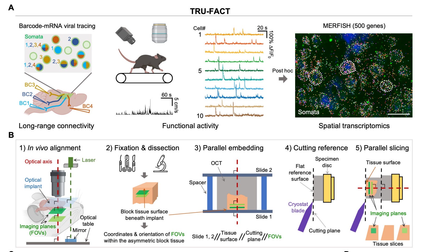
7: In a new study, living brains were profiled via two-photon calcium imaging of their cellular activity, then the tissue was fixed and sectioned for spatial transcriptomics. This allowed them to measure the RNA content of the same individual cells whose activity had been recorded in the live animal, with high spatial resolution.
8: A few years ago I looked into rapamycin for the purpose of trying to live longer. It was the hottest drug in longevity and, let’s be real, sometimes I follow fads. After some research, I sort of decided that aside from questions of efficacy, I was too concerned about infections, and I wasn’t sure whether I would also need (or be able to get) an antibiotic prescription as well. It seemed like some people prescribed them and some didn’t. I also felt bad about the idea of potentially contributing to antibiotic resistance without an actual medical indication (like in transplant patients who receive prophylactic antibiotics when they take it at a higher dose). It made the calculus worse for me. So I just never really pursued it, even though I always sort of had it in the back of my mind as something I “probably should have been doing.” Y’know, the whole death problem thing. But now some clinical trial evidence suggests that my subconscious was actually onto something!
Specifically, we got results from a randomized trial of adults aged 65-85 (n = 20 placebo, n = 20 of 6 mg rapa) for 13 weeks alongside exercise, with outcomes mostly related to exercise capacity. The primary outcome had no significant difference, but it leaned towards placebo (p = 0.089) and some of the prespecified sensitivity analyses significantly favored the placebo group. Granted this study doesn’t really measure what people tend to care about most with rapamycin, like the potential to slow aging or prevent some cases of cancer, but it’s not good if it worsens exercise capacity.
More troublingly in my book, the safety burden also seemed to favor placebo. There was one case of community-acquired pneumonia after a single dose of rapamycin, after which that participant was hospitalized and withdrew. More generally, adverse events attributed as potentially related to the drug were more common in the rapamycin group (35% vs 15%) and included upper respiratory tract symptoms, headache, and fatigue. Anyway, this data point seems to corroborate my priors for now, although I’m always open to changing my mind again in the future.
9: Mid-life elevated BMI is generally associated with an increased risk of Alzheimer’s disease. However, the “obesity paradox” in Alzheimer’s disease is the finding that late-life elevated BMI is often associated with lower rates of Alzheimer’s disease.
This is generally thought to be due to prodromal effects of Alzheimer’s disease leading to lower weight, but there hasn’t been all that much research on it.
A new study re-analyzes data from old Alzheimer’s trials with n = 1,663 participants to build a model to address this. They found that at lower amyloid levels (using the 25th percentile of the model for visualization), lower BMI was associated with better cognition (PACC) scores at baseline and over time. But at higher amyloid levels (e.g. at the 75th percentile), lower BMI was associated with slightly better cognition scores at baseline but a much faster decrease over time.
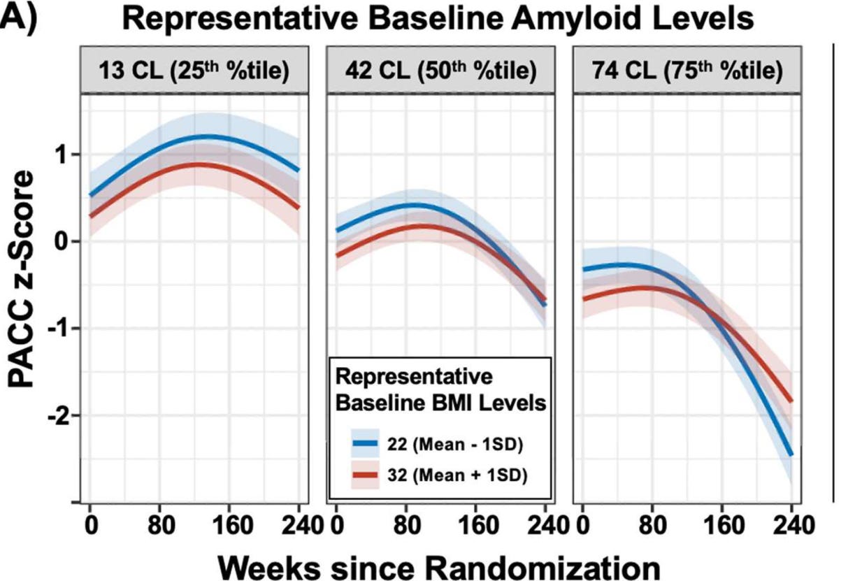
So the data seems to suggest that obesity really might be protective in adults with high amyloid, perhaps because of something like leptin signaling being beneficial in some way. However, I find their mechanistic explanations uncompelling and I still think it is most likely residual reverse causation.
This is because I think amyloid is not the whole story. Instead, amyloid is an imperfect proxy for disease stage. My explanation is that within the high-amyloid group, the people with lower BMI are more likely to be further along on the non-amyloid axes of disease burden (e.g. tau, neurodegeneration, hypothalamic dysfunction), which leads to lower appetite and feeding behavior, who would then be expected to have faster decline.
Whereas if people have low amyloid at baseline then they’re not having the amyloid cluster of problems in their brain, and lower BMI seems to be somewhat protective against cognitive decline for normal reasons like vascular function etc.
10: Obituary for Edna Foa, who helped establish exposure and response prevention (ERP) as the gold-standard therapy for OCD.
11: Study of an Italian cohort of n = 300 people with OCD finds that 18.6% (n = 56) were also diagnosed with obsessive compulsive personality disorder (OCPD). The group with comorbid OCPD was found to have a longer duration of untreated illness, of 9.2 ± 12.4 years vs. 6.1 ± 8.1 years.
12: A nice article with skepticism that whole brain emulation will matter for the AI transition.
I agree with a lot of it, but here are some disagreements because those are more interesting. The article seems to generally assume (a) rapid timelines, (b) somewhat rapid takeoff (to the extent that AI capabilities cannot be used to accelerate progress very much in the “wet lab” realm needed for emulation) and (c) no partial emulations.
The possibility of partial emulations is in my opinion the aspect that is most underappreciated here. An example would be only emulating the circuits for one or more particular neural functions, and simulating other inputs and outputs as needed. This might be useful for certain circuits that we value a lot, like those related to prosocial behavior, if that is ever possible.
Another example would be a broad category of “functional” emulations that capture higher-level properties of brain operations (such as those visible with neuroimaging) and use scale separation to abstract away from the underlying substrate. (On an even higher level, arguably this is what LLMs are already doing as simulators, and arguably there is a spectrum between emulation and simulation.)
Partial emulations would not qualify as identity-preserving for many people (even less so than full emulations!), but they could still be useful for other things, such as making a given AI system work in a more human-like manner.
I’m obviously not sure whether some sort of partial emulations will actually be relevant in the medium term, but I think it’s more complicated than the binary “full richly molecular annotated nanoscale-level human WBE or bust because of AI timelines” story.
I also predict eventually people will have debates about whether emulation has happened yet, in the same way that people currently can debate whether we have already achieved AGI.
13: Nice discussion by Nikki Olson on long-term monitoring systems for detecting cardiac arrest for the purpose of biostasis.
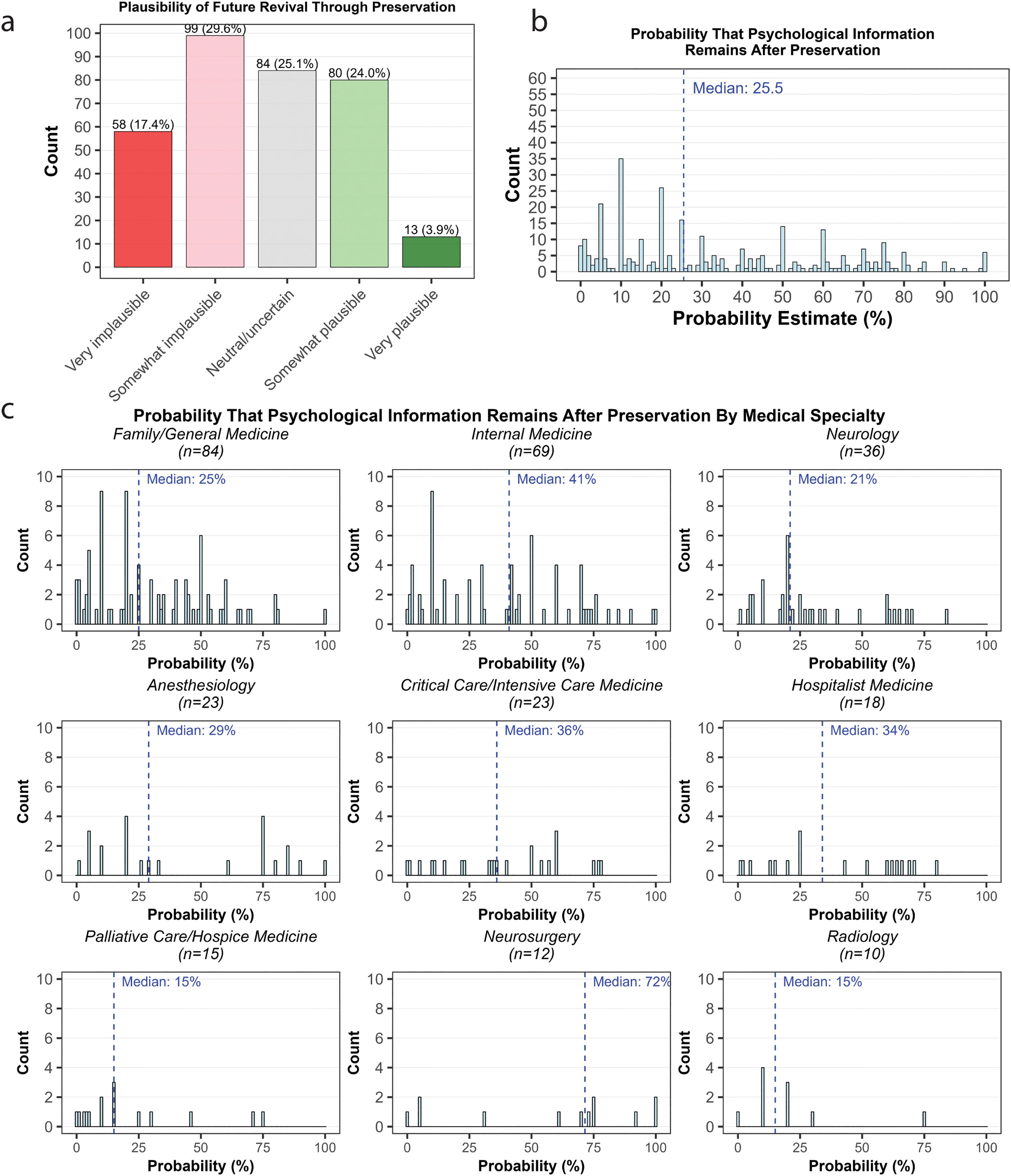
14: Our article “Physician estimates of the feasibility of preserving the dying for future revival,” has now been published. PLOS kindly did a press release for it, which is available here. Here’s one of the main figures, perhaps sort of self-explanatory:
No posts
Comments
Nothing yet. Say the first thing.
Sign in to join the conversation.