
It’s George.
The brain contains a vast neural architecture where billions of cells continuously exchange information.
This complex signaling is driven by neurotransmitters, the molecular messengers responsible for cellular communication.
So by attempting to decode neurotransmitters, we are attempting to map the specific language the brain uses to send instructions and process our thoughts and emotions.
Overall, in case you are new here, neurotransmitters are chemical substances that neurons release to send signals across synapses, the tiny gaps between nerve cells.
These molecules are categorized based on their effects.
We have:
Excitatory neurotransmitters that increase the likelihood of a neuron firing an action potential.
Inhibitory neurotransmitters that decrease the likelihood of a neuron firing, providing a calming effect.
Modulatory neurotransmitters that influence the overall activity of groups of neurons, modulating their function over time.
In the previous article, we talked about glutamate.
You can read it here:
In this one, we will talk about gamma-aminobutyric acid (GABA).
This is mainly the one that counterbalances the excitatory actions of glutamate and helps maintain the excitation-inhibition (E/I) balance, which is essential for proper brain function.
Reduced GABAergic tone frequently disrupts this equilibrium and leads to neuronal hyperexcitability (and a widespread over-excitation in different brain regions overall), which is a state implicated in numerous neuropsychiatric and neurological disorders.
This is why if you experience symptoms and conditions such as:
Generalized anxiety disorder
Panic disorder
Racing thoughts
Sleep-onset insomnia and fragmented sleep architecture
Muscle tension and physical restlessness
Heightened startle response
Bipolar disorder
ADHD
Emotional reactivity / irritability
Difficulty focusing / sensory overload
Major Depressive Disorder
Autism Spectrum Disorder (ASD)
Schizophrenia
Epilepsy (various syndromes)
Obsessive-Compulsive Disorder (OCD)
Chronic pain and central sensitization
Fibromyalgia
Alcohol withdrawal syndrome
Post-traumatic stress disorder (PTSD)
then you likely need to explore GABA.
Of course, low GABA tone/dysfunctional GABA signaling is rarely the sole cause of any of these and it usually interacts with inflammation, genetics, stress, and a few other factors besides glutamate excess.
But many of these symptoms/conditions improve when GABA tone is supported through lifestyle, nutrition, or targeted therapies.
So it’s a valuable puzzle piece towards resolving any of these.
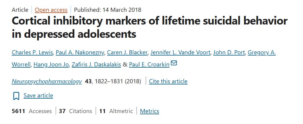
Overall, if GABA was irrelevant, benzodiazepines wouldn’t be so popular and abused or GABAB receptor-mediated inhibition is wouldn’t be so dysregulated in depressed adolescents (especially the ones with histories of suicidal behavior).
Now GABA (γ-aminobutyric acid) is a non-proteinogenic amino acid(*) that serves as the primary inhibitory neurotransmitter in the mammalian CNS and was isolated and identified as a major component of brain tissue by Eugene Roberts and Sam Frankel around 1950.
(*) Proteinogenic amino acids (the 20 standard ones + a few others like selenocysteine) are used by ribosomes to build proteins according to the genetic code.
GABA is non-proteinogenic, so it is not incorporated into proteins during protein synthesis.
It has a different structure (the amino group is on the gamma carbon instead of the alpha carbon), so it cannot be used in the standard translation process.
So GABA functions almost exclusively as a signaling molecule (neurotransmitter) and metabolic intermediate, not as a structural building block of proteins.
This is why it is rarely just called an “amino acid” without the qualifier “non-proteinogenic” in scientific literature.
It mediates inhibitory neurotransmission at roughly 20–50% of all synapses in the brain with particularly dense representation in (*)the cerebral cortex, hippocampus, amygdala, basal ganglia, and spinal cord.
You may find the range 30%-40% in some textbooks, but usually, most agree that’s roughly 20%-50%.
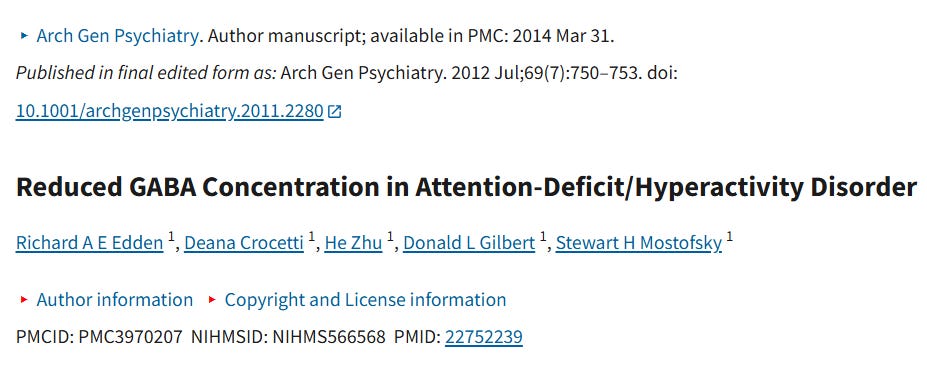
(*) Meta-analyses of magnetic resonance spectroscopy (MRS) studies show significantly reduced GABA concentrations in these exact key brain regions in individuals with active major depression, anxiety disorders, panic disorders and ASD.
GABAergic neurons and terminals are also dense in several other regions worth naming individually, since each maps onto a specific function:
The thalamus (relays sensory and motor information to the cortex).
The hypothalamus (regulates hormones, body temperature, hunger, and the stress response).
The brainstem (controls breathing, heart rate, sleep, and other vital automatic functions).
The cerebellum (coordination, balance, posture, and motor learning)
The olfactory bulb (processes smell).
The retina (processes visual information before it reaches the brain).
The superior colliculus (coordinates eye and head movements in response to visual stimuli).
The inferior colliculus (processes auditory information).
The vestibular nuclei (maintain balance and spatial orientation).
The suprachiasmatic nucleus or SCN (controls circadian rhythm and the sleep-wake cycle).
This widespread distribution is why GABA touches nearly every major aspect of brain function, not just mood and anxiety:
Attention
Memory
Movement
Sensory processing
Pain perception
Motor coordination
The sleep-wake cycle
….are all downstream of it.
In MDD for example postmortem studies show decreased expression of GAD67 and parvalbumin in prefrontal interneurons (we will talk about both of these in a moment).
Low GABA basically impairs prefrontal regulation of limbic structures, so the person has a very hard time getting out of negative thought loops.
Notably, successful antidepressant treatment (pharmacological or neuromodulatory) often normalizes cortical GABA levels, suggesting state-dependent rather than purely trait-related changes.
In schizophrenia, postmortem and imaging data also reveal deficits in GABAergic interneurons (particularly parvalbumin-positive cells that we’ll briefly talk about in a moment) and altered GAD expression, contributing to impaired gamma oscillations and sensory gating and in epilepsy, loss of inhibitory tone, whether through reduced GAD activity, interneuron loss, or chloride homeostasis disruption, lowers the seizure threshold.
Also, in generalized anxiety disorder, positron emission tomography (PET) studies demonstrate reduced benzodiazepine-binding site density in the amygdala, prefrontal cortex, and insula, consistent with diminished GABAA receptor availability and benzodiazepine sensitivity.
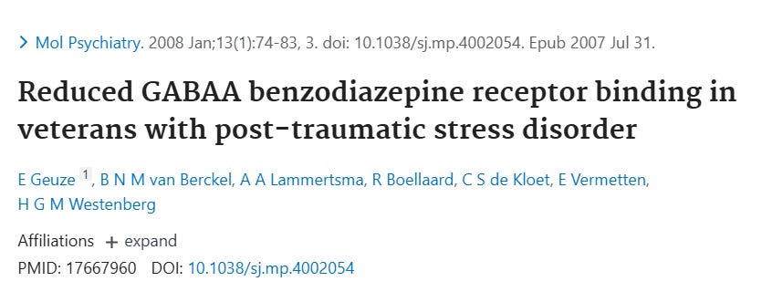
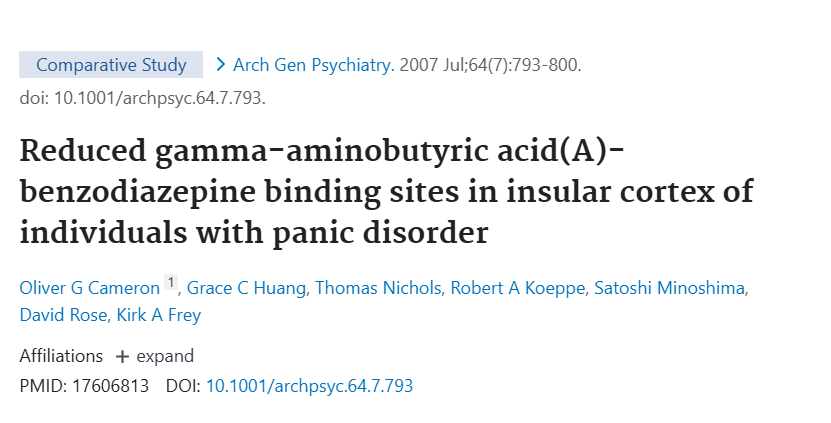
There also seems to be reduced GABAA-benzodiazepine binding sites in the insular cortex of individuals with panic disorder and PTSD.
All of these make sense even if we didn’t have any studies ont them, since dense GABAergic innervation of the amygdala, prefrontal cortex, and hippocampus enables top-down control of fear and stress responses.
So the hypo-inhibition experienced in MDD, GAD and so on, permits excessive amygdala reactivity to neutral or mildly threatening stimuli for example, producing persistent worry, autonomic hyperarousal, and even panic attacks.
At the cellular level, GABA released into the synaptic cleft binds primarily to two receptor classes:
Ionotropic GABAA receptors
These are ligand-gated ion channels (pentameric structures made of five protein subunits).
When GABA binds, they open and allow chloride ions (Cl⁻) to flow into the neuron.
In mature neurons, this causes hyperpolarization (the inside of the cell becomes more negative), making it harder for the neuron to fire.
To put a number on that: a neuron typically rests at around −70 mV.
Once chloride rushes in, the membrane potential drops further, often to around −80 mV or lower, while the threshold needed to fire an action potential sits at roughly −55 mV.
So a hyperpolarized neuron needs a much bigger excitatory push just to get back to where it can fire at all.
This is what “raising the bar” for glutamate-driven signals actually looks like in voltage terms.
They can also produce shunting inhibition, which dampens excitatory signals even without major voltage change, and by keeping the membrane well below threshold for long enough, GABA effectively prevents the voltage sensors on nearby sodium channels from activating in the first place, blocking the propagation of a signal before it can spread.
Comments
Nothing yet. Say the first thing.
Sign in to join the conversation.