
It’s George.
For most people with chronic conditions, addressing oxidative stress (OS) by supporting the Nrf2 pathway, glutathione system, mitochondrial function, and related antioxidant defenses is usually a reasonable supportive strategy.
Of course, oxidative stress is rarely the sole root cause of a chronic disease, but it frequently acts as a common downstream feature and self-perpetuating driver, amplifying inflammation and causing mitochondrial dysfunction, for example.
This is why it is a common downstream feature of many chronic diseases such as:
1. Neurodegenerative diseases
The brain is exceptionally vulnerable to oxidative stress since despite making up only about 2% of total body mass, it consumes roughly 20% of the body’s oxygen at rest.
This high oxygen demand stems from the intense metabolic activity required for maintaining neuronal membrane potentials, synaptic transmission, and axonal transport.
This creates a perfect storm for oxidative stress because:
Most of this oxygen is used in mitochondrial oxidative phosphorylation, where electron leakage naturally generates superoxide and other reactive oxygen species (ROS).
Neurons are rich in polyunsaturated fatty acids (PUFAs) such as docosahexaenoic acid (DHA) in their cell membranes.
These lipids are essential for fast signaling but extremely susceptible to lipid peroxidation by ROS.
Compared with other tissues (such as liver, muscle, or kidney), neurons possess relatively modest antioxidant capacity.
They have lower baseline levels of enzymes like catalase, rely heavily on glutathione and the thioredoxin system, and have limited ability to rapidly upregulate defenses when under sustained stress.
Together, these factors make oxidative stress a defining hallmark of neuronal vulnerability as we see in:
Alzheimer’s disease (AD)
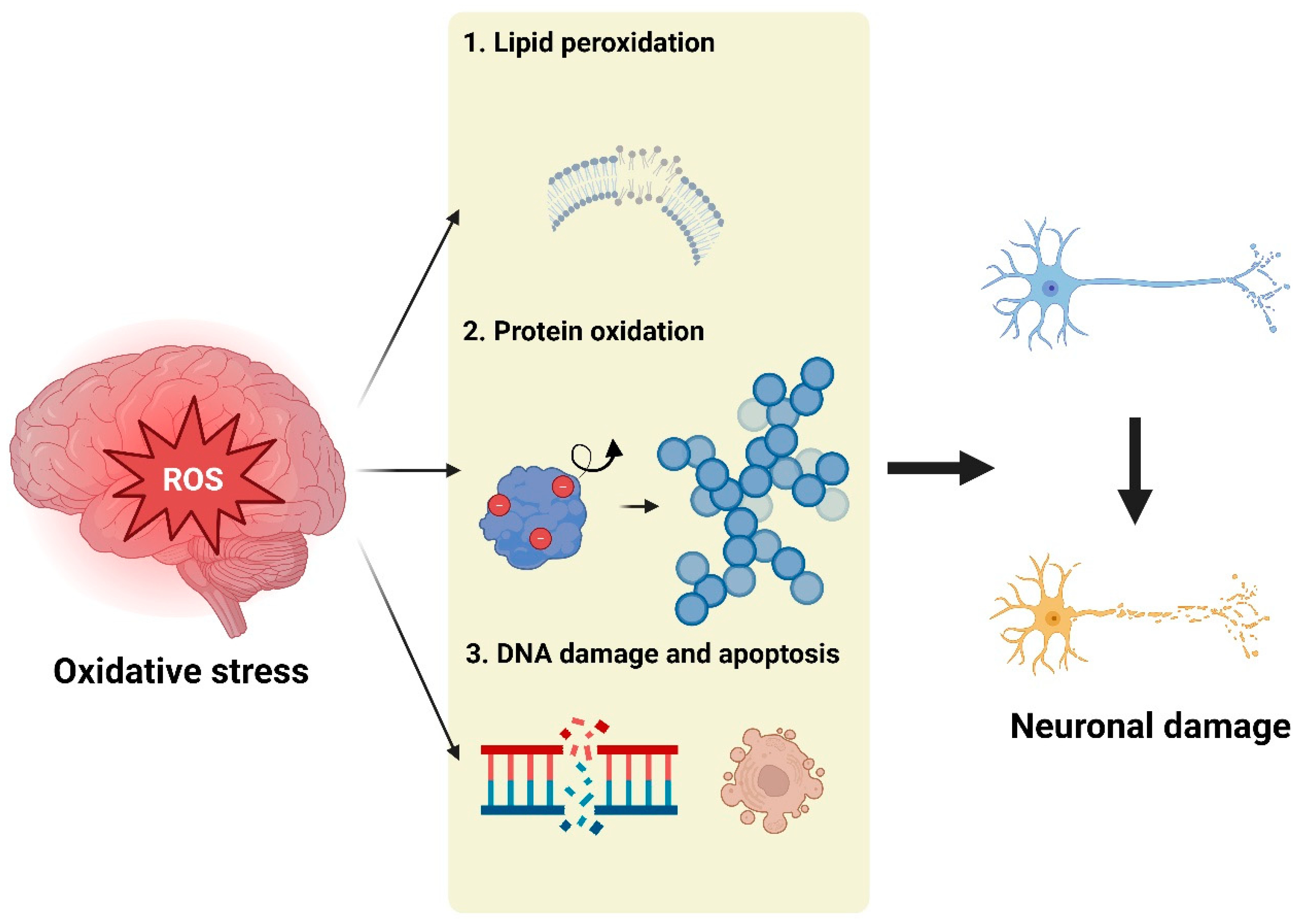
In Alzheimer’s disease, oxidative stress plays a central role in driving key pathological processes.
Reactive oxygen species (ROS) promote the clumping (oligomerization) of amyloid-β proteins and the abnormal hyperphosphorylation of tau protein.
This occurs largely through the over-activation of enzymes called kinases, particularly GSK-3β and CDK5.
At the same time, ROS produced inside mitochondria impair the cell’s ability to generate energy through oxidative phosphorylation.
This mitochondrial damage also triggers the release of cytochrome c, which activates caspases (enzymes that execute apoptosis in neurons).
Oxidative stress further weakens the blood-brain barrier by oxidizing and disrupting tight-junction proteins, allowing harmful substances to enter the brain more easily.
It also impairs the ability of microglia (the brain’s immune cells) to clear toxic amyloid-β and tau aggregates.
Also, ROS strongly activate the NF-κB pathway in microglia, leading to sustained neuroinflammation that further damages neurons and creates a self-reinforcing cycle.
Parkinson’s disease (PD)
In Parkinson’s disease, dopaminergic neurons in the brain region that produces dopamine (the substantia nigra pars compacta) are especially vulnerable to oxidative stress because:
Dopamine itself can undergo auto-oxidation and is broken down by the enzyme monoamine oxidase, both of which generate reactive oxygen species (ROS).
Mitochondrial dysfunction, particularly inhibition of Complex I in the electron transport chain, leads to increased ROS leakage.
Aggregates of the protein α-synuclein (a hallmark of PD) further damage mitochondria and promote ROS production.
Excess iron accumulation in these neurons triggers the Fenton reaction, producing highly damaging hydroxyl radicals.
When ROS react with nitric oxide, they form peroxynitrite, which nitrates key proteins such as α-synuclein and tyrosine hydroxylase (the rate-limiting enzyme in dopamine synthesis).
These modifications cause protein misfolding, loss of function, and the formation of toxic aggregates.
The result is a vicious cycle: more oxidative damage, more protein aggregation, more mitochondrial impairment, and progressive death of dopamine-producing neurons.
Then we have other examples such as Huntington’s disease where mutant huntingtin impairs mitochondrial trafficking and increases ROS, ALS, which involves SOD1 mutations or aggregates that reduce antioxidant defense and promote protein misfolding and multiple sclerosis that features OS-driven oligodendrocyte damage, lipid peroxidation of myelin, and axonal degeneration via excitotoxicity and nitrosative stress.
2. Cardiovascular diseases (CVD)
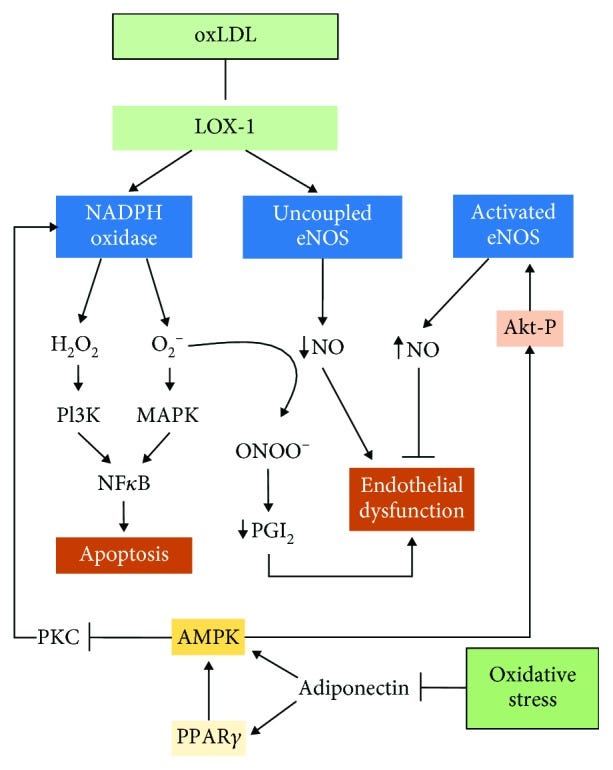
Oxidative stress is also a central and often neglected mediator of endothelial dysfunction.
In fact, it’s the earliest pathogenic step in atherosclerosis for a variety of reasons such as that:
ROS oxidize LDL particles, promoting scavenger receptor uptake by macrophages and foam cell formation.
Endothelial NADPH oxidases (NOX2, NOX4) and mitochondrial ROS activate NF-κB, upregulating adhesion molecules (VCAM-1, ICAM-1) and chemokines (MCP-1).
Superoxide reacts with nitric oxide to form peroxynitrite, reducing NO bioavailability and causing vasoconstriction.
Chronic OS also oxidizes BH4 (tetrahydrobiopterin), uncoupling eNOS and further increasing ROS.
Long-term OS promotes adverse ventricular remodeling through TGF-β–driven fibrosis and myocyte hypertrophy.
3. Metabolic disorders (type 2 diabetes, NAFLD etc)
Oxidative stress also plays a central role in the development and progression of metabolic diseases.
Here are a few basic examples.
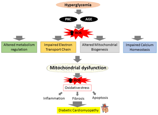
Chronic high blood sugar overloads mitochondria in cells, leading to excessive production of superoxide and other reactive oxygen species (ROS).
These ROS activate stress-sensitive kinases such as JNK and p38 MAPK.
These kinases add phosphate groups to IRS-1 (insulin receptor substrate-1) at serine residues instead of the normal tyrosine sites.
This modification blocks normal insulin signaling through the PI3K-Akt pathway, causing insulin resistance.
In the pancreas, oxidative stress damages insulin-producing β-cells, triggering their dysfunction and programmed cell death (apoptosis).
Over time, oxidative stress also drives the long-term complications of diabetes such as meuropathy, retinopathy, and nephropathy arise through multiple pathways, including the formation of advanced glycation end-products (AGEs), activation of the polyol pathway, and protein kinase C signaling (all of which are amplified by ROS).
And of course we can’t forget that in obese individuals, expanded and inflamed adipose (fat) tissue becomes a major source of ROS through overactive NADPH oxidases (NOX) and mitochondrial dysfunction within fat cells.
4. Cancer
Oxidative stress has a dual, context-dependent role in cancer.
Ιt can both promote tumor development and be harnessed to kill cancer cells.
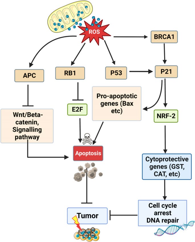
At moderate levels, ROS act as signaling molecules that drive many of the hallmarks of cancer since they cause direct DNA damage, including base modifications (such as 8-oxoguanine), strand breaks, and chromosomal instability.
This increases mutation rates in key oncogenes such as KRAS and MYC and tumor suppressor genes such as TP53.
ROS also activate several pro-survival and pro-tumorigenic pathways such as:
NF-κB, which promotes inflammation, cell survival, and resistance to apoptosis.
HIF-1α which drives angiogenesis and metabolic reprogramming (think the Warburg effect).
PI3K/Akt/mTOR pathway, which enhances cell proliferation, growth, and survival while inhibiting apoptosis.
In addition, ROS facilitate epithelial-to-mesenchymal transition (EMT), which increases cancer cell invasion and metastasis.
Many tumors basically adapt to high oxidative stress by upregulating antioxidant systems (particularly the Nrf2 pathway) to protect themselves from their own elevated ROS levels.
Now on the other hand, when ROS levels become excessively high, they can overwhelm the antioxidant capacity of cancer cells and trigger cell death.
This is the basis for many standard cancer treatments (and to be fair one of the tools we utilize for dealing with pathogen overgrowths as well).
5. Inflammatory and autoimmune conditions
Now over here, oxidative stress is both a consequence and an amplifier of chronic inflammation, creating self-sustaining cycles that drive tissue destruction in many autoimmune and inflammatory diseases.
In rheumatoid arthritis for example, oxidative stress is markedly elevated in the synovial fluid of affected joints.
Reactive oxygen species (ROS) and peroxynitrite directly damage cartilage by oxidizing and degrading proteoglycans and type II collagen, leading to loss of joint integrity.
They also activate the transcription factor NF-κB in synovial fibroblasts and immune cells, resulting in increased production of pro-inflammatory cytokines such as TNF-α, IL-6, and IL-1β.
This inflammatory milieu further stimulates ROS generation.
Additionally, oxidative stress promotes the differentiation and activation of osteoclasts (bone-resorbing cells) through RANKL signaling and NF-κB pathways, contributing to the characteristic bone erosions seen in RA.
Or in Crohn’s disease and ulcerative colitis for example, activated neutrophils in the intestinal mucosa generate large amounts of ROS through NADPH oxidase and produce hypochlorous acid via myeloperoxidase.
This results in damaged epithelial cell membranes, tight junction proteins (such as occludin and ZO-1), and the mucus layer, leading to increased intestinal permeability.
The resulting barrier dysfunction allows bacterial products like endotoxins to enter the tissue, further activating NF-κB and NLRP3 inflammasome pathways.
So there’s once again a vicious cycle of inflammation, oxidative damage, and impaired mucosal healing.
Another example is psoriasis where oxidative stress in the skin drives excessive keratinocyte proliferation.
ROS basically activate redox-sensitive transcription factors such as NF-κB and STAT3, which upregulate genes involved in cell survival and proliferation.
Lipid peroxidation products also stimulate the release of pro-inflammatory cytokines and chemokines that recruit T cells and dendritic cells, sustaining the plaques and chronic inflammation.
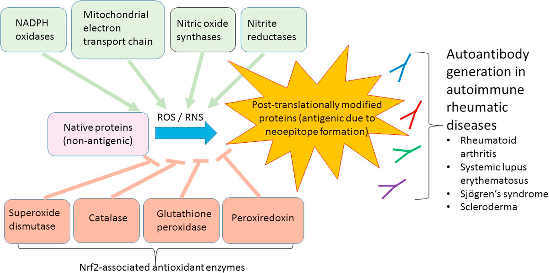
There are of course more examples of diseases that can demonstrate the impact of oxidative stress.
Even in eye diseases for example.
The eye is also particularly susceptible to oxidative stress due to its high oxygen consumption, constant exposure to light (which generates ROS), and presence of easily peroxidizable lipids.
In cataracts, oxidative stress causes direct oxidation of lens crystallin proteins, leading to disulfide bond formation, protein unfolding, and aggregation.
This results in lens opacification.
Lipid peroxidation of lens fiber cell membranes and depletion of lens antioxidants (especially glutathione) accelerate this process.
Or in age-related macular degeneration, in the retina, mitochondrial ROS and light-induced ROS cause extensive lipid peroxidation of photoreceptor outer segment membranes, which are extremely rich in docosahexaenoic acid (DHA).
This damage leads to the accumulation of toxic byproducts such as lipofuscin and contributes to drusen formation beneath the retinal pigment epithelium.
Chronic oxidative stress also activates the complement system and promotes chronic inflammation in the macula, driving progression from dry to wet AMD.
Oxidative stress is even a major driver of damage and progression in kidney disease for example.
ROS cause endothelial dysfunction in renal blood vessels by reducing nitric oxide bioavailability via peroxynitrite formation and increasing expression of adhesion molecules for example.
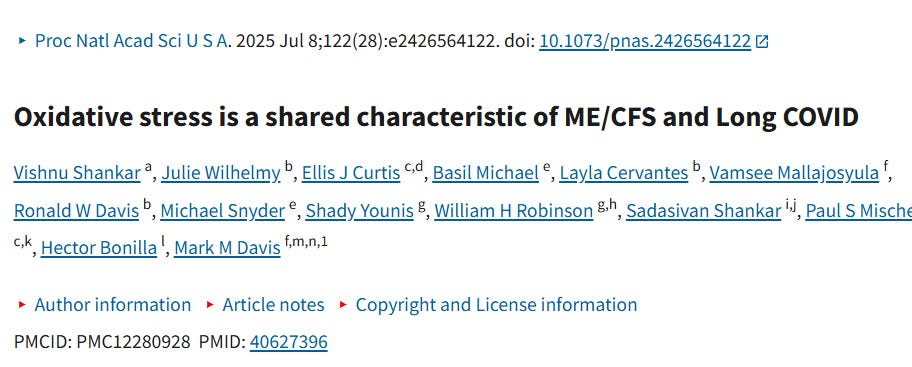
Other classic examples include reproductive disorders such as PCOS, ME/CFS, long Covid, depression, anxiety and schizophrenia.
Now oxidative stress is an imbalance where reactive oxygen species (ROS) and reactive nitrogen species (RNS) overwhelm antioxidant defenses.
It disrupts redox signaling, which is the process by which cells use controlled, reversible changes in the oxidation-reduction (redox) state of molecules to transmit signals and regulate biological functions.
It is a fundamental form of cellular communication, analogous to phosphorylation (adding phosphate groups) but based on electron transfer instead.
Oxidation = loss of electrons and reduction = gain of electrons.
Molecules that readily accept or donate electrons (ROS like hydrogen peroxide (H₂O₂), or antioxidants like glutathione) act as signaling messengers.
Low-to-moderate levels of reactive oxygen species (ROS) and reactive nitrogen species (RNS) are not just damaging “byproducts” but deliberate signaling molecules when produced in a controlled, localized, and transient way.
But oxidative stress is excessive, uncontrolled ROS that causes damage (lipid peroxidation, protein carbonylation, DNA breaks) while redox signaling is the physiological, regulated use of ROS at low concentrations for normal cell function.
Now the “bad guys” in this discussion about oxidative stress are the following (the ROS and RNS):
Superoxide (O₂⁻)
Hydrogen peroxide (H₂O₂)
Hydroxyl radical (OH)
Peroxynitrite (ONOO⁻)
Lipid peroxides
Comments
Nothing yet. Say the first thing.
Sign in to join the conversation.