
It’s George.
When we are trying to resolve a problem and get stuck after making some progress, what usually helps us is revisiting the “boring” basics we foolishly believe we’ve mastered and are above.
Even though it seems dumb when we realize it, we often overlook the key fundamentals that helped us experience progress in the first place.
For example, if you have symptoms such as:
Anxiety
Chronic pain
Restless mind
Cannot turn your brain off when you want to relax
Disorganized attention or inability to focus
Worry too much about things you should not
Gets overwhelmed easily
Depression
OCD
Complex motor stereotypies (CMS)
Migraine
Persistent symptoms after a traumatic brain injury
Seizures
Symptoms of PANDAS/PANS
Cannot handle folate supplements
Cannot handle L glutamine supplements
Experience massive benefits when it comes to your mental health and liver profile from P5P supplements
Then you probably have to look into the fundamental topic of glutamate.
Of course, every single one of these can have multiple drivers but let’s take chronic pain as an example.
While acute pain serves as a protective warning signal, excess glutamate shifts the entire nervous system from a state of normal transmission to a state of pathological hyper-reactivity across three interconnected levels:
The spinal cord
The brain’s cortex
Supporting glial cells
At the spinal level, specifically within the dorsal horn, glutamate acts as the critical gatekeeper for sensory signals entering the central nervous system.
During a brief or acute painful stimulus, presynaptic pain-sensing fibers release a controlled amount of glutamate.
This glutamate binds primarily to baseline, low-affinity AMPA receptors on the postsynaptic spinal neuron.
This triggers a rapid, fleeting electrical signal that alerts the brain to localized tissue damage.
However, when an injury is severe, poorly managed, or persistent, these presynaptic fibers enter a state of continuous, high-frequency firing.
This causes a massive, unremitting flood of glutamate into the synaptic cleft, overwhelming the baseline AMPA receptors.
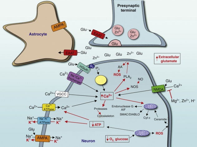
Under normal conditions, neighboring NMDA receptors remain dormant and silent because their ion channel pores are physically plugged by a protective magnesium ion.
As the excessive glutamate continuously stimulates the AMPA receptors, it causes a prolonged, severe electrical depolarization of the postsynaptic membrane.
This buildup of positive electrical charge acts like a magnet in reverse, electrostatically repelling and dislodging the protective plug from the NMDA receptor channel.
With the magnesium plug removed, the NMDA channel swings wide open.
This allows an unprecedented, massive influx of calcium to pour directly into the postsynaptic spinal cell. This calcium influx triggers an immediate intracellular emergency cascade:
It activates downstream protein kinases (such as PKC and PKA).
These enzymes phosphorylate the NMDA receptors, making them even more sensitive to glutamate and keeping them open longer.
So there’s a progressive increase in spinal cord excitability where subsequent pain signals are amplified exponentially.
The intracellular calcium surge does not stop at receptor activation.
It travels deep into the cell nucleus, activating transcription factors (like CREB) that alter gene expression.
The neuron begins manufacturing more pain receptors and inserting them into the synapse.
This permanently lowers the threshold for activation, meaning normal, harmless touch is suddenly misread as pain (allodynia) and mild discomfort feels excruciating (hyperalgesia).
Once these amplified, unchecked signals escape the spinal gate, they reach the brain, where glutamate drives the cortex-centric pain network to physically, functionally, and permanently reorganize.
The same NMDA-mediated calcium influx that hyper-sensitizes the spine occurs within the cerebral cortex (including the somatosensory cortex, insula, and anterior cingulate cortex).
Glutamate acts as the primary architect of this maladaptive neuroplasticity.
Essentially, neural pathways and brain circuits dedicated to processing pain undergo a structural widening.
Synapses are strengthened, dendrites branch out, and structural pathways become wider, stronger, and permanently open.
The brain effectively “learns” and memorizes the pain, creating a persistent neural echo that continues to generate the sensation of pain long after the original peripheral tissue damage has fully healed.
Also when cortical glutamate remains high, it spills out of the synapse and becomes toxic (excitotoxicity), damaging local inhibitory (GABA) interneurons that would normally quiet the system. Simultaneously, excess glutamate activates neighboring glial cells (astrocytes and microglia).
Instead of cleaning up the neurotransmitter, these inflamed glial cells release pro-inflammatory cytokines, which circle back to the neurons and force them to pump out even more glutamate.
This locks the brain into a self-perpetuating, hyper-excitable loop of chronic pain, emotional distress, and cognitive fatigue.
This precise cascade of glutamate-driven hypersensitivity is exactly why pregabalin is one of the common pharmaceutical treatments prescribed for chronic pain.
It binds tightly to the α₂δ (α₂-δ) subunit of these voltage-gated calcium channels.
By binding here, it restricts calcium from flooding into the presynaptic nerve terminal and without the calcium trigger, the neuron cannot release excessive amounts of excitatory neurotransmitters, effectively starving the synapse of excess glutamate.
But i probably digress.
We will break down how glutamate can lead to specific problems later in this article.
So let’s back up a bit and start from the beginning.
Glutamate (L-glutamic acid) is one of the twenty standard proteinogenic amino acids and the central nervous system’s predominant excitatory neurotransmitter that mediates the vast majority of fast (*) synaptic transmission in the mammalian central nervous system and underpins critical processes including:
Learning and memory (via LTP at glutamatergic synapses)
Cortical computation in general
Sensory processing
Motor coordination
(*) Fast means ionotropic receptors (ligand-gated ion channels) that produce rapid depolarization or hyperpolarization on the order of milliseconds.
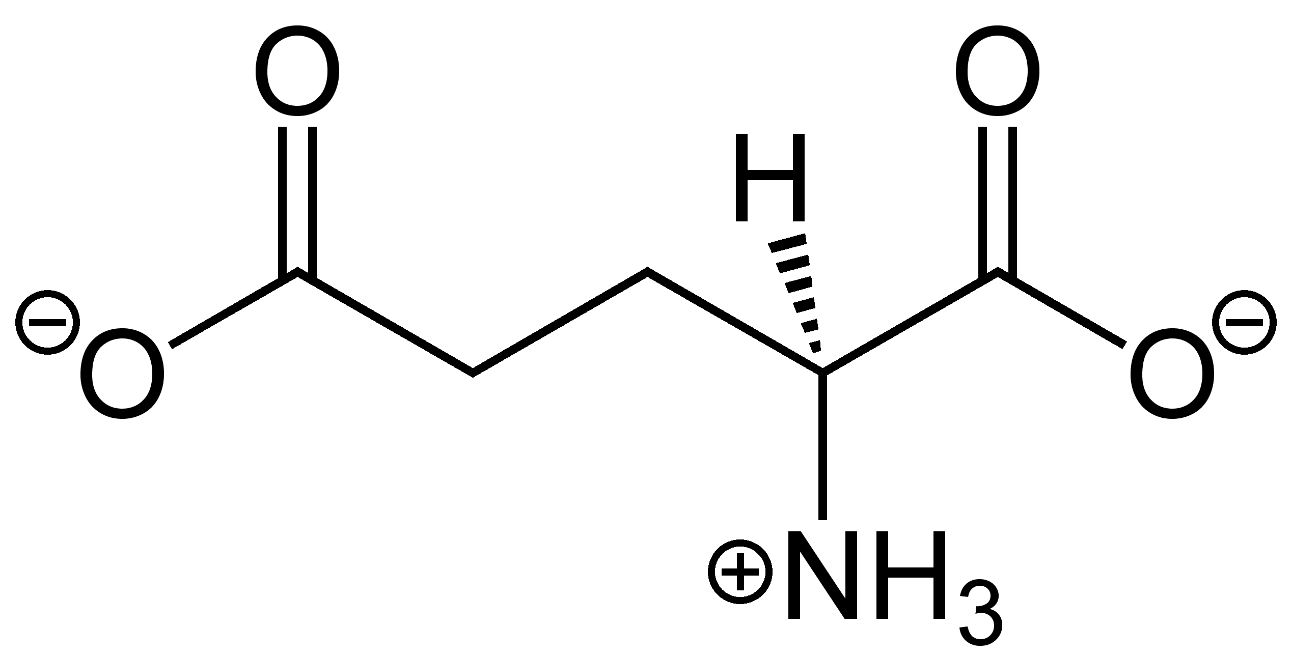
Glutamate is the dominant driver of the “go” signals that allow neurons to fire action potentials and propagate information quickly across circuits.
In the cortex, hippocampus, striatum, and most other forebrain regions, the great majority of excitatory synapses use glutamate.
From various anatomical and electrophysiological studies we estimate that glutamatergic synapses are 80–95% of all excitatory synapses in the mammalian brain (GABAergic synapses account for most of the remaining (inhibitory) fast transmission, creating the essential excitation-inhibition (E/I) balance).
It is a non-essential amino acid, meaning the body can synthesize it endogenously, but its importance extends far beyond neurotransmission.
Glutamate is integral to:
Nitrogen metabolism
Energy production via the tricarboxylic acid (TCA) cycle
Antioxidant defense through glutathione synthesis
Intercellular communication in both neural and peripheral tissues
Now before we move to discussing how glutamate impacts our bodies in detail, we must present a brief overview of how glutamate is produced, transported and regulated in the body.
As it was stated, glutamate is a non-essential amino acid synthesized endogenously in multiple tissues, with the brain having the most tightly regulated pools.
In the brain (CNS), there are two main ways to make glutamate.
The primary route is the glutamate–glutamine cycle.
This is the recycling system that supplies most of the glutamate used for normal day-to-day brain signaling.
Here’s the step-by-step loop:
1. Release & uptake
A neuron fires and releases glutamate into the synaptic cleft (the tiny gap between neurons).
This glutamate binds to receptors on the receiving neuron and transmits the excitatory signal.
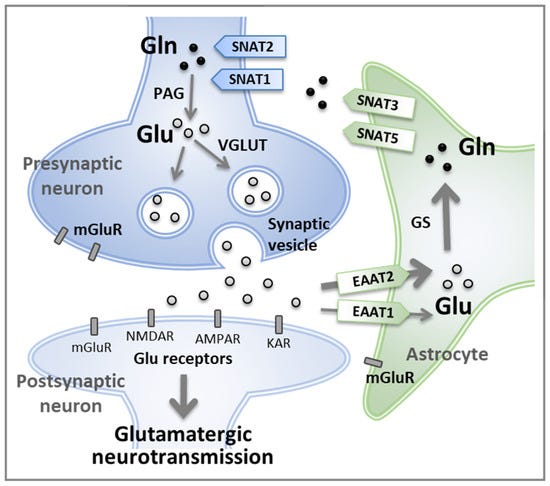
Immediately after, within milliseconds, nearby astrocytes remove most of the glutamate from the extracellular space using two high-affinity transporters:
EAAT1 (GLAST) that is more prominent in the cerebellum and developing brain.
EAAT2 (GLT-1) that is responsible for ~90% of glutamate uptake in most adult brain regions.
These transporters are sodium-dependent: they co-transport 3 Na⁺ ions + 1 H⁺ with each glutamate molecule, while counter-transporting 1 K⁺ ion.
This uses the sodium gradient (maintained by Na⁺/K⁺-ATPase) as the driving force.
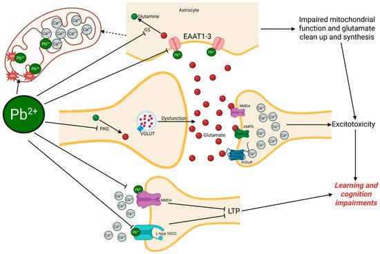
As a side note, these transporters are very sensitive to oxidative damage, inflammation, and energy failure/mitochondrial dysfunction.
Some diseases linked to step 1 impairment include:
Alzheimer’s that reduces EAAT2 expression and function.
ALS in which there’s a massive loss of EAAT2 in motor cortex and spinal cord.
Stroke / Ischemia where energy failure reverses the transporters (they start pumping glutamate out instead of in).
Epilepsy where reduced EAAT2 leads to seizure-prone networks.
Depression downregulates EAAT2 (mainly via glucocorticoids).
This first step is critical because glutamate is extremely potent.
If it stays in the synapse too long, it keeps stimulating receptors → over-excitation → excitotoxicity (cell damage or death).
Astrocytes act like a rapid cleanup crew, keeping extracellular glutamate at very low, safe levels (nanomolar range) which brings us to the next step.
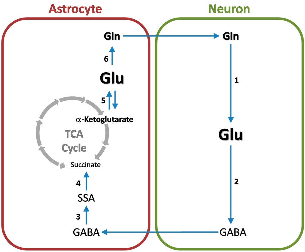
2. Conversion in astrocytes
Inside the astrocyte the reaction is: Glutamate + Ammonia (NH₄⁺) + ATP → Glutamine
This happens with the help of the enzyme glutamine synthetase (also called GS) and is one of the most important reactions in the entire brain.
As we stated, glutamate is the brain’s main accelerator.
When it stays outside neurons too long, it keeps firing receptors (especially NMDA (which we will break down in a moment)), causing excessive calcium entry.
This leads to:
Over-excitation (racing thoughts, anxiety, sensory overload)
Excitotoxicity (cell damage or death)
Astrocytes must remove it quickly.
But simply “sucking it up” isn’t enough, they must neutralize it.
This is where glutamine comes into play as the safe “shuttle” form since it is electrically neutral and much less reactive, it can be safely transported between cells without stimulating receptors and overall acts like a “disarmed” version of glutamate that neurons can re-arm later when needed.
This conversion is the brain’s way of saying: “We captured the dangerous signal — now let’s store it safely.”
Also, every time neurons use glutamate, some ammonia (NH₄⁺) is produced as a byproduct which is highly toxic to neurons and even small amounts can disrupt pH balance, cause swelling, and impair mitochondrial function.
Glutamine synthetase traps this ammonia by attaching it to glutamate, forming glutamine.
This is a major way the brain prevents ammonia toxicity (similar to how the liver uses the urea cycle in the body).
“This step sounds like it needs some ATP”.
Yes, you are right. But the brain is willing to spend energy here because keeping glutamate under control/this overall “cleanup” is more important than saving that ATP for other tasks.
This is also partly why step 2 is generally the most vulnerable point in the entire Glutamate–Glutamine Cycle.
Glutamine synthetase uses ATP directly so any drop in cellular energy (mitochondrial dysfunction, low magnesium, hypoxia) hits it hard.
Overall it is easily damaged by:
Oxidative stress / ROS
Nitrosative stress
Inflammatory cytokines
Heavy metals
Ammonia overload (ironically, the very thing it’s supposed to detoxify)
Chronic high cortisol / glucocorticoids (besides EAAT transporters, they also downregulate glutamine synthetase)
Vitamin B6 (P5P) deficiency (indirectly affects related pathways and astrocyte health)
Zinc deficiency (glutamine synthetase is zinc-dependent)
Low protein or specific amino acid imbalances
Hyperammonemia (liver dysfunction, urea cycle disorders)
Ketosis or severe metabolic acidosis (in some contexts)
Hyperglycemia / advanced glycation end-products (AGEs)
Hypothyroidism (reduces overall metabolic rate)
Chronic sleep deprivation (impairs astrocyte function and glymphatic clearance)
Excessive screen time / blue light at night (disrupts circadian regulation of astrocytes)
Sedentary lifestyle
High intake of free glutamate + poor overall diet
Dehydration
But let’s move to the next step which is:
3. Export to neurons
Once the astrocyte has converted glutamate into glutamine, it must deliver this safe precursor back to the neuron so the neuron can regenerate glutamate for future neurotransmission.
The astrocyte exports glutamine into the extracellular space using specialized neutral amino acid transporters on its membrane, primarily SN1 and SN2 (also known as System N transporters).
These transporters are sodium- and pH-dependent, allowing precise regulation based on local metabolic conditions.
Nearby neurons then take up the glutamine via their own transporters, mainly SAT1 and SAT2 (System A transporters).
This step is also quite important since glutamate is dangerous if it accumulates outside cells but glutamine, by contrast, is biologically inert as a neurotransmitter meaning it does not activate glutamate receptors.
By converting glutamate to glutamine inside the astrocyte, the brain transforms a potentially toxic molecule into a safe, neutral shuttle.
This continuous recycling allows high-frequency signaling without flooding the brain with toxic glutamate.
And the final step is:
4. Conversion back to glutamate in neurons
Inside the neuron (mainly in mitochondria):
Inside the neuron (primarily in the mitochondrial matrix) the following reaction happens: Glutamine + H₂O → Glutamate + Ammonia (NH₄⁺) with the help of the enzyme phosphate-activated glutaminase (PAG, also called GLS or GLS1 in humans where PAG basically cleaves the amide group from glutamine, releasing free glutamate that the neuron can use immediately.
This step happens in mitochondria because well, PAG is located on the inner mitochondrial membrane and in the matrix.
PAG is tightly controlled so the neuron doesn’t produce excess glutamate:
Higher inorganic phosphate (a sign of energy demand) activates PAG.
Glutamate itself inhibits PAG.
It’s sensitive to local pH and ammonia/ion levels.
There are two main forms, GLS1 (kidney-type, widespread in brain) and GLS2 (liver-type). GLS1 is the dominant one in neurons for neurotransmission.
Levels of PAG can increase under high activity or stress.
So this regulation ensures glutamate is produced “on demand” rather than in constant excess.
Now when a neuron gets this glutamate, most of it is transported into synaptic vesicles via vGluT1–3 transporters (using a proton gradient).
Some is used locally in the mitochondria (converted to α-ketoglutarate for the TCA cycle to make ATP) and a portion can be converted into GABA (in inhibitory neurons) via the GAD enzyme.
This final step is important since without PAG, neurons would run out of glutamate during sustained activity/it sustains overall neurotransmission and allows rapid adjustment of glutamate levels based on demand for example.
But balance is once again critical since too much PAG activity:
Can lead to excessive glutamate buildup → higher release → risk of excitotoxicity.
Is seen in some epilepsy models and under chronic stress.
And too little PAG activity:
Causes weakened excitatory signaling.
Contributes to cognitive slowing, anhedonia, or negative symptoms in schizophrenia (NMDA hypofunction).
Now this was the primary route, but we also have de novo synthesis.
The glutamate-glutamine cycle (recycling) is very efficient for normal, everyday brain activity.
However, during intense mental periods such as learning something new, solving complex problems, high stress, or sustained mental effort, the demand for glutamate exceeds what recycling can provide.
The brain then switches on de novo synthesis: it builds fresh glutamate using glucose as the starting material.
It’s a perfect example of how energy metabolism and neurotransmitter production are directly linked.
Comments
Nothing yet. Say the first thing.
Sign in to join the conversation.