
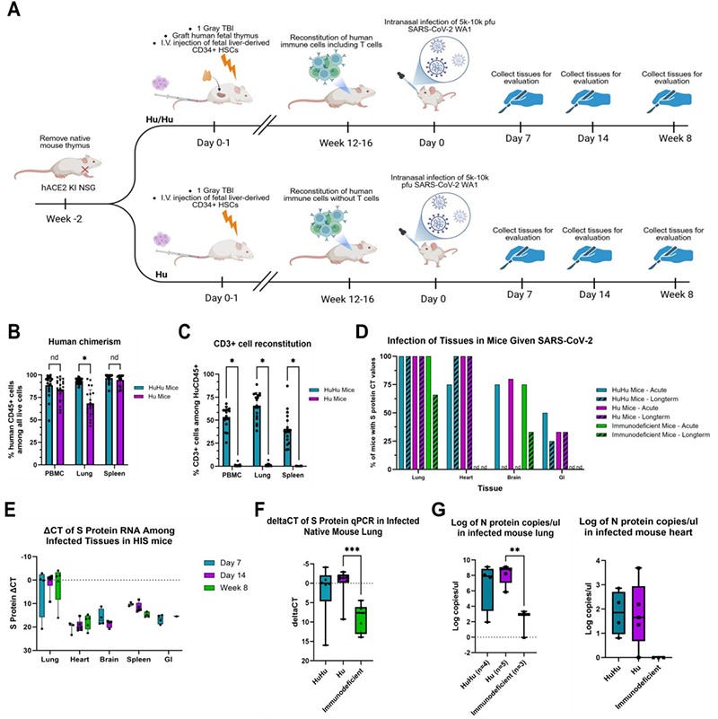
The presence of human immune cells promotes the dissemination of SARS-CoV-2 and increases viral levels in mouse lung.
a. Schematic of hACE2 KI NSG HIS mouse infection study. A subset of mice also expressed an HLA-A2 transgene. Homozygous hACE2 KI NSG mice were thymectomized and reconstituted with either fetal liver CD34+ HSCs (2x10⁵ cells) alone or both HSCs and a human fetal thymus graft under the kidney capsule or left unreconstituted. After 3-4 months to allow human immune cell reconstitution, mice were intranasally infected with the WA1 strain of SARS-CoV-2 (5x10³ pfu) or mock infected with PBS. Tissues were harvested at 7, 14 days, and 8 weeks post-infection. b,c. Reconstitution of human CD45+ cells (b) and human CD3+ T cells (c) in PBMC, lung, and spleen of HIS hACE2 mice at 16-20 weeks post-transplantation, as determined by flow cytometry. Hu/Hu n = 19, Hu n = 21. All displayed p-values < 0.0001. d. Percentage of mice from each infected group demonstrating SARS-CoV-2 spike protein-specific qPCR CT values across multiple tissues. PBS control mice (not shown) did not exhibit spike protein-specific CT values. nd denotes tissues in which spike protein-specific CT values were not detected. e. ΔCT values of spike-protein specific qPCR normalized to mouse GAPDH across multiple tissues and both timepoints. Box and whisker plots show min to max values. Individual values for each mouse are shown. Day 7 n = 3-4, Day 14 n = 4-6, Week 8 n = 1-5. f. Levels of viral spike protein RNA detected in the native mouse lung for all infected mice across all timepoints. ΔCT values of SARS-CoV-2 spike protein-specific qPCR were determined by normalizing the spike protein values against mouse GAPDH. Welch’s ANOVA test p-value = 0.0014, with an adjusted p-value = 0.005 between Hu and immunodeficient groups. Individual values for each mouse are shown. Hu/Hu n = 6, Hu n = 9, Immunodeficient n = 7. g. Log copies per microliter in RNA isolated from lung (left) and heart (right) tissue from nucleocapsid protein-specific qPCR. Copy numbers determined using a standard curve from provided nucleocapsid standards. Box and whisker plots show min to max values with adjusted p-values determined by one-way ANOVA. Individual values for each mouse are shown. Lung: Hu/Hu n = 4, Hu n = 5, Immunodeficient n = 3. One-way ANOVA p-value = 0.0142. Heart: Hu/Hu n = 4, Hu n = 5, Immunodeficient n = 3.
The new study reports that human immune cells can promote the dissemination of SARS-CoV-2 beyond the respiratory tract, with viral RNA persisting in multiple organs for weeks. Importantly, immune activation was also observed in tissues including the heart, intestine and brain.
The obvious question is:
How is the virus—or persistent viral material (Spike)—getting there?
One possibility deserves much attention: Could macrophages be acting as cellular Trojan horses?
This idea is not entirely new. In fact, a paper was written in 2021 discussing this very idea.
In conclusion, MN (monocytes) and MDM (monocyte-derived macrophages) are pivotal innate immune cells with a crucial role in defensive activities against various antimicrobial agents such as viruses. Besides this beneficial activity, they can exert unfavorable effects, extending viral infection within the body. However, their role has not yet been definitely established. Here, we showed that SARS-CoV-2 infects MN and MDM without production of the infectious virus but preserving infectivity. Therefore, these cells may act as spreaders for the virus in different body districts as a “Trojan horse”.
Macrophages and Monocytes: “Trojan Horses” in COVID-19
https://pmc.ncbi.nlm.nih.gov/articles/PMC8624282/
What the new paper shows is that macrophage dissemination is dependent on ACE2 expression!
We have observed persistent viral RNA and protein across multiple organs for up to 8 weeks along with inflammation and features of PASC. We have demonstrated that human immune cells promote the dissemination of virus to extrapulmonary organs and increase the level of pulmonary infection. Persistent viral RNA was associated with strong, long-lasting anti-viral immune responses in the heart while clearance of virus over time resolved inflammation in the brain. Our model uniquely recapitulates both prolonged viral persistence signatures and a human adaptive immune response in the context of physiologic hACE2 expression, providing new opportunities to explore PASC pathogenesis and evaluate therapeutic interventions targeting the virus-immune interface.
A novel model demonstrating that human immune cells promote multiorgan SARS-CoV-2 dissemination and human T cells limit anti-viral innate immunity
https://www.biorxiv.org/content/10.64898/2026.06.18.733232v1.full
And the presence of these SARS RNA-laden (Spike) macrophages are of the Smoldering variety.
To further investigate the transcriptional response to infection, we performed pseudobulk differential expression analysis and identified alveolar macrophages as the primary responder to infection, with 217 genes differentially expressed between infected and uninfected mice (Fig. 9c). Within alveolar macrophages, complement (Cfb), chemokines (CXCL2), and stress response genes (Mt1) were upregulated in response to infection, with significant transcriptional upregulation of NFkB1 (padj < 10-22) and PPARG (padj < 10-21), indicating a strong murine inflammatory macrophage response to infection
A novel model demonstrating that human immune cells promote multiorgan SARS-CoV-2 dissemination and human T cells limit anti-viral innate immunity
https://www.biorxiv.org/content/10.64898/2026.06.18.733232v1.full
So, there is the Spike-induced loop:
Spike enters/associates with monocytes → monocytes traffic to tissues → differentiate into macrophages → macrophages attempt to respond and repair → persistent Spike/inflammatory signaling prevents resolution → macrophages remain activated → chronic repair/remodeling continues → SMS.
How can we be confident in this assessment? Because the Spike abides – in macrophages.
Recent evidence suggests that immune cell persistence and reprogramming play a central role in these non-organ damaged related syndromes. Patterson et al. found that the SARS-CoV-2 S1 spike protein persisted in CD16+ monocytes up to 15 months post-infection in individuals with Long COVID, despite the absence of ongoing viral replication [7]. Complementary work by Simonis et al. revealed that mRNA vaccination induced durable epigenetic reprogramming in monocyte-derived macrophages, characterized by H3K27ac modifications and elevated IL-1β, TNF-α, and other inflammatory cytokines [8]. These findings demonstrate that spike protein exposure, whether through infection or vaccination, can lead to long-lasting macrophage activation [9].
The Central Role of Macrophages in Long COVID Pathophysiology
https://www.preprints.org/manuscript/202511.0998
The macrophage comes to heal—but it arrives altered. It contains Spike. It encounters a tissue that still carries the molecular signature of the original injury. And instead of completing the healing process and switching off, it remains activated. It continues to repair. It continues to remodel. It continues to recruit. It continues to signal. It overheals. And the healing response itself becomes pathological.
Smoldering Macrophage Syndrome.
Thank you, as always, for keeping me going. I appreciate all of your support and am grateful to everyone in our community. Please have a blessed week.
One-Time Donation
https://buy.stripe.com/6oUbJ0auFaWkcLs2ZXeUU00
No posts
Comments
Nothing yet. Say the first thing.
Sign in to join the conversation.