
Some people take thiamine correctly and don’t feel what other people describe.
Their blood-work comes back normal. Their B-complex is solid and their diet is reasonable. By every textbook measure, they are not deficient. But the fatigue and the post-meal heaviness don’t really change.
They are doing the thiamine work properly, by every protocol the literature describes, and they are getting back perhaps a third of the response other people report.
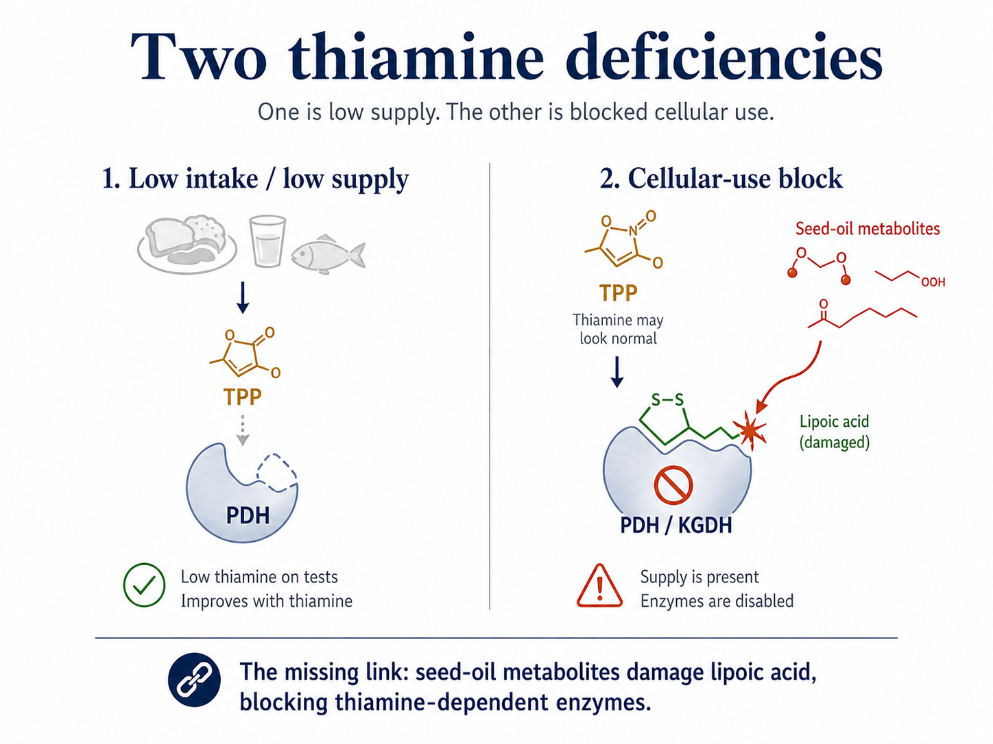
The first is one that medicine has been treating since beriberi was characterized. It is a problem of inadequate intake, malabsorption, alcoholism and food devoid of nutrition. The body isn’t getting enough thiamine, the deficiency is detectable on standard tests, and supplementation reliably fixes it. This is the deficiency every textbook describes and every writer talks about.
The second deficiency is a problem of cellular use rather than intake. The cells are depleted because something inside the cell is destroying thiamine as fast as the supply can replace it, and, more critically, disabling the enzymes thiamine depends on, regardless of how much thiamine is present.
That something has been sitting in plain sight for decades. It is the seed oil that fills adipose tissue, gets stored in cell membranes, and accumulates over years of modern diets. The molecular link between seed oils and thiamine status has never been described as a unified picture in mainstream nutritional medicine.
It exists in the biochemistry literature, scattered across three unrelated fields. Mitochondrial enzymology, lipid peroxidation chemistry and salmon ecology. Each field carries part of the answer yet very few have asked the full question.
The answer runs through a single molecule attached to two enzymes, attacked by a class of metabolites from seed oils.
For some readers, applying the metabolic advice to your specific case (your phenotype, your labs, your history, your medications, your dietary baseline, your life situation) is a lot of work, and you’d rather have someone with experience do that work with you.
The Comprehensive Metabolic Analysis is the coaching product I run. It is an analysis of your metabolic state, your bioenergetic state, and the upstream patterns that produce whatever you’re presenting with.
You complete a comprehensive intake covering your case, and I produce an in-depth written analysis specific to you, with your phenotype identified, the mechanism story behind your pattern, your levers ranked, a phased implementation roadmap, a supplement protocol matched to your phenotype, a curated reading list, tracking templates, and a 30-day follow-up to check in on implementation.
Pyruvate dehydrogenase (PDH) converts pyruvate into acetyl-CoA at the entrance of the TCA cycle. It is the gate between burning sugar through glycolysis and burning it cleanly through the mitochondria. When PDH works, carbohydrates feel clean. When it doesn’t, fatigue and brain fog follow food.
Most discussions of PDH focus on thiamine. Thiamine pyrophosphate (TPP), the active form of vitamin B1, sits inside the enzyme and handles the first catalytic step. Without TPP, PDH cannot do its job. Low thiamine means a broken PDH, which leads to energy collapse and various conditions with fatigue.
But PDH has a second cofactor. A small sulfur-containing molecule called lipoic acid, attached to the enzyme itself.
Lipoic acid is what physically moves the reaction forward. TPP starts the catalysis, but lipoic acid carries the substrate to the next step. If lipoic acid is damaged, the enzyme stalls even with perfect TPP, magnesium, and every other cofactor in place.
This is where the second thiamine deficiency exists. A cell can show normal serum thiamine and still have PDH activity that is measurably impaired, because the problem is not the substrate. Instead, the problem is the cofactor next to it.
Two other major thiamine-dependent enzymes; alpha-ketoglutarate dehydrogenase (KGDH) inside the TCA cycle, and branched-chain α-ketoacid dehydrogenase (BCKDH) for amino acid breakdown, are built the same way. Same TPP requirement with the same lipoic acid vulnerability.
If something can damage lipoic acid, it can shut off three of the most important thiamine-dependent enzymes in the body at once, regardless of how much thiamine is available.
That something is what we’ve missed.
The polyunsaturated fatty acids (PUFA) in seed oils; primarily linoleic acid in soybean, corn, sunflower, safflower, cottonseed, and canola, are absorbed, transported, and stored. Some go into adipose tissue as triglycerides, some incorporated into cell membranes. The composition of stored fat and cell membranes is, over years, a record of your dietary fat intake.
The half-life of linoleic acid in human adipose tissue is roughly 600–700 days. A person minimizing seed oils today is still mobilizing the oils they ate three years ago. Tissue composition turns over slowly and won’t show huge day-to-day benefits.
The structural feature that makes PUFA “essential” (its multiple carbon-carbon double bonds) is also the feature that makes it chemically unstable. Each double bond is a site where free radicals can abstract a hydrogen and start a chain reaction called lipid peroxidation. The more double bonds in a fatty acid, the more vulnerable it is. Linoleic acid has two, arachidonic acid has four, DHA has six, saturated fats have zero, which is why butter on the counter doesn’t go rancid the way fish oil does.
When PUFA peroxidizes inside cells, it fragments into a small family of reactive aldehydes including:
4-hydroxy-2-nonenal (4-HNE) is generated from omega-6 PUFA. The most reactive of the group. Nine carbons.
4-hydroxy-2-hexenal (4-HHE) is generated from omega-3 PUFA, especially DHA. Six carbons.
Malondialdehyde (MDA) is generated from PUFA generally. The marker most labs measure as a proxy for oxidative stress.
Acrolein is three carbons. Among the most aggressive electrophiles in human biochemistry.
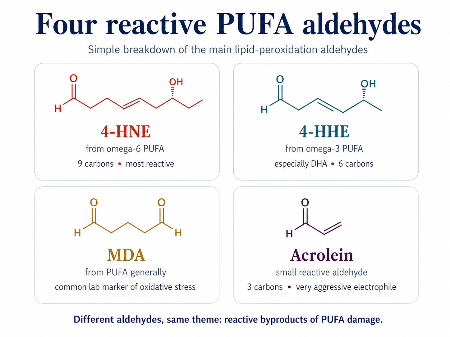
These are reactive electrophiles; molecules that grab electrons from other molecules within reach. They form covalent adducts with proteins, DNA, and small thiol-containing cofactors. They are the same chemistry that causes a bottle of fish oil left in sunlight to taste rotten, except they are also being generated continuously inside human cells proportionally to how much PUFA is sitting in tissue.
A person with high tissue PUFA load is producing these aldehydes at low levels every day, in every tissue. The flux is slow but constant, and it scales with stored PUFA. PUFA in tissue is a slow-release source of mitochondrial poisons.
Atlantic salmon in the Baltic Sea feed mostly on small, oily forage fish (herring and sprat). When the fat content of these prey fish is high, particularly when they’re rich in long-chain omega-3 PUFA like DHA, the salmon develop a condition called M74 syndrome. The spawning females become severely thiamine-deficient. Their offspring die at the yolk-sac stage. The condition has been studied for decades.
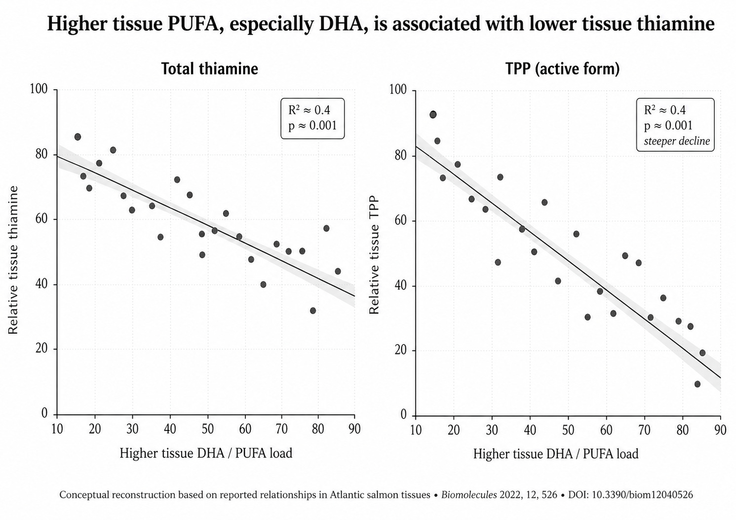
Researchers have measured thiamine concentrations directly against fatty acid profiles in salmon liver, muscle, ovaries, and eggs. The relationship is direct, in that the higher the tissue PUFA, especially DHA, the lower the tissue thiamine. The strongest version of this finding (DOI: 10.3390/biom12040526) reports correlation strengths around R² = 0.4 and p-values in the 0.001 range across multiple measurements. The active form of thiamine (TPP) collapses faster than total thiamine, which is the signature of a system that can no longer activate the substrate it does have.
The authors of one paper concluded directly that the cause of thiamine deficiency in these salmon is “especially an excess of dietary supply of highly unsaturated fatty acids, of which more than half is DHA (omega-3)” Their proposed mechanism is lipid peroxidation. The salmon’s spawning fast (when they stop eating and rely on their fat stores for the migration) accelerates the process. They burn their PUFA, generate peroxides faster than their antioxidants can handle, their thiamine collapse, and many of them die in the river.
Fish are not mammals so the metabolic context differs, but PUFA peroxidation generates the same aldehydes in salmon as it does in a human. Lipoic acid in mammalian PDH is the same molecule as lipoic acid in fish PDH. Thiamine pyrophosphate is structurally identical. The biochemistry is the same.
The salmon are showing us what happens when tissue PUFA load exceeds the antioxidant and thiamine reserves available to handle it.
A research group at UC Riverside has been publishing on soybean oil and metabolic disease for over a decade. Their 2015 paper showed that soybean oil is more obesity-causing than coconut oil and more than fructose in mice. Their 2017 paper traced the obesity-causing effect to specific compounds generated from soybean oil PUFA. In 2024, their focus moved to the effects on thiamine.
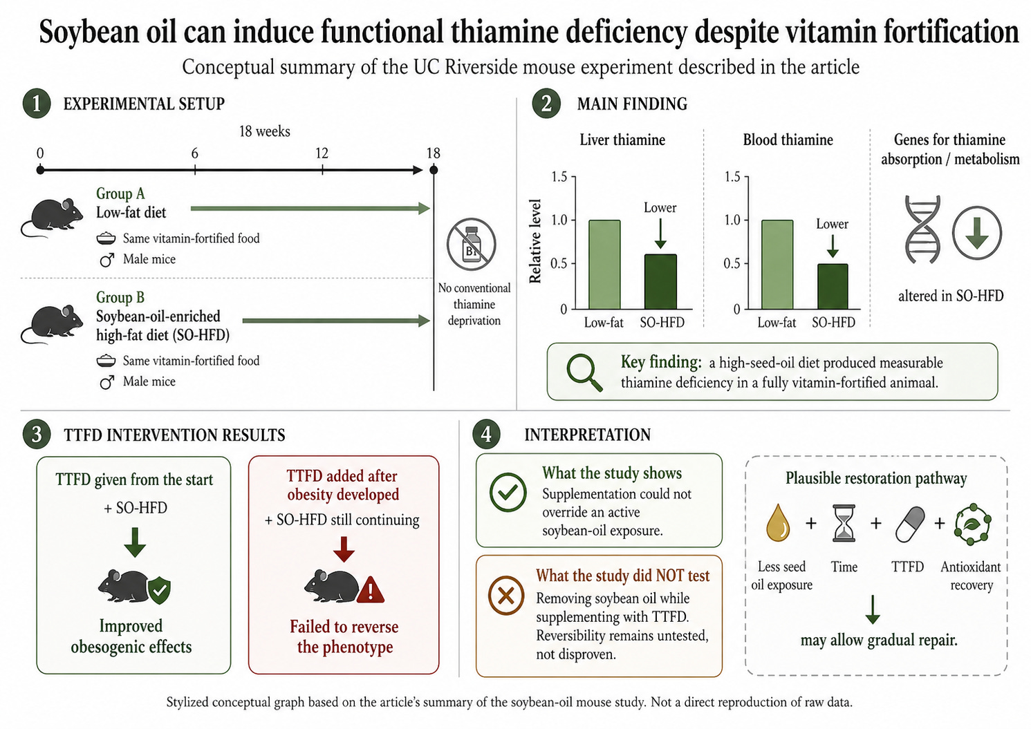
Male mice were fed either a low-fat diet or a soybean-oil-enriched high-fat diet (SO-HFD) at fat percentages comparable to the average American diet. Eighteen weeks, both groups got the same vitamin-fortified food. Neither was thiamine-deprived in any normal way.
After eighteen weeks, the mice fed soybean oil had developed MEASURABLE thiamine deficiency. Low thiamine in liver and blood, and had specific alterations in the genes responsible for thiamine absorption and metabolism. None of this was caused by inadequate intake, rather it was caused by the soybean oil itself.
The researchers tested whether TTFD (the most bioavailable thiamine derivative) could rescue the mice. When TTFD was given alongside the SO-HFD from the start, it improved the obesity-causing effects. When it was given *after* obesity had already developed, it failed to reverse it.
The obvious reading is that the soybean-induced damage is permanent. That is what the authors raised as a possibility. However, after reading it carefully, the experiment doesn’t actually support that conclusion. The mice were still being fed soybean oil while the TTFD was added. What was tested was whether supplementation can override an active dietary problem, and the answer is unsurprisingly no. You can’t out-supplement a negative exposure that is still ongoing. The same would be true if trying to out-supplement active alcoholism.
The experiment that would actually test reversibility by removing the soybean oil while supplementing with TTFD, has not been run.
Though it’s plausible to suggest that the system is restorable, given enough time and the right conditions. Mitochondria carry machinery to cleave damaged lipoic acid off and reinstall fresh cofactors. Seed oils in fat cells turn over with a half-life of roughly 600-700 days. Antioxidant systems can be rebuilt, and none of these processes are blocked. They are merely slow, and they require that the source of damage be reduced first.
In any case, the study legitimately demonstrates that a high-seed-oil diet can generate functional thiamine deficiency in a FULLY vitamin-fortified animal, and that deficiency cannot be supplemented away while the dietary load continues. This is an incredible finding and can explain lots of modern metabolic dysfunction.
If the findings hold up under full peer review, what’s being described is the mammalian version of the salmon study.
But why would a high-PUFA diet generate thiamine deficiency in a vitamin-fortified animal? There are three independent answers, each well-evidenced on its own.
In 1998, Humphries and Szweda published a paper in *Biochemistry* (37, 15835–15841) describing what 4-HNE (the major aldehyde from omega-6 PUFA peroxidation) does when added to isolated heart mitochondria. Two enzymes get selectively inhibited.
PDH and KGDH, which are the two enzymes that both require TPP and both carry covalent lipoic acid.
4-HNE effectively immobilizes the cofactor, and the whole complex suffers.
The enzyme can have perfectly normal TPP, normal magnesium, normal FAD, normal NAD+, and every cofactor, yet it still doesn’t function, because the lipoic acid has been chemically modified.
The inhibition is *increased* when the enzyme is actively using substrate. Working enzymes are more vulnerable than idle ones, since catalysis briefly reduces the lipoic disulfide to its dithiol form, exposing the sulfhydryls that 4-HNE attacks. The more cells are burning glucose, the more vulnerable PDH and KGDH are to aldehyde damage. A person with high metabolic flux is also a person at high risk if their tissue PUFA is loaded and aldehydes are high.
Acrolein produces the same effect in a dose-dependent manner, as well as 4-HHE from omega-3 peroxidation. The same mechanism has been documented in cardiac mitochondria and in animal studies.
This is functional thiamine deficiency at a cellular level. Cofactors and thiamine levels are normal, yet the cell is still failing to burn fuel properly, because the enzyme that’s supposed to do it has chemically modified lipoic acid.
Modern medicine measures the micronutrient levels but not whether the enzyme can use them.
Lukienko and colleagues (Bull Exp Biol Med, 2000, 130, 874–876) demonstrated that thiamine has direct antioxidant properties. It inhibits lipid peroxidation in rat livers. When thiamine is used as an antioxidant, it does not retain the original biochemical function.
The same mechanism that lets thiamine handle peroxides is the mechanism that destroys thiamine when peroxides are present. Each antioxidant event consumes a thiamine molecule. When there is more peroxidation from more seed oils, there’s less thiamine to be used by enzymes.
Gary Gibson and Hui Zhang (Neurochem Int, 2002, 40, 493–504) describe thiamine as a site-directed antioxidant, meaning it works locally, near sites of free radical generation, rather than acting as a broadly circulating defense like vitamin E. Their full review documents the bidirectional relationship where oxidative stress depletes thiamine, and thiamine deficiency itself raises oxidative stress markers (notably MDA), creating a loop a tissue can fall into and struggle to climb out of.
A cell with high PUFA in its membranes has more peroxidation events. Each event consumes a small amount of thiamine. Multiplied across tissues and across years, you get a chronic, low-grade depletion that diet does not compensate for, because the recommended daily average was decided with an assumption of low background peroxidation.
The mainstream thiamine intake recommendations were calibrated for a population whose tissue PUFA load was a FRACTION of what it is today.
The third mechanism is more indirect, but important nonetheless.
Transketolase is another thiamine-dependent enzyme of the pentose phosphate pathway. That’s the rate-limiting step controlling how much glucose-6-phosphate gets diverted toward NADPH production. NADPH is the cofactor that regenerates glutathione which is the cell’s primary water-soluble antioxidant (called the master antioxidant of the body).
PUFA in membranes → peroxide formation → glutathione consumption → glutathione regeneration requires NADPH → NADPH production requires functional transketolase → transketolase requires TPP.
A high-PUFA tissue has heavier demand on every step. The transketolase requirement scales with the antioxidant need, because the antioxidant tax scales with how much PUFA is being stored. The total thiamine demand of a high-PUFA cell is structurally higher than the same cell would have on a low-PUFA diet, before any of the direct destruction mechanisms are accounted for.
Therefore there are three mechanisms operating simultaneously:
The first is direct destruction of thiamine through the antioxidant work it does.
The second is direct inactivation of the thiamine-dependent enzymes through aldehyde adduction of lipoic acid.
The third is inflated demand on the thiamine system through transketolase’s role in regenerating the antioxidant defenses.
A person carrying a heavy tissue PUFA load is being attacked at three independent points at the same time.
If the model is approximately correct, the practical implications are the following.
Lowering tissue PUFA load is the most important move, but it works on a multi-year timescale. Linoleic acid has a 600–700 day half-life. Two years of lower PUFA intake will move the needle significantly while six months may not. This rewards patience.
Thiamine support is a more immediate lever, but only if the form bypasses normal transport pathways. Standard thiamine HCl uses the SLC19A2/3 transporters and is absorbed slowly. Functional doses for someone with a high oxidative tissue burden likely exceed what fortified foods can provide. Fat-soluble derivatives like TTFD and benfotiamine raise intracellular TPP more reliably, particularly in tissues with high demand. They might also bypass any aldehyde-mediated transport interference that may operate in heavily affected tissues, though this last point is not directly proven, only a plausible extension.
Vitamin E is the membrane defense. It is the antioxidant that lives inside the lipid bilayer and protects against peroxidation. Vitamin E does not fix the underlying PUFA problem, but it reduces the per-day damage rate while the load is being reduced.
Magnesium is the necessary partner for thiamine activation. Without adequate magnesium, intracellular thiamine cannot be phosphorylated to TPP. The two function as a team. Treating one without the other is incomplete. Magnesium is also one of the easier nutrients to under-dose, especially for people with high baseline stress, since they excrete more of it.
So reduce PUFA intake, provide vitamin E to lower current peroxidation rate, provide thiamine, preferably in a fat-soluble form, at doses that meet the ongoing demand. Provide magnesium adequately, and build your metabolism in the broader sense (body warmth, glycogen, low chronic stress, sleep).
There are two types of thiamine deficiency, and the second one is very underexplored.
The first thiamine deficiency is an input problem. Caused by inadequate intake, malabsorption, alcoholism, excessive refined-sugar intake without micronutrients, or medications. It is often detectable on serum testing, and fixes rapidly with supplementation of virtually all thiamine forms.
The second thiamine deficiency is a consumption problem. Caused by a high tissue seed oil load generating chronic aldehyde load, which destroys thiamine directly, inactivates the thiamine-dependent enzymes through lipoic acid modification, and increases demand through an antioxidant pathway. It is often invisible on serum testing. It responds partially and inconsistently to standard supplementation alone, and responds more reliably to a holistic approach that addresses the upstream cause.
This is PUFA reduction over time, vitamin E for membrane protection, fat-soluble thiamine forms at higher doses, and adequate magnesium for activation. It takes months to years to fix this, scaling with how long the tissue has been carrying the PUFA load and how aggressively that load is reduced.
The reason it has gone unexplored for so long is that no single tissue marker captures it.
A modern person, with a modern dietary history, presenting with modern symptoms of fatigue, metabolic inflexibility, early diabetes patterns and worsened cognition, is more likely to be in the second category than the first. Treating only for the first will produce only partial responses (though I’ve seen incredible transformations on thiamine alone).
It is the same story we have explored in “The Seed Oil Bible”. PUFA is destabilizing as it remodels tissue into an oxidation-prone state. Thiamine is as because it sits at the most crucial points for energy production.
No posts
Comments
Nothing yet. Say the first thing.
Sign in to join the conversation.