
By Dr. David Traster, DC, MS, DACNB
Co-owner, The Neurologic Wellness Institute
Boca Raton • Chicago • Waukesha • Wood Dale
www.neurologicwellnessinstitute.com
When we think about attention disorders, developmental disorders, or the lingering neurological effects of concussion, we often think first about behavior. A child cannot sit still. A student loses their place while reading. Someone with ADHD becomes distracted during a conversation. A patient recovering from concussion cannot tolerate a computer screen for more than a few minutes. A person with autism may interact with the visual world differently, including differences in eye movements, visual attention, and how visual information is prioritized.
But attention is not simply a psychological concept. Attention is a biological process produced by networks throughout the brain, and those networks are intimately connected with the systems controlling the eyes.
Before the brain can efficiently attend to something in the environment, it often must first determine where to look, what to stabilize, what to ignore, and what information deserves priority. This makes gaze fixation and visual tracking fascinating windows into brain function—and potentially important targets within a broader neurological rehabilitation program.
Imagine trying to read this sentence while your eyes continuously drift away from the words. Every time they move, you must relocate the sentence, determine where you were, suppress irrelevant information, and begin processing again.
The task suddenly requires substantially more neurological effort.
Gaze fixation is the ability to maintain the eyes on a particular location or object. Although fixation appears simple, the eyes are never completely motionless. Even during normal fixation, tiny microsaccades, tremor, and drift occur. The nervous system must continuously regulate these movements so that important visual information remains appropriately positioned for high-resolution processing.
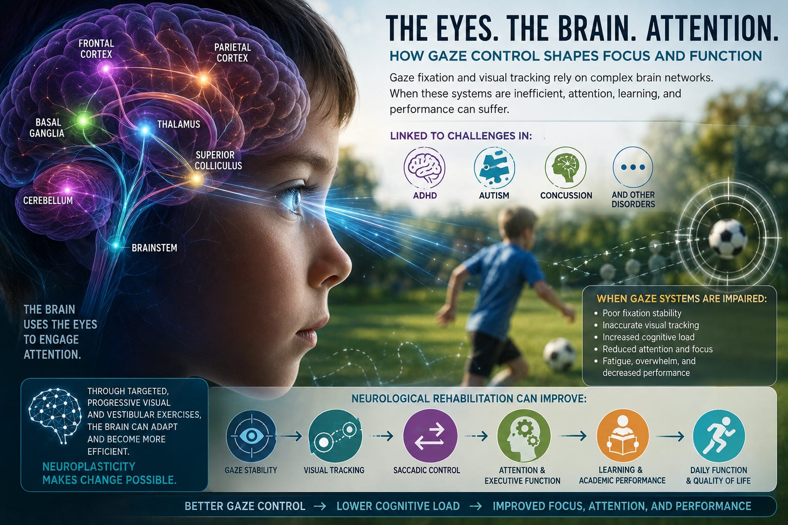
This requires communication among the retina, brainstem, cerebellum, superior colliculus, thalamus, basal ganglia, parietal cortex, frontal cortex, and multiple visual cortical regions. Fixation is therefore not merely an “eye exercise.” It is a neurological behavior involving sensory processing, motor control, prediction, inhibition, and attention.
Following a moving target requires another complex neurological operation.
Smooth pursuit eye movements allow the eyes to follow a moving object. The visual cortex must first detect the object and its motion. Cortical areas involved in motion processing help determine its direction and velocity. Parietal and frontal networks contribute attention and prediction. The cerebellum continuously compares expected movement with actual movement and helps correct errors. Brainstem ocular motor nuclei ultimately coordinate the extraocular muscles that move the eyes.
The brain is therefore performing a continuous cycle:
See the target. Predict where it is going. Move the eyes. Determine whether the movement was accurate. Correct the error. Continue following the target.
All of this happens within fractions of a second.
Saccades—the rapid eye movements that shift gaze from one location to another—require another overlapping network. These movements are especially important during reading, searching a room, navigating an environment, playing sports, and rapidly shifting attention between different sources of information.
Together, fixation, pursuits, and saccades provide an observable expression of how efficiently multiple brain networks are communicating.
One reason eye movements are particularly interesting in patients with attention problems is that the neurological systems controlling attention and eye movements substantially overlap.
The frontal eye fields participate in voluntary eye movements and attentional orientation. The parietal cortex helps determine where attention should be allocated in space. The superior colliculus participates in orienting the eyes and head toward important stimuli. The basal ganglia help determine which potential movements should be permitted and which should be suppressed. The cerebellum contributes timing, prediction, calibration, and error correction.
The prefrontal cortex contributes executive control and suppression of inappropriate responses. Attention therefore does not occur independently of the motor system. When you decide to look at one object instead of another, your brain has already performed a form of selection. Something has been chosen. Something else has been suppressed.
This relationship between attention and eye movements has produced an enormous field of research examining eye tracking as an objective measurement of cognition, neurological disease, developmental differences, concussion, and psychiatric conditions.
Research in attention-deficit/hyperactivity disorder has identified differences in several aspects of oculomotor control in at least some individuals with ADHD. One particularly interesting finding involves inhibitory eye movement control.
In an antisaccade task, for example, a stimulus suddenly appears on one side of a screen, but instead of looking toward it, the person must suppress the automatic response and intentionally look in the opposite direction. This sounds like an eye test. Neurologically, however, it is also an executive-function test. The individual must perceive the stimulus, inhibit a reflexive response, remember the instructions, generate a voluntary motor response, and monitor whether the response was correct.
Studies of ADHD have reported differences in antisaccade errors, saccadic inhibition, fixation stability, and other oculomotor measures, although findings vary among individuals and experimental paradigms. This does not mean abnormal eye movements cause ADHD. It means the neural networks responsible for attention, inhibition, executive control, and eye movements partially intersect. Dysfunction within these networks can therefore potentially appear both behaviorally as attention difficulties and physiologically as altered ocular motor performance.
Autism spectrum disorder provides another important example.
Eye-tracking research has identified differences in how some individuals with autism visually explore faces, social scenes, objects, and their surrounding environment. Differences have also been reported in fixation patterns, saccadic behavior, smooth pursuit, visual attention, and the amount of time spent examining particular components of social information.
Importantly, autism is extraordinarily heterogeneous. There is no single eye movement pattern that defines autism, and an eye-tracking abnormality should never be interpreted as a diagnostic test by itself. But these findings reinforce an important neurological principle: how the eyes explore the environment can provide information about how the brain is processing that environment.
Where someone looks, how long they remain there, how quickly they disengage, how accurately they move toward another target, and how efficiently they suppress distractions can all provide information about larger neurological networks.
There is another way to think about fixation that I find particularly useful clinically. Fixation is not simply the ability to look at something. It is the ability not to look at everything else. A child staring at a small target is simultaneously being exposed to hundreds of competing visual stimuli. Objects exist in the peripheral visual field. People move around the room. Sounds occur. Internal thoughts arise.
Maintaining fixation requires the nervous system to prioritize one target while suppressing competing information. Attention requires something remarkably similar. To listen to a teacher, read a book, complete homework, or participate in a conversation, the brain must continuously determine what information is relevant and what information should temporarily be ignored.
This does not make fixation and attention identical processes. They are not. But it helps explain why their neurological networks can be so closely related.
The relationship becomes particularly obvious after concussion. Patients following concussion frequently describe difficulty reading, concentrating, driving, watching television, working on computers, or being inside visually complicated environments such as grocery stores.
They may say:
“I can see the words, but I cannot process them.”
“I keep losing my place.”
“My eyes get tired.”
“I can’t concentrate on the computer.”
“Everything feels overwhelming.”
Some of these symptoms may involve abnormalities in ocular motor function, vestibular processing, accommodation, convergence, visual motion processing, autonomic regulation, migraine physiology, cervical function, cognitive fatigue, or combinations of these mechanisms.
Concussion can disrupt distributed networks connecting the frontal cortex, parietal cortex, cerebellum, brainstem, vestibular system, and visual system. Consequently, fixation, saccades, pursuits, convergence, vestibulo-ocular reflexes, visual motion tolerance, and attention may all become impaired simultaneously. The patient may perceive the problem simply as “brain fog” or poor concentration. The neurological problem may be considerably more complicated.
Every neurological task has a metabolic cost. If an automatic process becomes inefficient, higher brain regions may have to compensate. Consider reading. Normally, the eyes perform extremely rapid sequences of fixation and saccadic movements. The brain extracts information during brief periods of relative stability between movements. If ocular motor control becomes inefficient, reading may require substantially greater conscious effort.
The person may reread sentences. They may lose their position. Words may appear uncomfortable. Visual motion may provoke dizziness. Headaches may develop. Eventually the individual stops reading—not necessarily because they cannot understand the material, but because the neurological cost of obtaining that information has become too high.
The same principle may apply to classroom attention, computer work, sports, driving, and other visually demanding environments. Improving the efficiency of the underlying neurological systems may therefore reduce some of the processing burden associated with these activities.
Comments
Nothing yet. Say the first thing.
Sign in to join the conversation.