
By Dr. David Traster, DC, MS, DACNB
Co-owner, The Neurologic Wellness Institute
Boca Raton • Chicago • Waukesha • Wood Dale
www.neurologicwellnessinstitute.com
Translation is one of the most fundamental forms of human movement, yet it is often overshadowed by discussions of rotational movement and the vestibulo-ocular reflex. Every step we take, every elevator ride, every acceleration in a vehicle, and every shift of our body across space requires the nervous system to continuously detect, interpret, and respond to linear motion. Translation represents far more than simply moving from one location to another. It is a complex neurological event requiring continuous integration of vestibular, visual, proprioceptive, somatosensory, cerebellar, autonomic, and cortical information. The ability of the brain to accurately interpret translation is essential for balance, gaze stabilization, cardiovascular regulation, spatial orientation, navigation, and movement efficiency. When these systems fail to integrate appropriately, patients may develop dizziness, imbalance, visual motion sensitivity, dysautonomia, anxiety, cognitive fatigue, chronic pain, and persistent post-concussion symptoms.
Understanding how the nervous system processes translation provides clinicians with one of the most powerful tools for promoting neuroplasticity. Translation exercises can selectively challenge nearly every sensory system in the brain while simultaneously modifying sensory weighting and enhancing multimodal integration. The specific characteristics of the movement—including speed, posture, visual conditions, cognitive load, and gaze stabilization demands—determine which neural networks become activated and how plasticity develops.
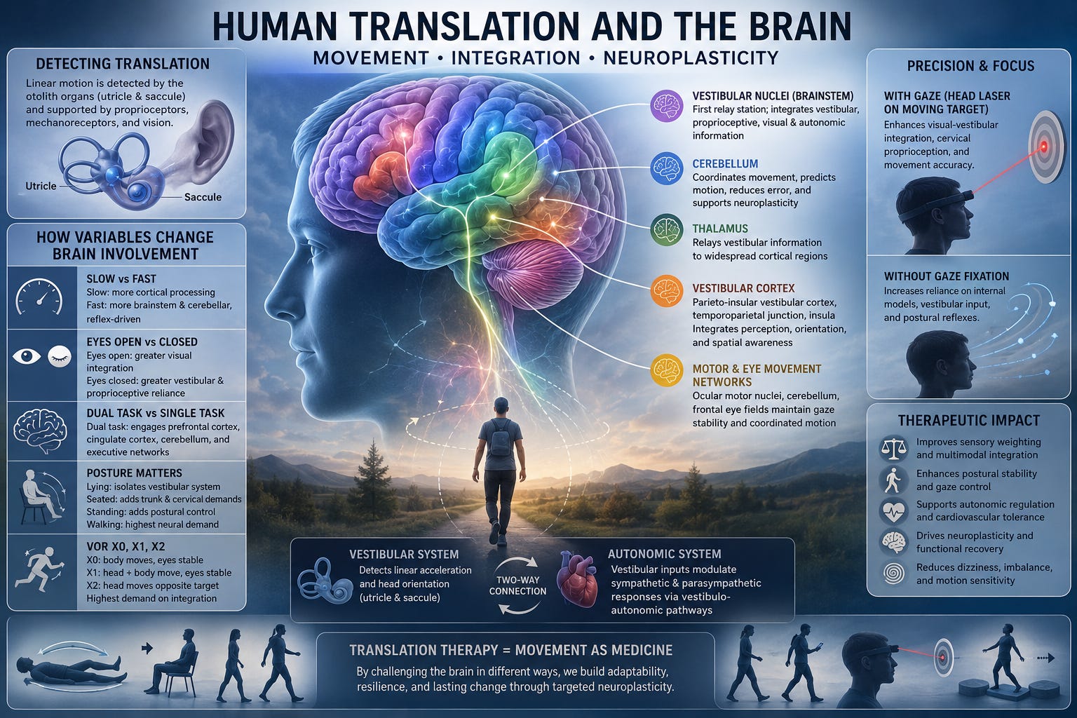
The first structures responsible for detecting linear motion are the otolith organs of the vestibular labyrinth: the utricle and the saccule. Unlike the semicircular canals, which detect angular acceleration, the otolith organs detect linear acceleration and changes in head orientation relative to gravity.
The utricle primarily responds to horizontal linear accelerations such as walking, running, riding in a car, or side-stepping. The saccule is more sensitive to vertical accelerations such as jumping, riding an elevator, or standing up from a chair. Hair cells embedded within the gelatinous otolithic membrane are displaced by inertial forces acting upon calcium carbonate crystals known as otoconia. This mechanical deflection alters the firing rate of vestibular afferents, providing continuous information regarding both acceleration and gravitational orientation.
Translation, however, is never interpreted solely by the otolith organs. Every movement also activates muscle spindles, Golgi tendon organs, joint mechanoreceptors, plantar cutaneous receptors, visceral mechanoreceptors, and cervical proprioceptors. These peripheral inputs provide essential information allowing the brain to distinguish between active self-generated movement and externally imposed motion.
Translation information ascends through the vestibular nuclei of the brainstem before diverging into numerous parallel pathways. Rather than existing as a single translation center, the brain utilizes a distributed network that continuously integrates sensory information.
The vestibular nuclei serve as the first major integration hub. Here, vestibular signals combine with cervical proprioception, spinal somatosensory information, cerebellar modulation, and visual inputs. From the vestibular nuclei, translation signals project to numerous regions including the cerebellum, thalamus, superior colliculus, reticular formation, ocular motor nuclei, spinal cord, autonomic centers, and multiple cortical regions.
Within the cerebellum, the flocculus, nodulus, uvula, vermis, fastigial nucleus, and vestibulocerebellum continuously refine movement accuracy, predict future motion, reduce sensory error, and optimize postural responses. The cerebellum compares expected movement with actual sensory feedback and updates internal models to improve future performance.
The thalamus relays vestibular information toward widespread cortical regions involved in conscious perception of motion. Cortical vestibular processing involves the parieto-insular vestibular cortex (PIVC), posterior parietal cortex, temporoparietal junction, insular cortex, superior temporal cortex, cingulate cortex, supplementary motor area, premotor cortex, hippocampus, and frontal eye fields. Together these structures generate our perception of movement, body position, navigation, attention, and spatial memory.
Comments
Nothing yet. Say the first thing.
Sign in to join the conversation.