
By Dr. David Traster, DC, MS, DACNB
Co-owner, The Neurologic Wellness Institute
Boca Raton • Chicago • Waukesha • Wood Dale
www.neurologicwellnessinstitute.com
Most people think of oxygen as the most important gas for brain function. While oxygen is certainly essential for survival, carbon dioxide (CO₂) is arguably the most powerful moment-to-moment regulator of cerebral blood flow. Small changes in CO₂ can dramatically alter how much blood reaches the brain within seconds. This is why capnography has become an increasingly valuable tool in emergency medicine, anesthesia, intensive care, concussion management, autonomic testing, vestibular rehabilitation, and functional neurology.
For clinicians interested in cerebral physiology, capnography provides much more than a respiratory measurement. It offers a window into cerebrovascular function. When combined with transcranial Doppler ultrasound (TCD), blood pressure monitoring, or functional neuroimaging, capnography allows clinicians to evaluate two distinct—but often confused—physiologic systems: cerebral autoregulation and cerebrovascular vasoreactivity. Although these concepts are closely related, they measure different properties of the cerebral circulation and should never be used interchangeably. Research consistently demonstrates that autoregulation and vasoreactivity can become uncoupled in many neurological disorders.
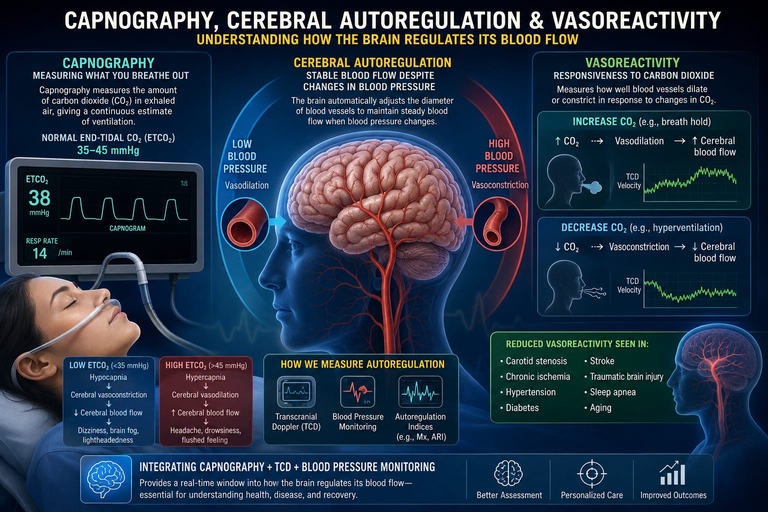
Capnography is the continuous measurement of carbon dioxide in exhaled air throughout the respiratory cycle. Unlike a pulse oximeter, which measures oxygen saturation, capnography measures ventilation.
The measurement most clinicians use is the end-tidal carbon dioxide (EtCO₂), which represents the concentration or partial pressure of CO₂ measured at the very end of exhalation. Under normal physiological conditions, EtCO₂ closely approximates arterial CO₂ (PaCO₂), usually differing by only 2–5 mmHg in healthy individuals.
Capnography provides several important measurements including:
End-tidal CO₂ (EtCO₂)
Respiratory rate
Respiratory pattern
Shape of the capnogram waveform
Breath-by-breath ventilation changes
Unlike intermittent arterial blood gases, capnography provides continuous real-time monitoring.
Normal EtCO₂ generally ranges between 35 and 45 mmHg.
Values below approximately 35 mmHg indicate hypocapnia. This most commonly results from hyperventilation, excessive respiratory drive, anxiety, pain, dysautonomia, panic attacks, traumatic brain injury, or compensation for metabolic acidosis.
Values above approximately 45 mmHg indicate hypercapnia. Causes include hypoventilation, COPD, obesity hypoventilation syndrome, neuromuscular disease, opioid use, excessive sedation, sleep apnea, or severe pulmonary disease.
The absolute number is important, but equally important is how EtCO₂ changes during different physiological challenges such as standing, exercise, breath holding, hyperventilation, cognitive tasks, or rehabilitation exercises.
Carbon dioxide is one of the strongest regulators of cerebral vascular tone.
An increase in arterial CO₂ lowers extracellular pH around cerebral arterioles. This causes smooth muscle relaxation and cerebral vasodilation, increasing cerebral blood flow.
Conversely, reducing CO₂ through hyperventilation raises pH, producing cerebral vasoconstriction and reducing cerebral blood flow.
A classic physiological approximation is that cerebral blood flow changes by approximately 2–5% for every 1 mmHg change in PaCO₂ within the normal physiologic range. Consequently, even modest hyperventilation can substantially decrease cerebral perfusion, while modest hypercapnia can significantly increase it.
This explains why patients who hyperventilate during panic attacks frequently develop:
Lightheadedness
Dizziness
Blurred vision
Brain fog
Tingling
Near syncope
Difficulty concentrating
These symptoms are often consequences of transient cerebral vasoconstriction rather than inadequate oxygenation.
Comments
Nothing yet. Say the first thing.
Sign in to join the conversation.