
For over six decades, human evolutionary biology has been anchored by a single, seductive narrative: the Obstetrical Dilemma Hypothesis (ODH). First formalized by Sherwood Washburn (1960), the ODH asserts that human pelvic evolution represents an uneasy, antagonistic compromise between two mutually exclusive evolutionary vectors. On one hand, the shift to obligate bipedalism required a narrow, compact pelvis to keep the hip joints—specifically the femoral head centers—close to the sagittal midline, thereby minimizing the mechanical bending moment of body weight and reducing the metabolic cost of walking and running. On the other hand, encephalization required a wide, spacious pelvic canal to permit the passage of secondary altricial, large-brained neonates.
Under the classic ODH framework, female pelvic anatomy is constrained from expanding to an ideal obstetrical width because doing so would penalize locomotor efficiency. Consequently, childbirth in humans became notoriously difficult, dangerous, and dependent on assisted delivery—a biological sacrifice paid on the altar of bipedal efficiency.
However, a growing body of biomechanical, metabolic, and clinical evidence has chipped away at the foundation of this paradigm. Experimental studies tracking metabolic energy expenditure during walking and running across diverse human pelvic shapes have repeatedly demonstrated that a wider distance between the femoral head centers does not increase the oxygen cost of locomotion (Warrener et al., 2015; Kramer and Sylvester, 2023). Simultaneously, metabolic research suggests that the timing of human birth is governed primarily by maternal energetics—the Energetics of Gestation and Growth (EGG) model—rather than an absolute skeletal boundary of the pelvic inlet (Dunsworth et al., 2012).
Despite these critiques, testing the evolutionary dynamics of the pelvis across deep time has been severely hindered by the scarcity of intact fossil pelves. Pelvic bones consist of delicate cancellous core structures encased in thin cortical shells; they are among the first elements to crush, fragment, or weather in paleoanthropological contexts. Nowhere has this limitation been more acutely felt than in the study of our closest extinct relatives, the Neanderthals (Homo neanderthalensis).
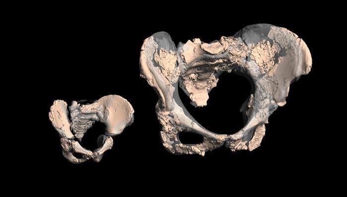
A landmark study published in the Proceedings of the National Academy of Sciences by Christoph P. E. Zollikofer, Marcia S. Ponce de León, and an international team of collaborators (Zollikofer et al., 2026) delivers a transformative, empirically grounded critique of the Obstetrical Dilemma. By integrating high-resolution computed tomography (CT) with advanced virtual reconstruction techniques, the authors present complete 3D pelves of two extraordinarily preserved Neanderthal specimens: a 1.5-year-old infant from Dederiyeh Cave, Syria (Dederiyeh 1), and a young adult female from Sima de las Palomas del Cabezo Gordo, Spain (Palomas 92, colloquially known as “Paloma”).
By comparing these fossils against a broad ontogenetic and adult sample of modern humans (Homo sapiens), as well as previously reconstructed Neanderthals (Tabun 1 and Kebara 2) and early hominins (Australopithecus and early Homo), Zollikofer et al. (2026) provide an exhaustive quantitative evaluation of hominin pelvic evolution. In my view, their findings conclusively dismantle the classical Obstetrical Dilemma. Instead, the data align with a paradigm shift: the primary evolutionary trade-off shaping the human and Neanderthal birth canal was not between locomotion and obstetrics, but rather between obstetrics and the structural mechanics of pelvic floor stability.
To understand the analytical power of this dataset, we must first contextualize the skeletal remains at the center of the study. The Neanderthal evolutionary lineage represents a distinct, highly encephalized hominin trajectory that occupied Western Eurasia from approximately 430,000 years ago until their disappearance around 40,000 years ago. Throughout this duration, Neanderthals maintained large body masses and cranial capacities that equaled or exceeded those of contemporary Homo sapiens (Ponce de León et al., 2008). Understanding how Neanderthal females successfully delivered infants with cranial volumes comparable to modern human neonates—while retaining a postcranial skeleton adapted for hyper-robust, high-force physical demands—requires intact pelvic evidence.
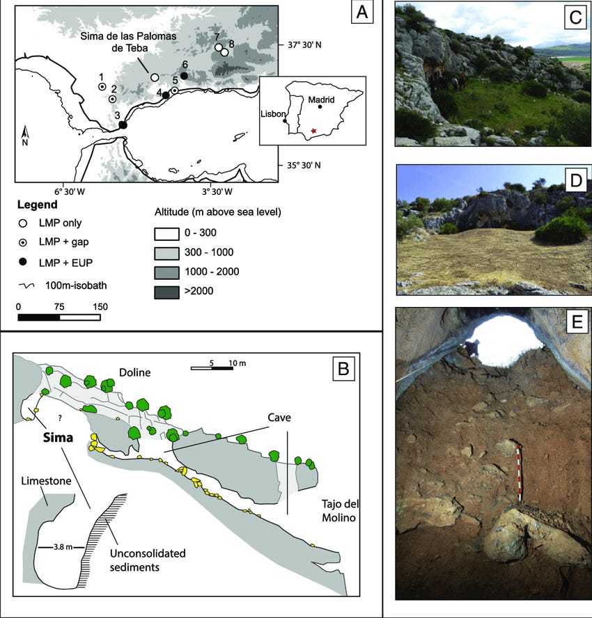
Located in southeastern Spain, Sima de las Palomas is a natural karstic shaft penetrating 18 meters into the Permo-Triassic marble hill of Cabezo Gordo. Excavations directed by Michael J. Walker have exposed a deep stratigraphic column rich in Middle Paleolithic Mousterian artifacts, faunal remains, and exceptionally preserved Neanderthal fossils (Walker et al., 2011).
In 2006–2007, excavators recovered a partial, articulated skeleton of a young adult Neanderthal female designated Palomas 92 (or “Paloma”) from Conglomerate A, an ancient brecciated scree layer positioned between the Upper and Lower Gray Layers, dated by radiocarbon and electron spin resonance (ESR) to between 40,000 and 60,000 years before present (Walker et al., 2011).
Palomas 92 was discovered embedded in a heavily cemented matrix of breccia. Taphonomically, while the skeletal elements were preserved in anatomical articulation—including the femoropelvic joints, rib cage, and hand bones positioned near the face—the pelvic girdle had suffered extensive in situ fragmentation and plastic distortion under sediment pressure. The matrix cementation was so severe that mechanical separation using traditional pneumatic air-scribes carried an unacceptable risk of bone destruction.
Thanks for reading The Deep History Refuge! This post is public so feel free to share it.
Situated in the Afrin region of northwestern Syria, Dederiyeh Cave is a massive karstic cavern excavated by a joint Syrian-Japanese team led by Takeru Akazawa and Osamu Kondo. In 1993, the site yielded one of the most complete infant Neanderthal skeletons ever discovered: Dederiyeh 1, buried in an intentional pit context within Middle Paleolithic Layer 11, dated to approximately 50,000 to 60,000 years ago (Akazawa et al., 1995; Kondo et al., 2000).
Dederiyeh 1 represents an infant with an estimated age at death of 1.5 years (39 months). The postcranial skeleton is exquisitely preserved. However, because the individual was an infant, the three main ossification centers of the os coxae—the ilium, ischium, and pubis—remained unfused, separated by broad cartilaginous synchondroses. Sacral vertebrae were likewise unfused. Consequently, analyzing the pelvic architecture of Dederiyeh 1 required an approach capable of virtually articulating delicate, unfused osteological elements within a realistic 3D anatomical space.
The methodology deployed by Zollikofer et al. (2026) represents the gold standard in contemporary digital paleoanthropology, combining high-resolution tomographic segmentation, anatomical mirror-imaging, comparative developmental baselines, and 3D Geometric Morphometrics (GM).
The brecciated pelvic fragments of Palomas 92 and the isolated pelvic elements of Dederiyeh 1 were scanned using high-resolution medical CT scanners. Digital segmentation was performed to separate fossilized bone from surrounding calcified sediment matrices without applying physical stress to the fossils.
For Palomas 92, individual cortical fragments were reassembled in virtual space using preserved fracture margins, structural continuity, and bilateral symmetry constraints. Where taphonomic crushing had destroyed localized regions on one side, mirror-imaged reconstructions of the undamaged contralateral counterparts were generated (SI Appendix, Zollikofer et al., 2026). To account for potential reconstruction uncertainties, the authors generated three distinct reconstructive variants for each fossil adult specimen (Palomas 92, Tabun 1, and Kebara 2), ensuring that analytical metrics were robust to slight variations in virtual positioning.
To reassemble the unfused pelvic girdle of the 1.5-year-old Dederiyeh 1 infant, the team utilized retrospective clinical CT data from a pediatric sample of modern human infants (aged 1 month to 8.5 years). By measuring soft-tissue cartilage gaps across developing human os coxae, the authors created an anatomically precise virtual envelope that constrained the spatial arrangement of the unfused Dederiyeh ilia, ischia, pubes, and sacral segments.
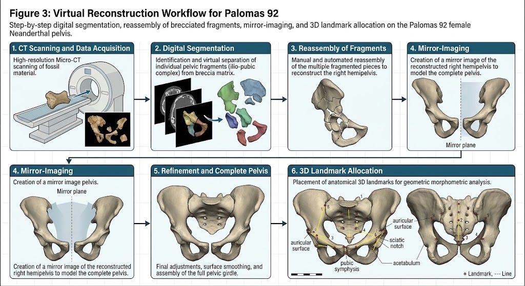
To quantify form and shape independently of size, landmark configurations were collected across the sample:
Infant Dataset (K_i = 153 landmarks): Designed to capture individual unfused anatomical units across N = 34 modern human infants and Dederiyeh 1.
Adult Dataset (K_a = 123 landmarks): Placed across N = 112 adult modern humans (56 females, 56 males, aged 14–49 years) and reconstructed Neanderthals.
Landmark arrays were subjected to Generalized Procrustes Analysis (GPA) to remove translation, rotation, and scale, isolating pure shape configurations in Procrustes shape space. Principal Component Analysis (PCA) was then used to identify major axes of variation.
To evaluate functional performance, the authors derived specific linear, angular, and areal metrics:
Obstetrical Canal Metrics: Elliptic cross-sectional areas and shape indices (Anteroposterior diameter / Transverse diameter × 100) for the pelvic Inlet, Midplane, and Outlet.
Locomotor Moment Arms: Mediolateral and anteroposterior spatial offsets from the Femoral Head Center (FHC) to muscle attachment points and body weight axes.
Abductor Moment Arm (AMA): Distance from FHC to the greater trochanter / iliac crest abductor insertion.
Body-Weight Moment Arm (BMA): Mediolateral distance from FHC to the sagittal midline.
Effective Mechanical Advantage (EMA): Ratio of AMA / BMA.
Flexor and Extensor Levers: Anteroposterior distances from FHC to the ischial tuberosity (extensors) and anterior inferior iliac spine / lesser trochanter (flexors).
Acetabular Orientation: Quantified via version angle (theta) and inclination angle (alpha) relative to the coronal plane intersecting both femoral head centers and the S1 superior midpoint.
Thermoregulatory Cylindroid Modeling: Extended traditional 1D cylindrical body models (Ruff, 1991) into 3D cylindroid models taking both anteroposterior (AP) and mediolateral (ML) diameters into account to calculate true Surface-to-Volume (S/V) ratios.
Unlock deeper insights into human evolution with full access to rigorous, multidisciplinary site breakdowns, technical data analyses, and critical evaluations of ancient hominin cognition. By upgrading to a paid subscription, you directly support independent, primary-source science journalism while unlocking exclusive deep-dives that challenge the dogmas of deep history. Join our community of researchers and enthusiasts to read the rest of this investigation and explore the full archive.
Comments
Nothing yet. Say the first thing.
Sign in to join the conversation.