
Xuewu Liu · Clinical Reports · Second injection performed 17 August 2026, Tianjin, China
This report follows a single canine patient first described in When the Tumor Took the Tongue: Five Days That Changed Everything (31 July 2026). The framework used to read the intraoperative colour change is set out in Fifteen Minutes Under Anesthesia: Knowing Whether an Oral Tumor Is Still Alive (14 August 2026).
The patient is a dog with an advanced tongue tumour measuring approximately 6.5 × 5.3 × 2.9 cm on CT, an estimated volume of about 47 mL. The tumour had invaded and largely replaced the tongue base. Before any treatment, the attending veterinarian judged that loss of the tongue was unavoidable, because the structures holding the tongue in place had already been destroyed by the tumour itself.
On 28 July 2026, guided by CT imaging, we performed a direct intratumoral injection of 10 mL of chlorine dioxide ablation solution. The tumour and the tongue rapidly turned black before the animal recovered from anaesthesia. Large volumes of necrotic tissue were shed the following day. On day 4, the tongue separated and detached spontaneously. By day 5, the visible tumour mass was largely absent. No incision was made, no tissue was surgically removed, and no sutures were required. Nutritional support was maintained through a gastric tube.
That report ended with an open question: what happens when you go back and inject the same animal again? This one answers it.
The first session was small in volume by design — 10 mL into a 47 mL tumour, roughly one fifth of the tumour by volume. That dose removed the great majority of the mass within five days. What a single low-fraction pass was never going to do is clear the tongue base completely.
Our own view of how this agent should be used in a tumour of this size and geometry is several sessions in close succession — on the order of four injections across three weeks, not one. In practice this dog received one, and the second session did not take place until day 20. Residual tissue at the tongue base was still present, and on the day of the second procedure it appeared somewhat larger than it had at day 5. That is what a three-week interval after a one-fifth dose fraction would be expected to produce. The interesting question in this case is not the residual tumour. It is whether the animal could be treated again at all.
In the same interval, two subcutaneous masses became palpable elsewhere on the body. Both were identified and marked before the procedure (Figures 3 and 4), and both were treated in the same session.
Date: 17 August 2026, day 20 after the first injection. General anaesthesia, mouth held open with a standard gag. Total dose: 25 mL of chlorine dioxide ablation solution across three targets — the residual tongue-base tumour and the two subcutaneous lesions. Recovery from anaesthesia was normal.
[Per-lesion volumes, needle counts and guidance details to be inserted from the operative record before publication.]
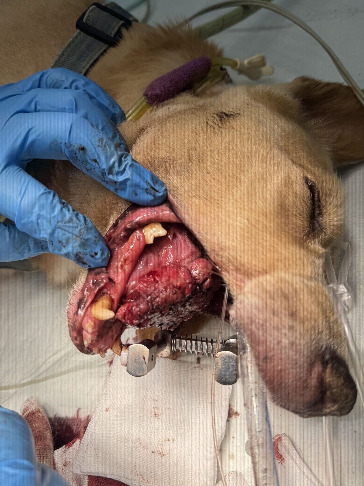
Figure 1 — During injection. The dog is in lateral recumbency under general anaesthesia with a mouth gag in place. The residual tumour occupies the tongue-base region; the anterior tongue detached spontaneously on day 4 after the first injection and is absent.
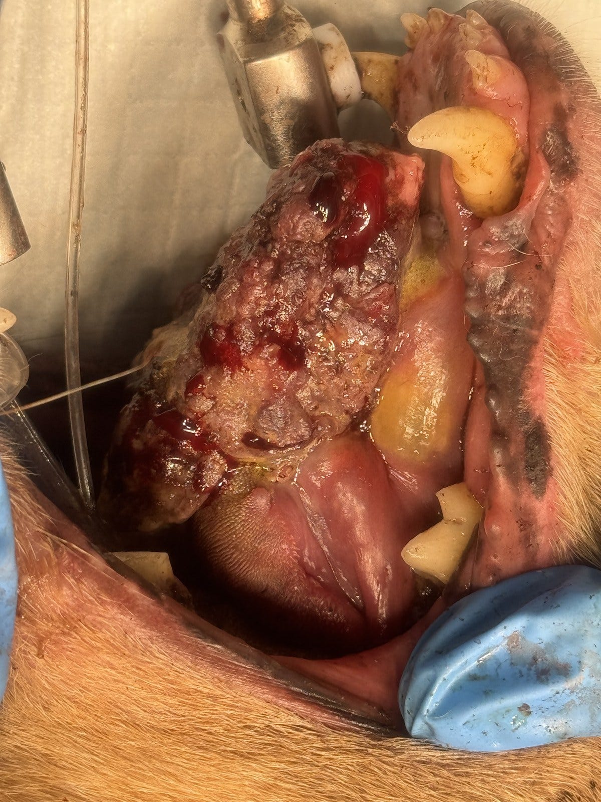
Figure 2 — Five minutes after injection. The treated region has lost the smooth, glistening surface of living tissue and shows a mottled grey-brown discoloration with dark red foci, clearly demarcated from the surrounding mucosa.
Comments
Nothing yet. Say the first thing.
Sign in to join the conversation.