
BUN-YO-MATIC! #NeoArmory:
We love it when the companies name their product so funky, that we don’t have to do anything creative and just add this as our title.
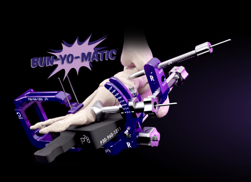
Again, right from the landing page…
Paragon28, one of our regulars here, have created what appears like a prop from ‘Total Recall’. This jig in combination with their SMART28 planning platform, allows intra-op targeted correction in both axial and rotational planes.
SMART28 works with weight bearing x-rays or CT and can create 3D planning images. Acclaimed as the first device to this and hels surgeons intra-op
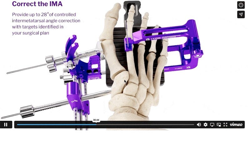
Seriously, keep up with the colour schema
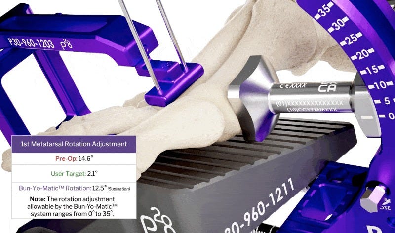
Millimeters and half degrees matter..
The tech seems to be from their recent acquisition DISIOR and Paragon28 have been acquired by Zimmer Biomet. There’s always a bigger fish eh…
As interesting as it looks, we do not have real world data or evidence on outcomes. There is no information on clearance for patient use as well.
From Curiosity to Classification: The Story Behind the Patel–Sharma Fragment #FromThePresent:
In orthopaedics, names carry weight. They’re not just labels — they’re stories, discoveries, and sometimes, a bit of legacy. As trainees, most surgeons grow up memorising names like Colles, Barton, Neer, and Volkmann, never really thinking they might one day contribute something of their own. For Dr. Patel and Dr. Sharma, this journey didn’t start with the intention of naming anything. It started, like most meaningful things in surgery, with simple curiosity.
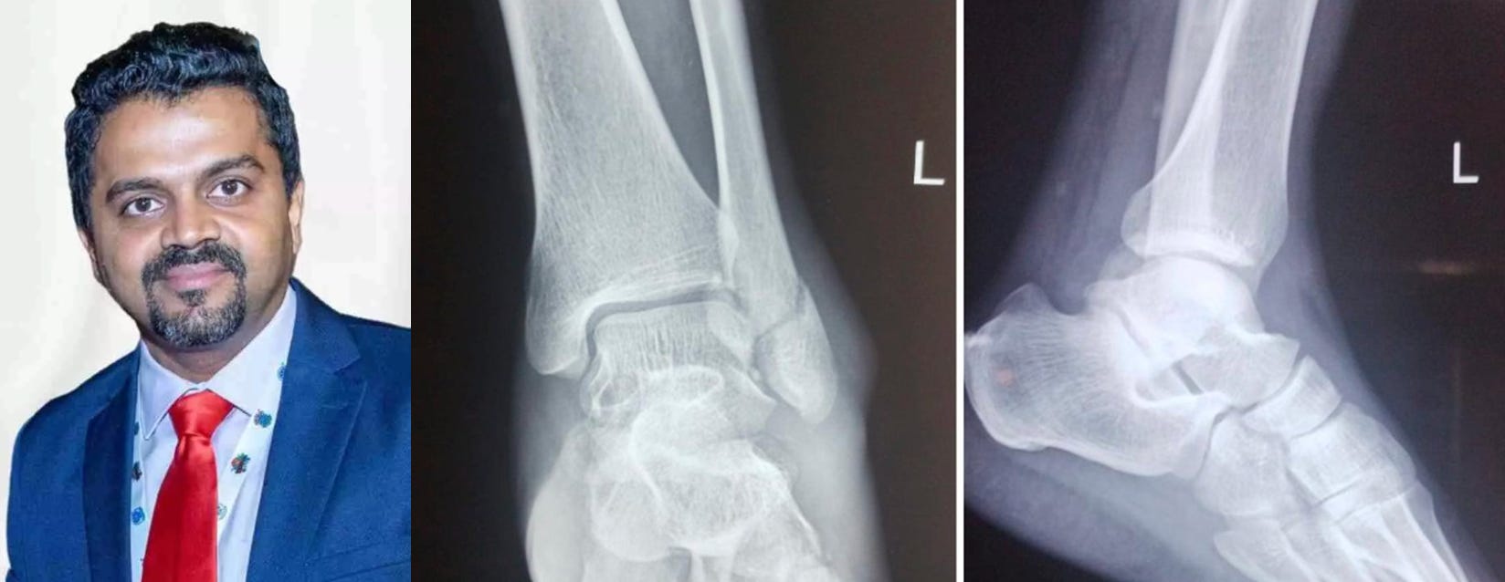
During training at PGI Chandigarh, exposure to a high volume of trauma cases began to change the way fractures were seen. They weren’t just isolated injuries anymore — patterns started emerging. Mechanisms made more sense. Fixation strategies became more intuitive. And occasionally, something didn’t quite fit. That “something” turned out to be a fibular-sided avulsion of the posterior inferior tibiofibular ligament (PITFL). It was subtle, easy to miss, and more importantly — it didn’t have a name.
Now, that’s unusual in orthopaedics.
The syndesmosis has been studied for over a century. The AITFL avulsions? Already named — Chaput-Tillaux and Wagstaff. The posterior side on the tibia? That’s the well-known Volkmann fragment. But the fibular counterpart of the PITFL? Strangely, it had been sitting there, unnamed and often ignored. Recognising this wasn’t just about spotting a “new” fragment. It was about identifying a missing piece in the syndesmotic puzzle.
Getting it accepted wasn’t easy. Reviewers needed convincing. The argument had to be clear — this fragment wasn’t just an anatomical curiosity; it had real clinical implications. Miss it, and you risk syndesmotic instability, poor reduction, and suboptimal outcomes. And that’s where the Patel–Sharma fragment earned its place — not as a name for the sake of it, but as a concept that sharpens how surgeons look at ankle injuries. Interestingly, this wasn’t a one-off contribution. The same line of thinking extended into anterior malleolus fractures as well, leading to newer pattern recognition and the Patel–Dhillon classification — again, with the same goal: simplify, clarify, and improve decision-making.
Because at the end of the day, this isn’t about having your name in a textbook.
It’s about making sure the next surgeon looks at an X-ray just a little more carefully.
Opening Pandora’s Box in Ankle Fractures: Rethinking Syndesmotic Reduction #SurgicalPearl:
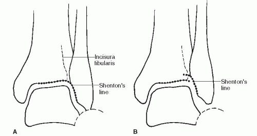
Syndesmotic reduction has been a point of concern in ankle fractures for years. Despite advencements about 50% are still malreduced. Tornetta et al. in a cadaveric study, have compared “articular surface reduction method” vs “incisura based redcution method”
Key takeaway
Look at the joint, not the incisura. Aligning the anterolateral tibial articular cartilage with the anteromedial fibular articular cartilage gives a more anatomical syndesmotic reduction.
Malreduction >2 mm:
7% with articular method
20% with incisura method
At the joint level, tibia and fibula have similar AP depth (~2 mm difference)
At the incisura (1 cm above joint), depth mismatch is large (~6 mm), making visual judgment unreliable
How to perform the Articular Surface Method?
Expose the anterolateral corner of ankle joint
Identify the anterolateral distal tibial articular cartilage (plafond).
Identify the anteromedial fibular articular cartilage.
Visually align these two cartilage surfaces perfectly:
Smooth continuation, maintenance of Shenton’s line of ankle
No anterior or posterior translation
Hold reduction manually or with a clamp without levering the fibula posteriorly or anteriorly.
While maintaining this alignment, insert provisional fixation (K-wire or reduction clamp).
Confirm that the cartilage surfaces remain flush during fixation.
Proceed with definitive fixation (screw or suture-button).
Fluoroscopy is used only as a secondary check, not the primary reduction guide.
Dont forget to check out the full paper and a surgical demonstration of articular method
Implant Wave Alert, Mg Screws in action now! #MostCited:
Metal plates and screws are used in the management of fractures, but they cause stress shielding and often require removal. In recent years, many studies confirmed that the biodegradable high-purity magnesium (Mg) screws exhibited sufficient mechanical strength with an adequate degradation rate for effective bone healing, avoiding the need for implant removal operations.
Zhao et al. tested it in action in a study of 24 patients comparing Mg screws to traditional Ti screws for ankle fracture management. All patients achieved good fracture alignment, and imaging examination showed that the biodegradable high-purity Mg screws degraded gradually without breakage or displacement. None of the patients experienced infection, failure of internal fixation, malunion or other complications in both groups. Hence, now we have an answer to those patients who ask for disappearing implants.
Events to check out:
The Association of Bone and Joint Surgeons (ABJS) 78th Annual Meeting - 15 – 19th April 2026 Napa, USA
The 99th Annual Meeting of the Japanese Orthopaedic Association 21 – 24th May 2026 - Kobe, Japan
46th SICOT Orthopaedic World Congress 29 September ‐ 1 October 2026 - Kyoto, Japan
Have a fabulous March guys! See you all next month.
Comments
Nothing yet. Say the first thing.
Sign in to join the conversation.