
New research suggests that your mild case of COVID-19 might have quietly damaged your brain’s dopamine system.
Dopamine is a key neurotransmitter in the brain that drives motivation and reward, facilitates smooth movement and motor speed, and supports cognition, attention, memory, and decision-making. But can Long COVID dysregulate dopamine signaling?
The first clues that a SARS-CoV-2 infection might cause long-term damage to the dopamine system came from a 2023 clinical study by researchers at the University of Toronto, revealing that persistent depressive and cognitive symptoms following COVID-19 were associated with neuroinflammation in a brain region enriched with dopamine-producing neurons.
However, the consequences of this neuroinflammation were unknown. Until now.
The same research group just published a sequel to their paper, revealing that this neuroinflammation might be driving a reduction in dopamine-producing neurons.
Specifically, a subgroup of those suffering from Long COVID report a lack of feeling, emotion, interest, and concern (apathy) in addition to the cognitive slowing and fatigue so many with Long COVID experience.
In this article, we will break down the discovery and tell you everything you need to know.
Let’s begin by understanding the study details.
Researchers from the Jeffrey H. Meyer, MD, PhD Lab at the University of Toronto measured the abundance of vesicular monoamine transporter 2 (VMAT2) in specific brain regions using Positron Emission Tomography (PET).
The amount of VMAT2 was measured by intravenously administering the radioligand (+)[11C]DTBZ to research participants. Once in the bloodstream, the radioactive isotope will circulate, cross the blood-brain barrier, and specifically bind to VMAT2 on the surface of dopamine-producing (dopaminergic) neurons. This causes the molecule to accumulate on the surface of VMAT2-expressing neurons, while the unbound radioligand will be cleared by the kidneys.
The radioligand (+)[11C]DTBZ will emit a positron that collides with a nearby electron, producing a gamma ray detected by the PET scanner. The amount of gamma rays detected is proportional to the amount of VMAT2 in the brain region of interest. In other words, lower VMAT2 is assumed to indicate fewer dopaminergic neurons.
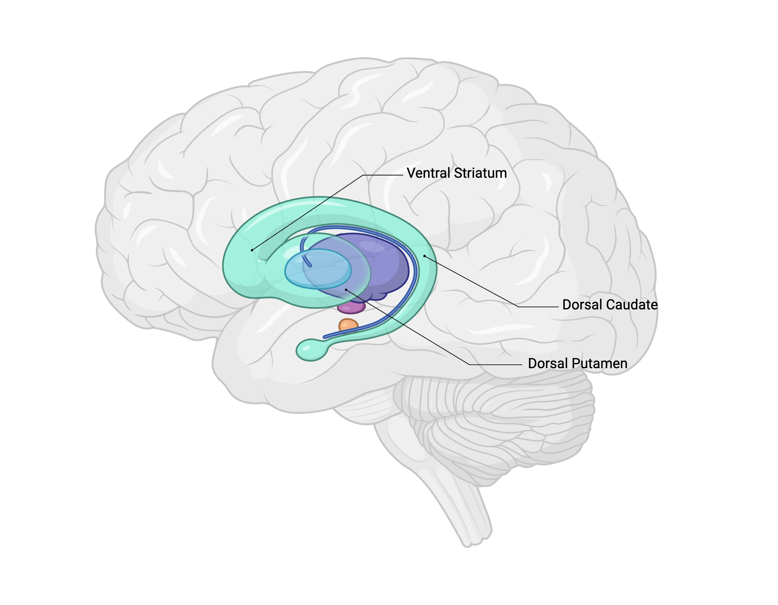
The imaging study focused on three main sub-regions of the striatum (pictured below):
Ventral Striatum (associated with apathy and motivation)
Dorsal Putamen (associated with movement speed)
Dorsal Caudate (associated with memory)
To participate in the study, individuals must have had their initial acute COVID-19 infection confirmed by PCR or rapid antigen testing and must have had a strictly mild or moderately severe case of COVID-19.
Participants were excluded if they had a history of neurological illness before getting COVID-19, or any recent cigarette or substance use.
A total of 67 participants had their brain dopamine transporter densities visualized:
24 individuals with Long COVID.
43 healthy controls, which are further divided into:
24 individuals who had a history of COVID-19 but had fully recovered.
19 individuals who had no history of COVID-19.
For the primary analysis, the 24 Long COVID participants were compared directly against 24 healthy, age-matched (+/- 5 years) controls.
To be included in the study, participants must have developed their neuropsychiatric symptoms from a previously healthy state within 3 months of their acute COVID-19 infection, and those symptoms must have lasted for at least 3 months prior to the study.
The participants with Long COVID exhibited a distinct profile of cognitive, neuropsychiatric, and physical symptoms outlined below.
Each of the 24 individuals with Long COVID experienced apathy (lack of interest, enthusiasm, or concern) and were required to meet the clinical criteria for at least five neuropsychiatric symptoms of a major depressive episode (determined using the Structured Clinical Interview for DSM-5), which included assessing:
Low concentration / cognitive slowing
Anhedonia (inability to feel pleasure)
Memory decline
Low energy
Low mood
Many participants also experienced active physical symptoms, recorded as follows:
Headache: 9 participants (37.5%)
Tiredness: 13 participants (54.2%)
Muscle aches or pains: 5 participants (20.8%)
General aches or pains: 3 participants (12.5%)
Comments
Nothing yet. Say the first thing.
Sign in to join the conversation.