
In Part I of this series, I covered why a “normal” ferritin level varies across research.
Now I want to zoom in on the situation where hemoglobin looks fine, serum iron is good, but ferritin level is low. How is that possible? Let’s dive into the labs!
Quick definitions:
Serum iron is the amount of iron in your blood.
Hemoglobin is the protein inside the red blood cells that uses iron to carry oxygen.
Ferritin is the protein that stores iron.
Transferrin Saturation is the percentage of carrier proteins that move iron.
Total Iron-Binding Capacity (TIBC) measures how well proteins (like transferrin) bind and carry iron in the blood.
It’s called iron depletion without anemia, also called latent or non-anemic iron deficiency (IDNA).
It’s the earliest stage of iron deficiency, where body iron stores are diminished (what ferritin actually reflects). But hemoglobin and serum iron remain normal because red blood cell production has not yet been compromised.
The staging framework researchers use lines up with that idea directly.
Stage 1 (IDNA-1) is serum ferritin roughly <30 ng/mL with transferrin saturation >16%.
Stage 2 (IDNA-2) is ferritin <20 ng/mL with transferrin saturation also dropping <16%.
Stage 3 (IDA-3) is when hemoglobin finally drops below 12 g/mL, and you have crossed into frank anemia.
Hemoglobin is the last domino to fall, not the first. By the time it drops, you have already been running on depleted stores for a while.
IDNA is also extremely common.
A 2025 systematic review and meta-analysis (Thompson and colleagues, Pediatric Blood & Cancer) pooled data from over 17,500 collegiate athletes across 23 countries and found that roughly 23% had ferritin below 20 ng/mL, and just under 54% fell below 50 ng/mL.
In other words, between 25 and 50% of collegiate athletes are walking around with depleted iron stores at any given time.
If ferritin is low, but hemoglobin and serum iron look normal. Here is what else to look at for a better picture of what’s going on:
A TSAT <16–20% confirms restricted iron supply to tissues even when serum iron appears "normal" on a single measurement, and it’s time to focus on dietary iron and supplementation.
Note: TSAT is determined by serum iron/TIBC x 100%. In true deficiency, TIBC tends to rise as the body wants to increase transferrin production so it can scavenge whatever iron it can find. The body is so wildly smart, it’s amazing.
Ferritin is an acute-phase reactant, meaning it reacts to inflammation. Whether that inflammation is from an infection, injury, or a really intense workout.
A CRP value helps confirm that the low ferritin is not being masked by inflammation (i.e., the true iron deficit may be even worse if CRP is elevated).
In addition to a standard CBC panel, checking a reticulocyte hemoglobin lab (CHr/Ret-He), if available, measures the amount of hemoglobin in newly produced red blood cells.
This lab is an early marker of whether the body has trouble supplying iron to make new red blood cells (iron-restricted erythropoiesis). And, before the size and volume of red blood cells decrease.
I’ll dive deeper into CRP and CBC later in this article.
The AGA and ACG recommend testing for celiac disease in patients with unexplained iron deficiency, as celiac disease impairs iron absorption in the small intestine.
Assess caloric intake, RED-S screening, iron-rich food consumption, vegetarian/vegan status, and menstrual blood loss if applicable.
If iron deficiency persists despite adequate supplementation, or in male athletes or postmenopausal women without a clear cause, an endoscopy should be pursued to rule out any hidden GI bleeding.
I asked the question: Why do CRP and CBC labs matter in relation to ferritin? Let’s dive into this together.
Ferritin rises with inflammation, which can mask true iron deficiency by making stores appear adequate when they are not.
So a CRP lab will provide context to the ferritin value in two ways:
A low CRP (<5 mg/L): The ferritin value can be taken at face value. In this case, a low ferritin reliably reflects depleted iron stores.
Elevated CRP: The true iron deficit is likely worse than the ferritin suggests.
Most standard labs would consider a CRP >10 mg/L as elevated. However, the BRINDA project that studied inflammation-adjusted ferritin cites >5 mg/L as the threshold.
A 2010 meta-analysis found that inflammation increases ferritin by approximately 30–90%. This is a big range, but basically, ferritin rises and falls with the phases of the inflammatory response.
In athletes, post-exercise inflammation (via IL-6) can temporarily raise both CRP and ferritin, so timing of blood draws relative to training matters.
Key Takeaway: The day (or two) before your blood is drawn for labs, plan recovery/rest days or a lighter training load to reduce that inflammatory response.
Standard red blood cell labs reflect iron-restricted erythropoiesis (the body has trouble supplying iron to make new red blood cells).
But they are late markers, meaning they change only after iron deficiency has persisted long enough to affect the mature red cell population (which has a ~120-day lifespan).
Standard red cell labs:
MCV (mean corpuscular volume): Falls below 80 fL in established iron deficiency anemia, but remains normal in early/latent iron deficiency.
MCH (mean corpuscular hemoglobin): Drops below 27 pg, paralleling MCV changes.
RDW (red cell distribution width): Often the earliest conventional CBC abnormality. It rises above 14.5% as a mixture of normal and iron-deficient red cells emerges, even before MCV drops.
Reticulocyte hemoglobin content is reported as CHr or Ret-He. It measures the hemoglobin content of newly released red blood cells and is the most sensitive peripheral blood marker for detecting iron-restricted erythropoiesis before anemia develops.
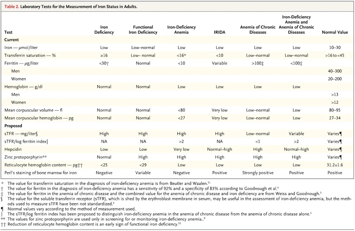
The following table from a New England Journal of Medicine review summarizes how these iron-related markers behave across the spectrum of iron deficiency states:
When you have an athlete with low ferritin, no GI symptoms, and the training load and diet are a reasonable explanation. An aggressive GI workup is not the first move.
That said, it is worth knowing where the line actually sits for further testing.
The 2020 American Gastroenterology Association (AGA) clinical practice guidelines on the GI evaluation of iron deficiency anemia lay out when investigation is warranted, though it is worth being precise about scope:
The recommendations apply once someone has crossed into iron-deficiency anemia territory (low hemoglobin, not just low ferritin).
For suspected celiac disease, the AGA recommends starting with serology instead of a small bowel biopsy.
For postmenopausal women and men with confirmed iron-deficiency anemia and no clear alternative explanation, the AGA recommends bidirectional endoscopy (upper endoscopy plus colonoscopy) to rule out a GI source, including malignancy.
For young endurance athletes and premenopausal women with IDA, the recommendation is softer and gives more room for clinical judgement. The doctor and patient can reasonably decide to hold off on GI testing and start with diet changes and iron supplementation.
If no cause of IDA is identified after endoscopy, testing for H. pylori is recommended. H. pylori impairs iron absorption independent of any bleeding it might cause.
Stay tuned for part 3 of this series as we dive into strategies to increase ferritin through diet and supplementation.
Normal hemoglobin with low ferritin is its own diagnostic stage, non-anemic iron deficiency (IDNA), not a lab error. It is extremely common, affecting an estimated quarter to half of collegiate athletes depending on the cutoff used, and it can impair aerobic capacity and energy well before anemia ever shows up. Athletes are more prone to it than the general population. Beyond ferritin, transferrin saturation, soluble transferrin receptor, reticulocyte hemoglobin content, inflammation markers, and a real diet and menstrual history all add useful context. A GI workup is not the default move for isolated non-anemic depletion. It becomes relevant when the numbers do not add up, or the deficiency will not resolve with treatment.
What resonated with you? What questions do you still have? Whether you’re an athlete, a coach, or a fellow RD, I want to hear your perspective. Leave a comment below and let’s keep the conversation going.
Peeling P, Dawson B, Goodman C, Landers G, Trinder D. Athletic-induced iron deficiency: new insights into the role of inflammation, cytokines and hormones. European Journal of Applied Physiology. 2008;103(4):381-391.
Clénin G, Cordes M, Huber A, et al. Iron deficiency in sports, definition, influence on performance and therapy. Swiss Medical Weekly. 2015;145:w14196.
Thompson AA, et al. The global prevalence of iron deficiency in collegiate athletes: a systematic review and meta-analysis. Pediatric Blood & Cancer. 2025.
Barney DE Jr, Ippolito JR, Berryman CE, Hennigar SR. Running increases hepcidin and decreases dietary iron absorption. Journal of Nutrition. 2022;152:2039-2047.
Ko CW, Siddique SM, Patel A, Harris A, Sultan S, Altayar O, Falck-Ytter Y. AGA clinical practice guidelines on the gastrointestinal evaluation of iron deficiency anemia. Gastroenterology. 2020;159:1085-1094.
Auerbach M, DeLoughery TG, Tirnauer JS. Iron deficiency in adults. JAMA. 2025.
Hamdeh S, Micic D, Hanauer S. Review article: drug-induced small bowel injury. Alimentary Pharmacology & Therapeutics. 2021.
ter Steege RW, Kolkman JJ. Review article: the pathophysiology and management of gastrointestinal symptoms during physical exercise, and the role of splanchnic blood flow. Alimentary Pharmacology & Therapeutics. 2012.
Bjarnason I, Scarpignato C, Holmgren E, et al. Mechanisms of damage to the gastrointestinal tract from nonsteroidal anti-inflammatory drugs. Gastroenterology. 2018.
Stewart JG, Ahlquist DA, McGill DB, et al. Gastrointestinal blood loss and anemia in runners. Annals of Internal Medicine. 1984;100:843-845.
Stoffel NU, Cepeda-Lopez AC, Cervantes-Gracia K, et al. The effects of reducing chronic inflammation in overweight women on serum hepcidin and iron absorption with and without supplemental ascorbic acid. British Journal of Nutrition. 2021;126(6):877-884.
Nolte S, Malhan D, Klemmer A, et al. Training in normobaric hypoxia induces hematological changes that affect iron metabolism and immunity. Scientific Reports. 2025;15:17757.
Garvican-Lewis LA, Vuong VL, Govus AD, et al. Iron insufficiency diminishes the erythropoietic response to moderate altitude exposure. Journal of Applied Physiology. 2019;127:1569-1578
No posts
Comments
Nothing yet. Say the first thing.
Sign in to join the conversation.