
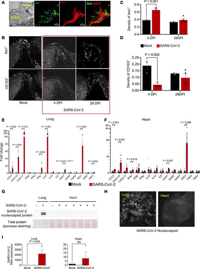
(A) Representative images showing the conduction tissue marker contactin 2 (red) and the distribution of Iba1+ macrophages (green) in the region. (B) Representative images showing Iba1+ and CD163+ macrophages in the conduction region in mock- or SARS-CoV-2–infected hearts at 4 and 28 dpi. (C and D) Density of Iba1+ cells and CD163+ cells in CCS region of the mock- or SARS-CoV-2–infected hearts at 4 and 28 dpi. (E and F) Gene expression of cytokines and IFN-stimulated genes in lung (E) and heart (F) at 4 dpi, evaluated with qPCR. Unpaired 2-tailed t test was used to test for significance in normally distributed lung data in E. Mann-Whitney U test was used as the nonparametric significance test in heart data in F. (G and H) SARS-CoV-2 nucleocapsid protein was detected in lung, but not in the heart, by Western blot (G) or by immunofluorescence (H) at 4 dpi. (I) Viral spike protein mRNA was detected in lung, but not in the heart, by qPCR at 4 dpi. Unpaired 2-tailed t test.
I referred to the Spike Protein shortly after the beginning of the Pandemic as being a “Swiss Army Knife of Death.” The graphic I created illustrating this is now ubiquitous with many claiming to have created it. I care not. The point is that the information is out there.
Today I would like to discuss another “tool” in this lethal multi-purpose weapon. This tool is the ability of the Spike Protein to remove (alter to a harmful state) cardiac resident macrophages.
To understand this, let’s look at what cardiac resident macrophages do. They prevent sudden cardiac death during heart stress by maintaining cardiac impulse conduction, preventing lethal arrythmias.
Cardiac arrhythmias are a primary contributor to sudden cardiac death, a major unmet medical need. Because right ventricular (RV) dysfunction increases the risk for sudden cardiac death, we examined responses to RV stress in mice. Among immune cells accumulated in the RV after pressure overload-induced by pulmonary artery banding, interfering with macrophages caused sudden death from severe arrhythmias. We show that cardiac macrophages crucially maintain cardiac impulse conduction by facilitating myocardial intercellular communication through gap junctions.
Cardiac macrophages prevent sudden death during heart stress
https://www.nature.com/articles/s41467-021-22178-0
In the context of the Spike Protein, we discover something very interesting. Patients with severe COVID have reduced numbers of these protective macrophages vs inflammatory macrophages.
Interestingly, patients with severe COVID disease have increased numbers of inflammatory monocyte-derived macrophages (v.v. resident macrophages); a macrophage subset that has been shown to express gene programs associated with tissue repair and fibrosis [15].
“Role of Cardiac Inflammation in the Pathology of COVID-19; relationship to the current definition of myocarditis”
https://pmc.ncbi.nlm.nih.gov/articles/PMC9061642/
Also, observe the extremely dramatic loss in density of these protective macrophages following COVID infection.
Following SARS-CoV-2 infection at 4 dpi, significant macrophage infiltration was observed throughout the heart, and the density of Iba1+ macrophages increased by 78% in the AV node/His bundle region of COVID-19 animals compared with mock-infected controls (Figure 3C). Despite the increase in macrophage infiltration, the density of CD163+ macrophages decreased by 74% in the AV node/His bundle region.
Innate immune activation and mitochondrial ROS induce acute and persistent cardiac conduction system dysfunction after COVID-19
https://insight.jci.org/articles/view/193164
Yet, what is most important here is that COVID infection (the complete virus) need not be present to induce this potentially catastrophic situation. The Spike Protein alone can induce it.
Cardiac arrhythmias are a common complication in severe and fatal cases of COVID-19, with increased arrhythmia incidence correlated with severity of viral infection2,3. These patients also have heightened activation of resident macrophages and elevated expression of inflammatory cytokines and show evidence of vascular damage and cardiac remodeling. Identifying the pathophysiologic origins of cardiac arrhythmias in severe and fatal COVID-19 has inspired much debate. A number of studies, using in vitro heart tissue and human induced pluripotent stem cells, have suggested that cardiac damage may result from direct infection of myocytes by SARS-CoV229,30. Yet, others have proposed cardiac arrhythmias as a consequence of the systemic cytokine storm following viral infection9,10,11. We have previously provided strong evidence, using human cardiac tissue from fatal COVID-19 cases, that acute insult from viral spike protein triggers inflammation-induced vascular leak and consequent proarrhythmic disruption of NaV1.5-rich ID nanodomains7. Briefly, histologic and molecular studies in cardiac tissue from 11 fatal COVID-19 cases demonstrated presence of perivascular viral spike protein, elevated expression of pro-inflammatory cytokines, considerable microvascular damage including perivascular edema, and remodeling of key ID components essential for proper impulse propagation. SARS-CoV2 RNA was not detected with an ultrasensitive in situ hybridization method, consistent with other findings that note the lack of viral RNA despite clearly evident cardiac abnormalities in fatal COVID-197,9,12,15. These findings were consistent with results from our previously defined arrhythmia mechanism, whereby inflammation-induced vascular leak, and consequent edema, disrupts ID nanodomains and slows conduction, thereby generating a proarrhythmic substrate16.
SARS-CoV-2 spike protein-induced inflammation underlies proarrhythmia in COVID-19
https://www.nature.com/articles/s41598-025-12807-9
If persistent spike antigen maintains a chronic, low-grade inflammatory state—as proposed in the Smoldering Macrophage Syndrome hypothesis—the continual replacement of resident macrophages by inflammatory monocyte-derived macrophages could impair the specialized support these cells provide to the cardiac conduction system. This possibility has not yet been directly tested but deserves investigation.
At the postsynaptic level, we previously showed that infiltrating monocyte-derived macrophages increased and resident macrophages decreased in the cardiac conduction system during the acute phase of SARS-CoV-2 infection.20 The early loss of resident macrophages was prevented by mTEMPO or Ruxo, without significant effects on the infiltration of monocyte-derived macrophages.20 Thus, the loss of important supporting functions of resident macrophages50 in the conduction system could contribute to altered neuron-pacemaker interactions. Activation of the innate immune system has been shown to induce chronic inflammation or neuropathy via neuroimmune cross talk,51 and there is increasing evidence that SARS-CoV-2 infection is associated with neuroinflammation and disorders of the cranial and peripheral nervous system,34,52 which could contribute to ANS dysfunction in patients with long COVID.
Dynamic changes in cardiac autonomic function persist in the postacute phase after SARS-CoV-2 infection in a hamster model of COVID-19
https://www.sciencedirect.com/science/article/pii/S2666501826002515
The evidence now paints a compelling picture. Cardiac resident macrophages are essential components of the heart’s electrical conduction system, helping maintain normal impulse propagation and protecting against lethal arrhythmias. Independent studies have shown that SARS-CoV-2 infection depletes these specialized macrophages while replacing them with inflammatory monocyte-derived populations, and that the Spike Protein alone can provoke inflammatory remodeling of cardiac tissue even in the absence of detectable viral RNA. If persistent Spike antigen continues to sustain low-grade innate immune activation, as proposed in the Smoldering Macrophage Syndrome hypothesis, then the chronic loss or dysfunction of these protective resident macrophages could represent one previously overlooked mechanism linking persistent spike exposure to long-term cardiac electrical instability and sudden cardiac death.
Thank you, as always, for keeping me going. I appreciate all of your support and am grateful to everyone in our community. Also, a great thank you to the generous donor who contributed over the weekend. Please have a blessed week.
One-Time Donation
https://buy.stripe.com/6oUbJ0auFaWkcLs2ZXeUU00
No posts
Comments
Nothing yet. Say the first thing.
Sign in to join the conversation.