
Peter from the Hyperlipid blog has recently retired, in which he shared his nickname “The Wizard”. (I recall that he posted a picture somewhere with this, but can’t find it now. Peter has left X, and seems to have deleted his account, unfortunately.)
It’s an appropriate nickname, IMHO. I’ve learned an enormous amount from him, and I’ve read every post he ever did on his blog.
Peter and I both agree that excess linoleic acid (LA) is a problem, causing obesity, diabetes, and probably many other ills.
However we disagree to some extent about the mechanism. (The nice thing is we’re both winning from this discussion, and often shared information back and forth, as we both share the same goal: the truth.)
In a nutshell, Peter thinks it’s related to how LA is handled in the mitochondria, specifically the electron transport chain (ETC); while I think it’s related to LA’s ability to peroxidize and become toxic, negatively affecting many aspects of cell functioning.
When it enters the body LA has, basically, three fates:
Disposal, via a variety of mechanisms.
Incorporation into cell membranes throughout the body.
Oxidized (different from peroxidized) for fuel.
(Oxidation is when a fuel like LA is, essentially, combusted, in the body to CO2 and water. Peroxidation is when an oxygen atom is added to a fuel molecule, often with a hydrogen atom, known as “hydroxy”, hydrogen + oxygen. Two entirely different fates.)
Peter’s position revolves around 3, mine around 2. In another nutshell, Peter thinks LA is inherently bad, due to it’s effect on intra-cellular insulin signalling via inadequate ROS generation in the ETC; while I think that while he’s probably correct as far as it goes, but that is overwhelmed by the primary mechanism for badness, which depends on LA’s tendency to turn into toxins like HNE (4-hydroxynonenal).
So while doing some Google Scholar queries spurred by Peter’s recent post:
“Insulin resistance (16) Yes. Vitamin E can cause weight loss.”
I came across this paper:
This paper is looking at the effect of increasing the oxidation of fats to resolve metabolic disease, specifically non-alcoholic fatty liver disease (NAFLD, now also called MAFLD—Metabolic-Dysfunction Associated Fatty Liver Disease—maybe some other time I’ll explain why I’m not using that term…)
Chaperonin is the heat-shock protein 60 (HSP60), and while it has a number of roles, it turns out that one of them is to facilitate the oxidation of fats. In diseases like obesity, diabetes, or NAFLD, impaired oxidation of fats is often present, as is a reduced quantity of HSP60.
“Decreased HSP60 expression correlated with severe steatosis in human NAFLD biopsies and murine DIO….
Impaired fatty acid oxidation is a hallmark of obesity development [36], while promotion of lipolysis prevents increased adiposity and body weight gain [37, 38]. (Weng, 2023—All subsequent quotes will be bolded)
One of the effects of the diabetes drug metformin is to upregulate HSP60 production (Docrat, 2020).
So in (Weng, 2023) they fed the mice a high fat diet, D12331.
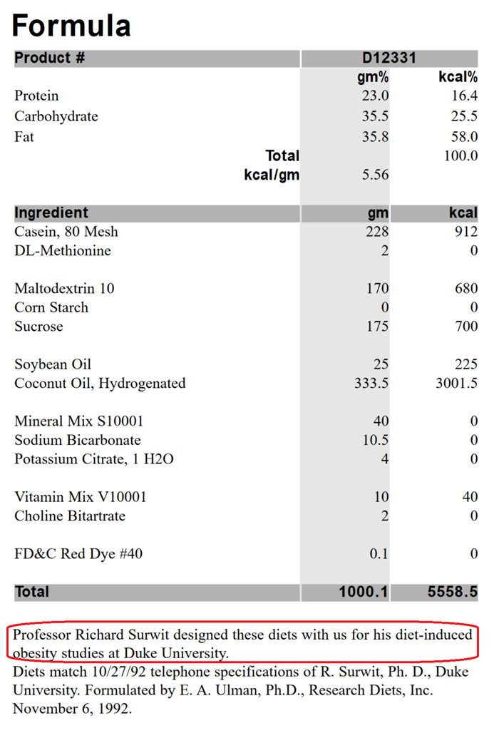
Annoyingly, the methods in (Weng, 2023) lists D12331 as coming from LabDiets, while in fact it is produced by Research Diets (Research Diets, Inc., 2006); although LabDiets produces a diet called 58R3, about which they say, “Originally manufactured as “D12331”. One of four diets in the Surwit Series” (TestDiet, Land O’ Lakes. 2023). So I’m just assuming this is D12331, and that the apparent misattribution is a typo, but even if they used 58R3, it appears to be a clone of D12331. It’s one of the diets produced by the obesity researcher Richard Surwit, is pretty commonly used in obesity research, but it has minimal LA, so is an interesting case study in what actually is causing diabetes.
D12331 is the diet Peter discusses in his post above (Dobromylskyj, 2026):
“The Surwit diet does this.”
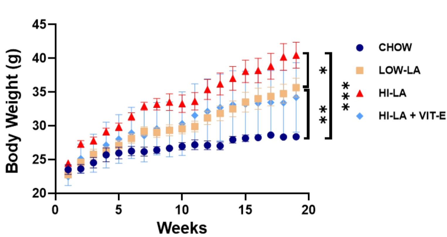
What Peter is referring to is this graph, from (Graham, 2023):
(Graham, 2023) is a paper we discussed with the first author here:
Dalton designed this paper based on a number of my posts and ideas, and—thank God!—it worked as expected.
In Peter’s post he observes that there seem to be two obesogenic triggers in the diet used in (Graham, 2023)—it’s not exactly D12331, but it’s close. Medium-Chain Triglycerides (MCT), from the coconut oil; and LA. Vitamin E blocks the LA-induced obesity, but not the MCT-based obesity. Adding Vitamin E to the Surwit-derived diet + 8% LA (red triangles) reduces obesity to the level of the 1% LA Surwit diet (yellow squares).
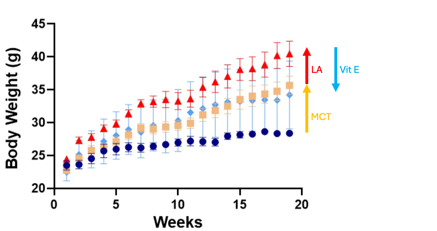
It appears to cancel out most of the LA-induced obesity.
(We’re not going to worry for the moment about MCT-induced obesity here, as a) I have never seen evidence that coconut fat causes obesity in humans, and b) mice aren’t humans.)
In the comments to (Dobromylskyj, 2026) I observed that this effect is similar to that seen in (Chang, 2023), also fed D12331, but via an entirely separate mechanism—or is it?
How do we reconcile your comments about the diets above with this paper (Chang, 2023), which is able to similarly reduce the fat effect of LA in the 1% LA Surwit diet?…
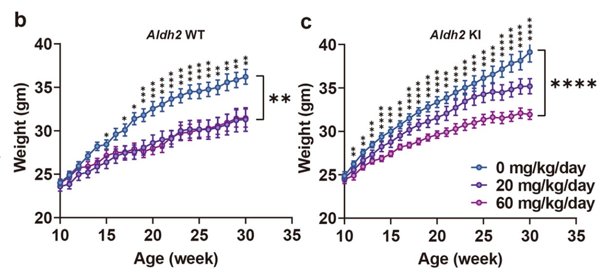
Comparing the outcomes, it looks like there are two distinct obesogenic effects of Surwit (D12331): MCT and LA. Either vit. E or increased ALDH2 will ameliorate the effect of LA on obesity, while leaving the effect of MCTs untouched.
Both vit. E and ALDH2 are addressing the lipid peroxidation pathway...
In (Graham, 2023) Vitamin E reduces body weight from ~38 to ~32g (eyeballing it). In (Chang, 2023) the ALDH2 stimulator AD-9308 reduces body weight from ~35 to ~30g (again, eyeballing it) in the wild-type (WT) mice, and from ~38 to ~30g in the human genetic mutation knock-in (KI) mice.
We discussed (Chang, 2023) in this podcast:
What’s fascinating here is the common denominator. Vitamin E has an effect because it prevents LA from becoming peroxidized, and AD-9308 cleans up peroxidized LA via stimulating the detoxification pathway aldehyde dehydrogenase (ALDH2).
In (Graham, 2023), Dalton measured levels of malondialdehyde (MDA), which made me sad, because I prefer to look at HNE. HNE can only be produced from Ω-6 fats like LA, while MDA can also be produced from Ω-3 fats. HNE is a better signal for Ω-6-specific toxicity. Dalton didn’t have the budget for HNE testing.
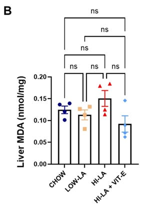
We can see that MDA is reduced by vitamin E. Depending on the details of how the TBARS test for MDA is done, it can also produce a positive result for oxidized LA.
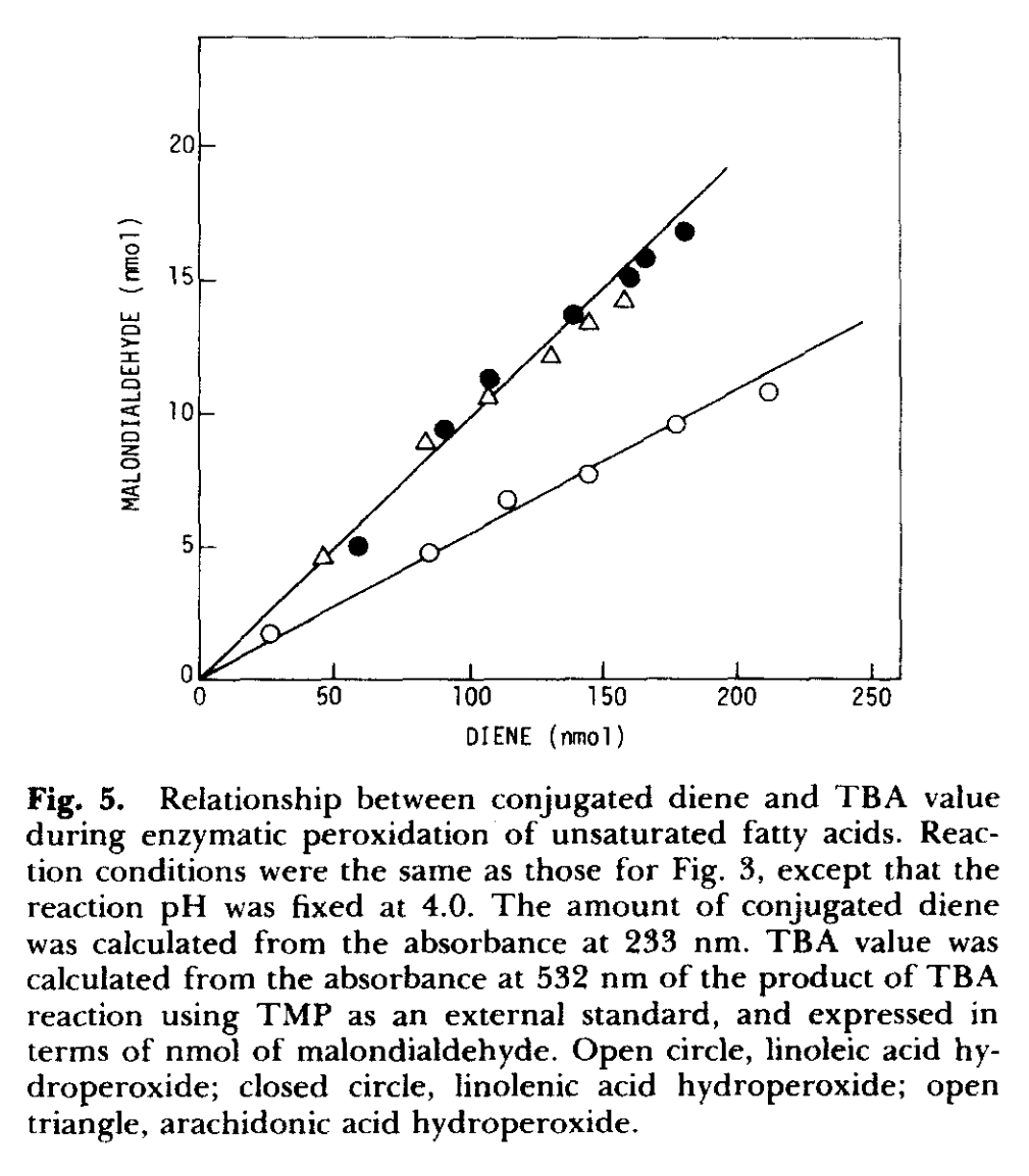
“These data show that the substances reacted with TBA in our system are unsaturated fatty acid hydroperoxides.” (Ohkawa, 1978)
We know from other work that vitamin E can prevent the production of lipid hydroperoxides (L-OOH, such as hydroxyoctadecadienoic acid (HODE), a peroxidized LA:
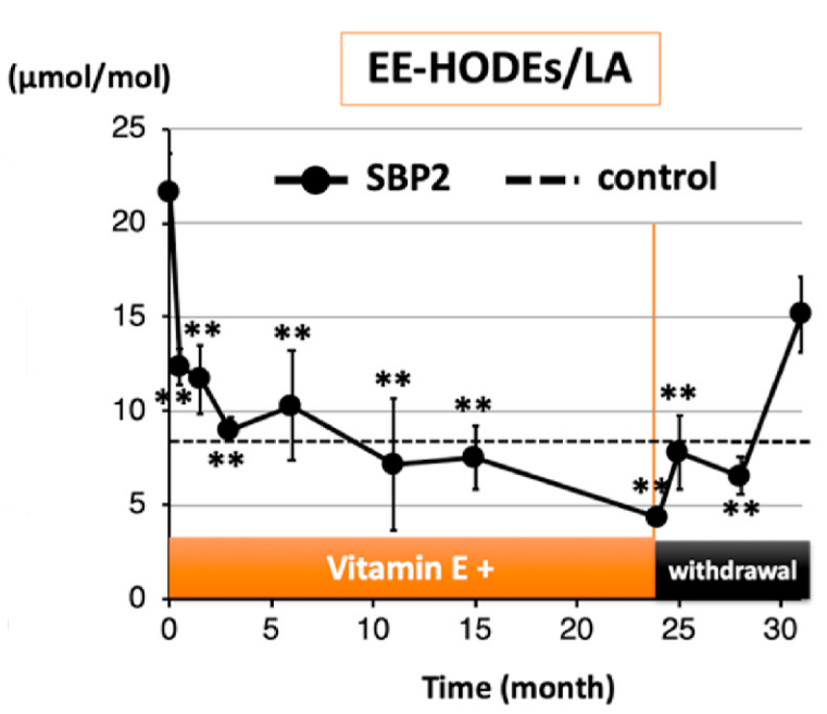
Which are precursors to HNE:
“L-OOHs undergo further oxidation, e.g., as an iron-dependent Fenton reaction, which generates various oxidation products with electrophilic properties, such as 4-HNE…” (Saito, 2021)
We can thus safely infer that HNE is also being reduced in (Graham, 2023).
(Weng, 2023) in my opinion reconciles the two approaches above. It forces LA from the pathway 2 described above, “Incorporation into cell membranes throughout the body”, to the pathway 3, “Oxidized for fuel”. If the cause of obesity and insulin resistance is insufficient ROS signalling from LA, this should exacerbate it (confounded by the fact that more fat may be being oxidized for fuel).
The authors use a transgenic mouse that over-produces HSP60, and in these mice both obesity and insulin resistance are reduced, despite a higher flux of fat through the electron transport chain.
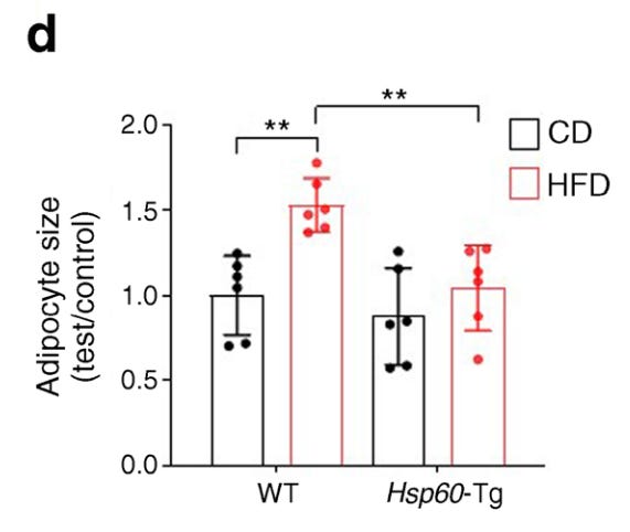
“Hsp60-Tg mice developed less body fat, had reduced serum triglyceride levels, lower levels of insulin resistance and higher serum adiponectin levels than wild-type mice upon HFD feeding.”
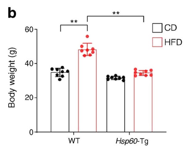
They additionally had smaller adipocytes.
The transgenic mice had a lower respiratory exchange ratio (RER), a measure of fat oxidation. So HSP60 is indeed promoting fat oxidation.
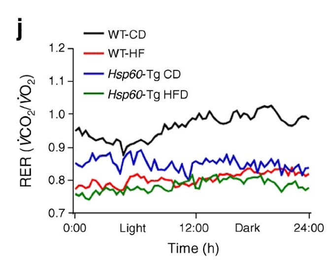
The control diet (CD) WT-mice had a high RER (black line) because they were burning mostly carbohydrates. Interestingly the biggest increase in fat burning was in the WT mice fed a high-carb diet.
And, significantly, they had a lower production of HNE.
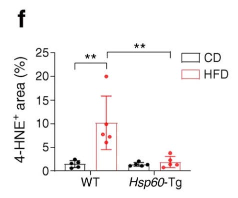
The electron transport chain is a significant source of HNE, due to being susceptible to dietary manipulation of fatty acids which alters the fat composition of the lipids holding the chain together, specifically the mitochondria-specific cardiolipin (Zhong, 2014).
HNE given alone produces larger adipocytes, through, it seems, damage to various proteins in the adipocyte during differentiation.
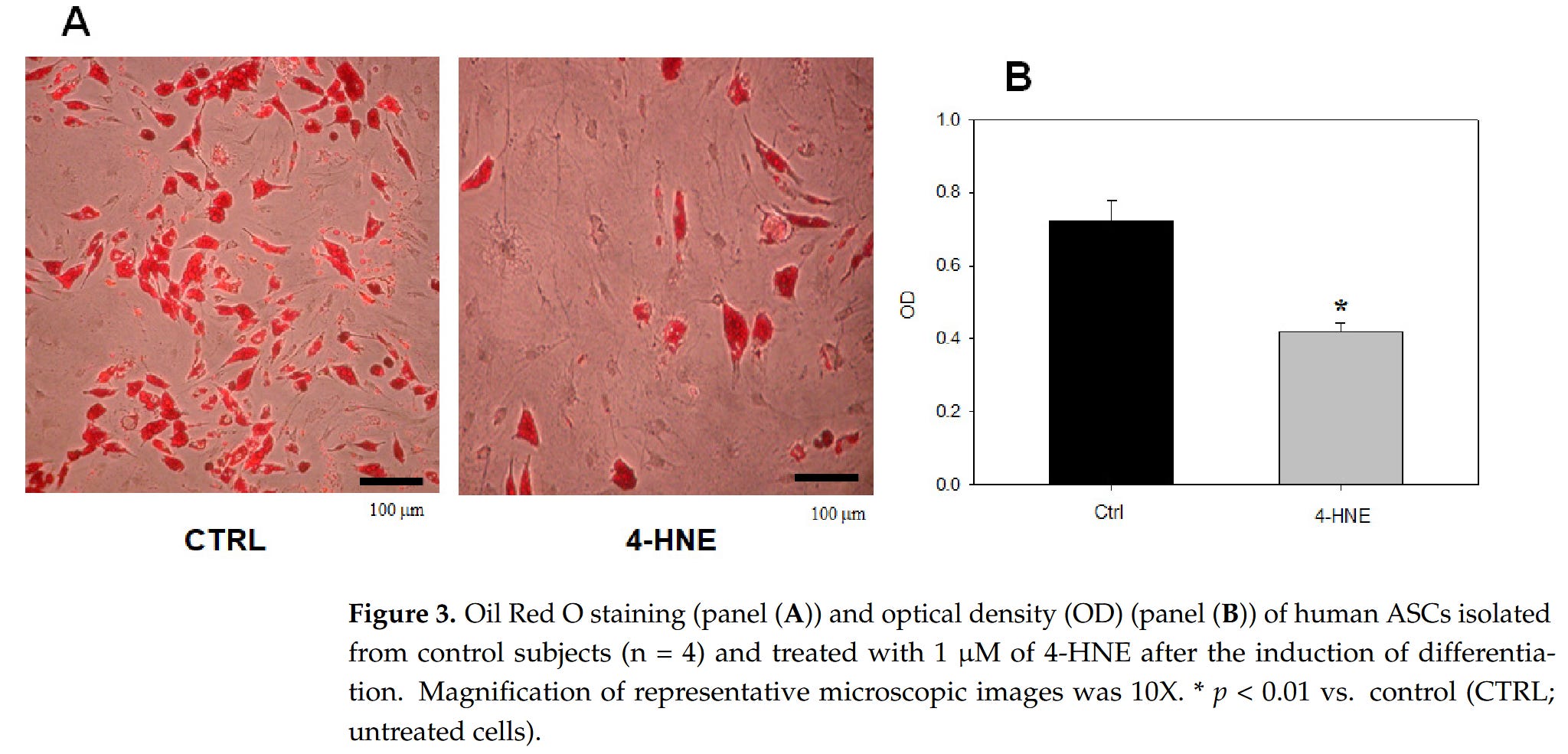
“As compared to the control (treatment with the 4-HNE vehicle DMSO), treatment with 1 μM 4-HNE inhibited the adipogenic differentiation of uncommitted ASCs, as ascertained by either contrast phase microscopy or Oil Red O staining by day 14 after the addition of a supplementation medium (Figure 3).” (Murdolo, 2023)
All the rest of the adipocyte disruption follows from excess HNE concentration—that’s a long post, for another time.
But, to make the point, (Murdolo, 2023) also look at HNE levels in normal and type-2 diabetic obese human adipocytes:
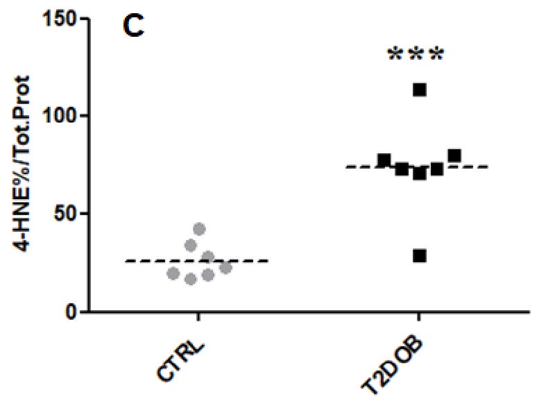
Another study looking at the same effect found similar results.
“Our data report for the first time impairment of the adipogenic capacity of OM preadipocytes taken from obese and morbidly obese IR individuals (including T2DM patients) compared to their IS counterparts, with a potential role of 4-HNE accumulation in this impairment.” (Jaganjac, 2017)
So how could HNE and HSP60 interact to cause these effects? The primary route of damage via HNE to cells is via binding to proteins forming adducts, which damage the protein and ultimately the cell (Ishimura, 2008).
One of the proteins HNE binds to is HSP60. Interestingly, in atherosclerosis, macrophages react to HNE-HSP60 adducts as they would to an antigen, stimulating the auto-immune reaction that typifies later atherosclerosis.
“Upon exposure to HNE, HNE-LDL, and LDL oxidized with CuSO4, but not to native LDL, HNE-HSP60 adducts were formed in THP-1 cells at levels well higher than in basal conditions.” (Arcaro, 2015)
Autoimmunity is typical of atherosclerosis, and macrophage infiltration into adipose tissue is another aspect in which the two processes share similarities.
Clearly one of the mechanisms by which HNE damages cells is by damaging this specific protein, which would explain why reduced levels are found in human disease related to HNE.
Comments
Nothing yet. Say the first thing.
Sign in to join the conversation.