
ENERGY SCIENCE
Full-Text Link to Primary Source
Blood and other fluids are essential for life. While scientists and physicians have studied blood for centuries, we’re still making new discoveries about what is in our blood and how the contents of our blood support our biology and our health. In this article, we cover a new paper that gives us a closer look at different types of particles found in blood and saliva, including particles that resemble mitochondria.
Blood flowing through your body is like a mass transit system that delivers oxygen, nutrients, and chemical messages to all of your cells. It also plays an important role in moving waste products and mobilizing your defenses when your body is injured or is experiencing an infection.
There are many well-known passengers in this transit system. Red blood cells carry oxygen and some carbon dioxide. White blood cells have specialized immune functions. And billions of platelets travel along.
White blood cells and platelets contain mitochondria. Red blood cells don’t.
Recent work by our group highlights some more mysterious passengers. We still need to investigate further before drawing any conclusions, but some of them look remarkably mitochondrial.
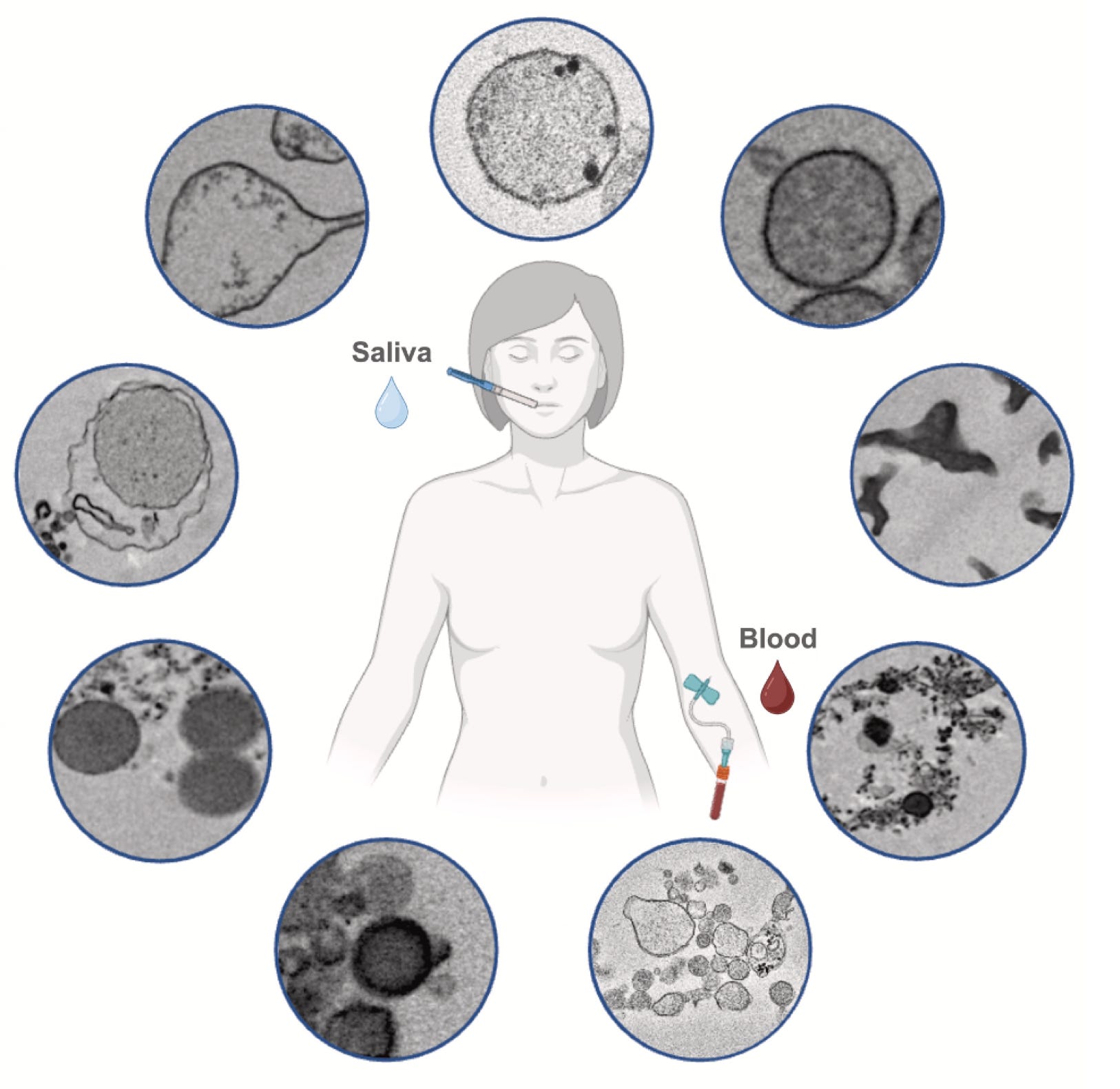
In addition to serving in delivery, clean-up, and protection roles, blood is the bridge between your breath and the transformation of energy in your mitochondria. It carries both the electron-giving food substrates, and the oxygen that accepts electrons in your mitochondria.
Mitochondria are multifaceted organelles. They perform a wide range of functions beyond transforming energy from food and oxygen into forms that your cells can use. For example, they produce signals that can influence inflammation, stress responses, gene activity, and cell survival. They are the processor of the cell.
As you may know, they also have their own separate genome—the mitochondrial DNA, or mtDNA.
Scientists have known for years that mtDNA can be found outside of cells, circulating in blood, saliva, and other bodily fluids. Circulating mtDNA is often called cell-free mtDNA (cf-mtDNA). The name brings to mind loose fragments of mtDNA drifting freely through the bloodstream.
Five years ago, we wrote a grant to study the effects of mental stress on cf-mtDNA. And the pro-inflammatory effects of cf-mtDNA. Turns out that most cf-mtDNA in our blood isn’t naked DNA capable of triggering inflammation.
Our hypothesis was wrong.
But what if some of that mtDNA is not actually free? What if it is packaged up inside pouch-like vesicles, fragments of mitochondria, or intact mitochondria traveling between your cells and tissues?
Mitochondria are usually described as structures that live inside our cells. But they do not always remain confined to the cells where they originated.
Work by Al-Amir Dache a few years ago suggested that normal human blood contained about one million mitochondria per milliliter. Mitochondria, interestingly, that could respire and contained intact mtDNA.
A growing body of research also suggests that different cell types can release mitochondria or mitochondrial components into the surrounding environment, which can then enter other cells. Mitochondria transfer to other cells via direct connections between the cells, inside extracellular vesicles, or possibly as free-standing extracellular structures.
Through these transfers, mitochondria may directly participate in communication between cells and tissues, or even transfer energetic resources from one cell to another.
So, we wanted to know, if we looked closely enough at what is in blood and saliva samples that contain cf-mtDNA, could we actually see evidence of mitochondria in transit?
To answer this question, I had the chance to work with Alexandra Volos, Soah Grace Franklin, Jeremy Michelson, Shannon Rausser, and Jonathan Brestoff, co-authors on the new paper. This project started as one of Jeremy’s PhD thesis projects.

Jeremy and Shannon collected blood and saliva from ten healthy adults, processing the samples using established methods (check out our paper for all the details). This left us with the small particles from samples of plasma, serum, and saliva.
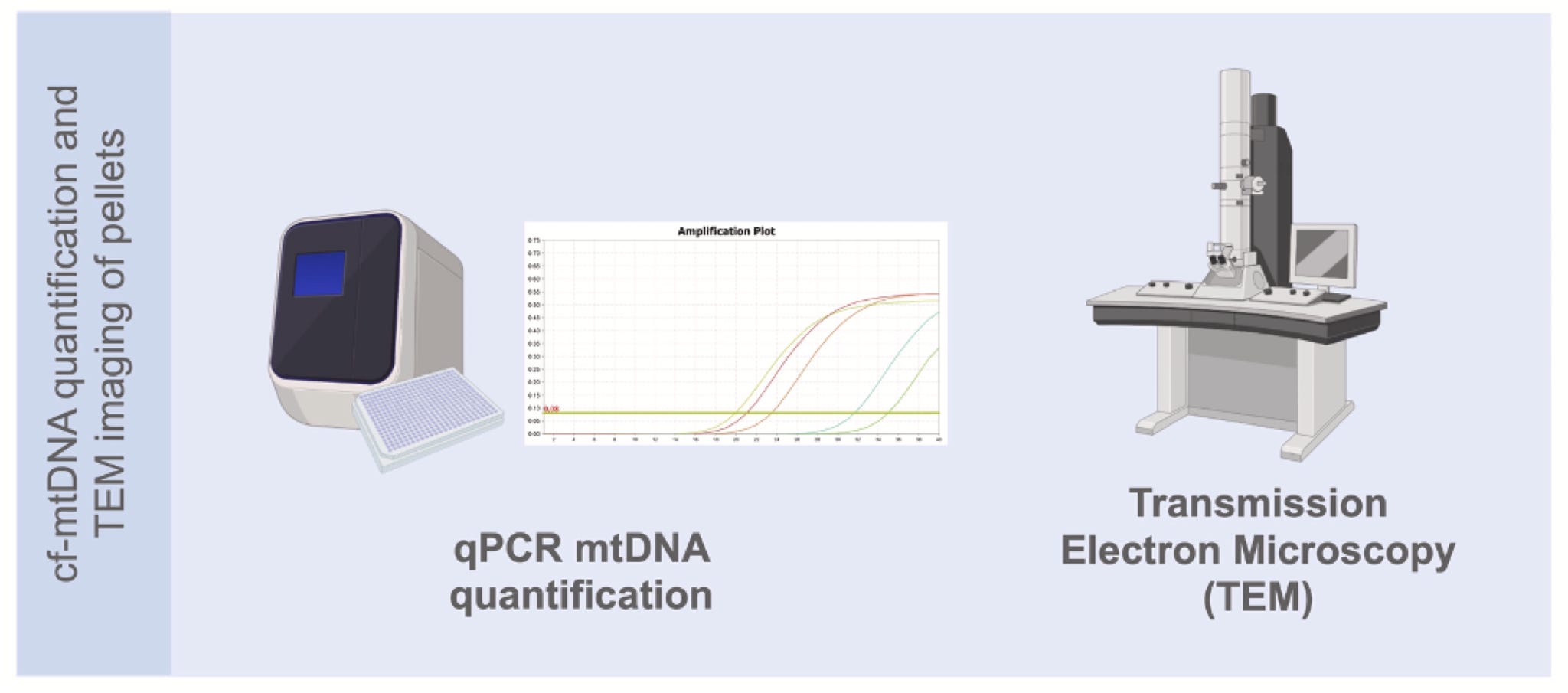
In each sample, we quantified how much mtDNA they contained with quantitative polymerase chain reaction (qPCR).
To get high-resolution images of the tiny particles in the blood and saliva samples, we used transmission electron microscopy. This involves shooting an electron beam through very thin slices of each sample.
With electron microscopy, the dense things, like DNA and proteins with metals, stop the electrons. The electron-lucent structures like the watery inside of the mitochondrial matrix let most electrons pass through. The contrasts reveals the cellular and sub-cellular structures in detail.
There was a lot to see. We collected thousands of single images, some stitched into large, high-resolution tiles. From these images, we classified particles into 14 different types based on their size, structure, and electron density. Plasma, serum, and saliva samples each had a distinct mix of particles.
Some particles were large, membrane-bound structures containing smaller vesicles. Others were diffuse and irregular or small and granular. The image below shows the wide range of particles that likely float around in your blood.
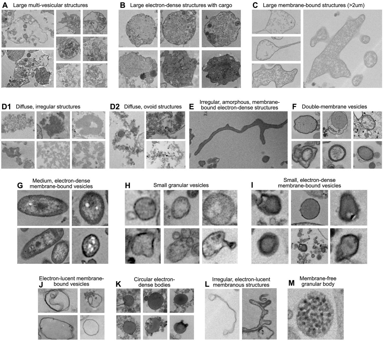
While we can’t say with certainty what each of these kinds of particles are or what they do from the images alone, it’s likely that we’ve captured cellular debris; biological packages carrying proteins, lipids, and other molecules; and particles involved in signaling.
Of particular interest to us, some of the particles had features consistent with extracellular mitochondria.
In each of our cells, the mitochondria and nucleus are the only organelles with a double membrane. Unlike the nucleus, the inner mitochondrial membrane folds inward to form cristae.
Several particles from our image catalog appeared to have this same general architecture. They had an outer boundary, an inner membrane, and, in some cases, internal folds resembling cristae. These particles were present in plasma, serum, and saliva, but they were more common in plasma.
Here are some from healthy participants (two on the left, one on the right):
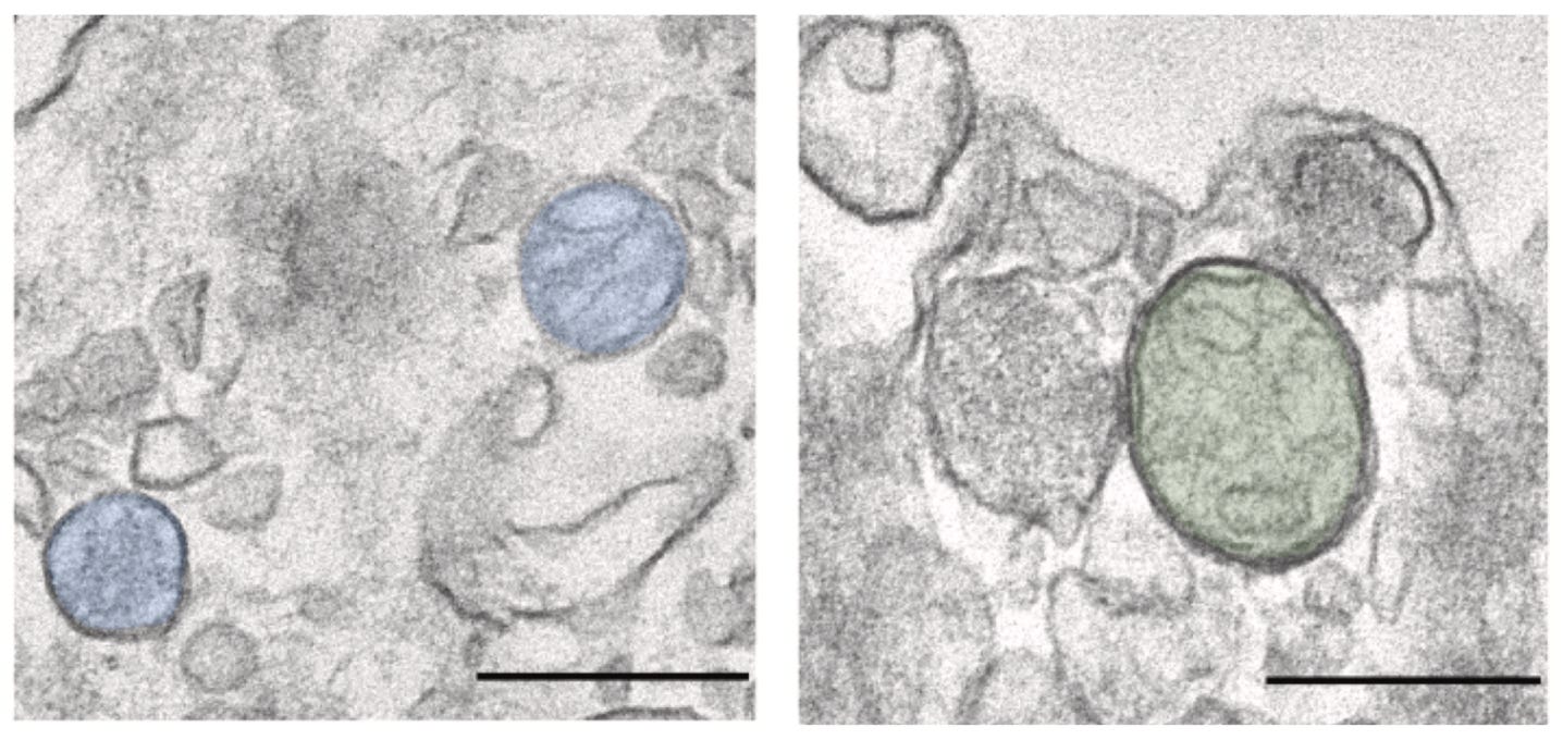
The images are compelling, but it’s important not to jump to conclusions. Until further studies can be done to determine if these particles are actually pieces of mitochondria, whole mitochondria, or something else entirely, we’re calling them mitochondria-like particles.
Whether mtDNA is exposed or enclosed inside a membrane-bound particle can change the kinds of biological effects that the mtDNA can have.
When released during injury or stress, mtDNA activates immune and inflammatory pathways. As a result, elevated cf-mtDNA is often interpreted as a sign of cellular damage or physiological stress. But mtDNA sealed inside a vesicle or an intact mitochondrion may serve different purposes and have different biological effects.
Stepping back to look at the big picture, cf-mtDNA may not be just one biological signal.
When we measure cf-mtDNA using typical methods, we are likely capturing the mtDNA that is truly cell-free and released from damaged cells, as well as mtDNA that is within vesicles or mitochondria-like particles. And a mitochondrion likely carries much more information than just mtDNA would.
Separating these potentially distinct sources of mtDNA is important because one may signal injury, while the others may be involved in communication, adaptation, or repair and healing.
If we confirm that the particles are mitochondrial in nature, we should examine if they are able to perform some of the many important tasks that mitochondria do. For example, can they maintain a membrane potential and transform energy?
Also, where do the particles come from and where are they going? Future research could track labeled mitochondria from their original cells to follow their movement and identify their destinations.
We could then evaluate whether the transfer of mitochondria changes things like energy resistance (éR). If so, incoming mitochondria could alter metabolism, inflammation, resilience to stress, and tissue repair. How might the nature or dynamics of these mitochondria-like particles change with exercise, aging, sleep, stress, infection, injury, or disease?
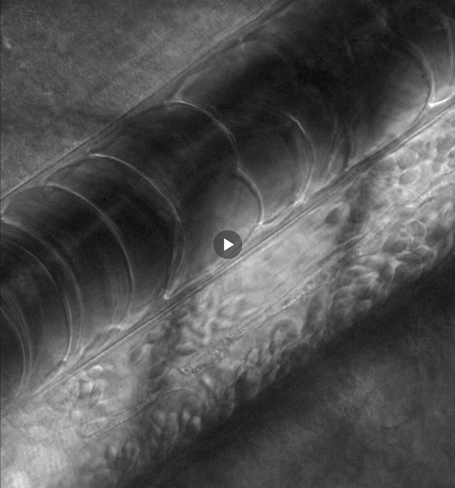
Through this study, we created an open-access catalog of high-resolution electron microscopy images that anyone in the world can use for free. This will serve as a resource to guide future studies of mitochondrial transfer, extracellular mitochondrial biology, and cf-mtDNA.
We don’t yet know if these intriguing particles are cellular debris, distress signals, delivery vehicles, a mode for reallocating metabolic capacity, or perhaps all of these things depending on the context.
For now, these mitochondria-like particles remain mysterious travelers whose journeys remain to be further explored.
Curious about mitochondria and how they move, connect, and support our energy needs?
Visit MitoLife to learn more and discover your energetic self through mitochondria.
No posts
Comments
Nothing yet. Say the first thing.
Sign in to join the conversation.