
Phew, it’s been awhile for a Field Notes! This one’s a bit longer than usual!
Get your notebooks ready :)
In this issue:
Where the alarm system sits and what sets it off
Why tissue swells, and what leaves the vessel first
The loop that keeps inflammation escalating
How your body shuts the whole thing down on purpose
What chronic inflammation looks like at the tissue level
I had to memorize this piece by piece, cell by cell, for a course conference (more like an oral exam) for my immunology course this semester. It was interesting that I was studying this the same week I got stung by a bee on my finger (DIABOLICAL). It was fun to actually kind of SEE it happening in realtime, even though I know all of us have experienced a cut, a bug bite, a scratch, etc.
We all know what it looks like, but ever wonder what’s happening inside that cut? What our immune cells are doing? How they’re acting? What causes all the swelling and itchiness?
Wonder no more, my friends, let me explain it all to you!
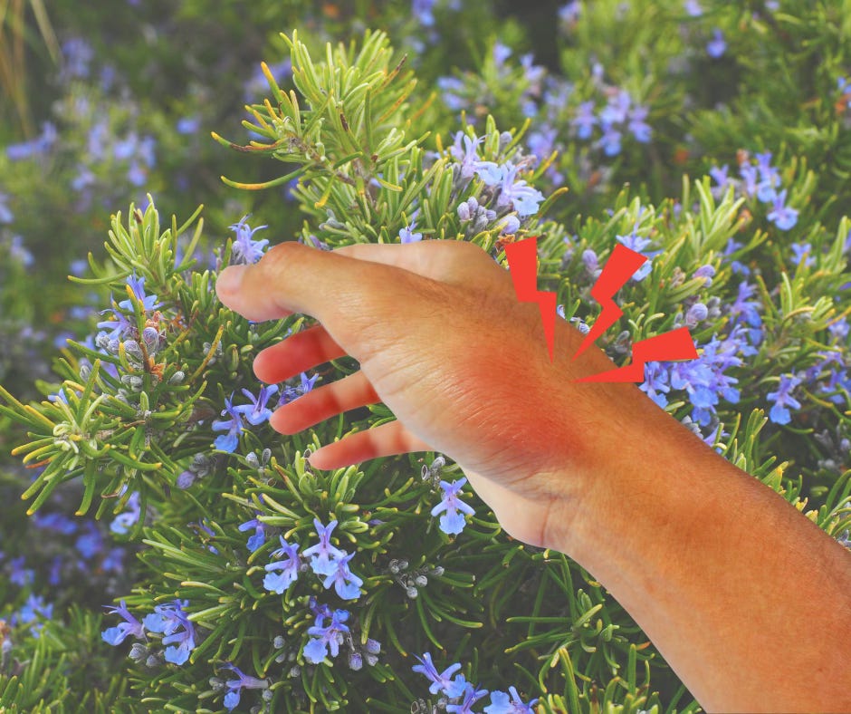
Okay, so let’s say you slice your finger open on the lid of a can. THE WORST.
Two things come in on that lid. Bacteria ride through the break in your skin. Right alongside them, a bunch of your own cells get crushed in the process, spilling their insides out into the surrounding tissue. Your body picks up on both of these using two separate sensing systems, each one watching for a different kind of trouble.
The cells doing the watching were already there before you even opened the can. Macrophages, dendritic cells, and mast cells (often termed “resident cells”) sit parked in your tissues on guard.
What does this mean? Well, they’re kind of decked out in receptors called PRRs, or pattern recognition receptors, and these are what pick up on trouble and sound the alarm to get the inflammatory response started (Roh & Sohn, 2018).
So, what counts as trouble? The bacteria riding in on that can lid are covered in PAMPs, or pathogen-associated molecular patterns, and they’re pretty self-explanatory, right? The PRRs recognize molecular patterns that come from pathogens, which could be LPS (lipopolysaccharide) from gram-negative bacteria, or even the little tail some bacteria use to swim around (flagellin). These are things our human cells just don’t make!
Then we’ve got DAMPs (also self-explanatory), damage-associated molecular patterns, coming from all those cells that were just sliced open by the sharp lid. Here the PRRs recognize things like ATP or mitochondrial DNA :( , the normal stuff that’s supposed to stay INSIDE the cell but sets off the alarm the moment it turns up OUTSIDE (Roh & Sohn, 2018).
This is why you’ll see swelling when you sprain your ankle, for example. No visible damage, no way for any outside bacteria to GET IN, but you still end up with some inflammation.
In the scenario we’re working with, though, you get both....sorry :(
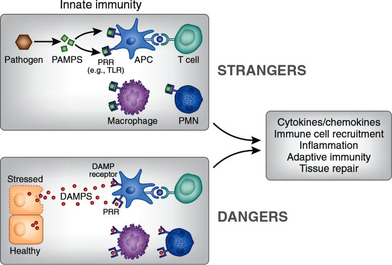
PAMPs and DAMPs
Remember those resident cells? Yeah? Okay, let’s focus on mast cells. And I know, you’ve probably heard all about them with the MCAS discourse online, but here, they’re not really our enemy. I’ll explain!!
So, the second the mast cell “senses” trouble, it degranulates, which is really just the fancy way of saying it dumps out everything inside of it: histamine, serotonin, leukotrienes, proteases, and cytokines like TNF-α and IL-1 (Granger & Senchenkova, 2010). And I literally mean the “second,” because this whole thing happens in seconds. Histamine is the one I want to zero in on here, because it reaches the endothelial cells lining your blood vessels and tells them to contract (Granger & Senchenkova, 2010).
Those cells are normally standing shoulder to shoulder, holding hands, sealing the vessel wall TIGHT. Now they’re scrunching up, letting go of each other, letting little gaps open up all along the blood vessel wall.
This leads to what’s called “increased vascular permeability,” meaning more things are able to get in and out of the blood vessel. And we want this to happen! (More on that coming up.) Histamine working on our vessels is actually what’s behind the swelling. Plasma proteins are the first thing to squeeze through those gaps into your tissue, and water follows right behind them (Granger & Senchenkova, 2010).
While the mast cell was doing its thing in a matter of seconds, those resident cells that first caught the PAMPs and DAMPs have been working on something a whole lot slower behind the scenes.
Their PRRs switched on a protein called NF-κB, which heads straight into the nucleus and starts flipping genes on (Liu et al., 2017). Those genes code for the next big wave of the response: pro-inflammatory cytokines and chemokines like IL-1, IL-6, IL-12, and TNF-α (Liu et al., 2017). None of this stuff was sitting pre-packed and ready like the mast cell’s stash. The cell has to build every single one of these from scratch, which is exactly why this part takes hours instead of seconds. The chemokines are basically a flare going up into the sky, and the neutrophils are the ones who come running.
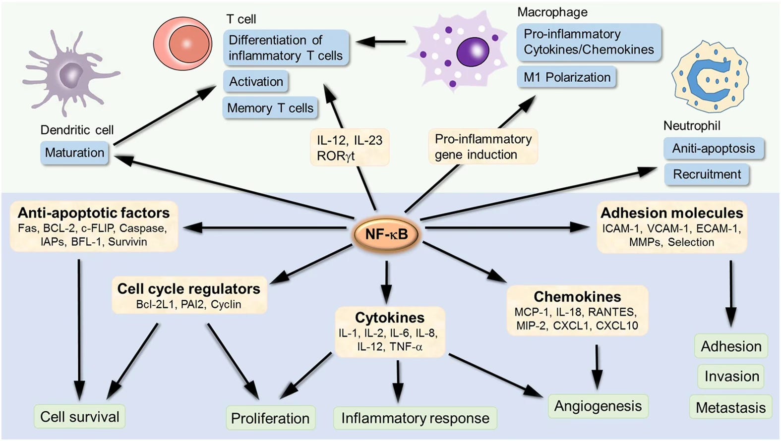
NF-κB target genes involved in inflammation development and progression.
Alright, so initiation got the ball rolling. Now amplification is what turns a small, contained response into something big enough to actually deal with the threat.
Those neutrophils we called in don’t just show up, do their job, and file out. As they arrive and get to work, they release their own batch of signaling molecules, the same kind of cytokines and chemokines that called them in the first place. So the first responders are basically radioing for backup, and that backup shows up and radios for even more. One cell calls in ten, those ten call in a hundred, and the whole thing snowballs. Yikes.
A feedback loop like this, a response that feeds itself and keeps building instead of staying flat, is what makes amplification its own phase. And it’s exactly what you want when there’s a real threat, because a handful of cells was never going to be enough to clear a wound full of bacteria. You need the crowd.
The neutrophils are the muscle. They swarm the site, gobble up bacteria, and dump out enzymes and reactive molecules that kill off invaders (Filippi, 2019). It’s messy, effective work, and it’s a big part of why an inflamed area gets hot, swollen, and tender. You’re feeling the crowd doing its job.
But a response that only knew how to grow would be a disaster, right? Something has to eventually tell everyone to stand down. Aaand that’s exactly where we’re headed next.
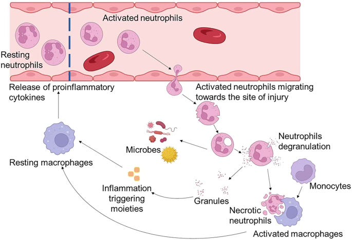
Amplification Loop
Okay so the threat is handled! The bacteria are cleared, the wound is under control, and now your body has the opposite problem on its hands: a site absolutely packed with neutrophils that showed up ready to fight, with nothing left to fight. You can’t just leave that crowd standing around, because all those enzymes and reactive molecules they’ve been flinging will start chewing through your own tissue if nobody calls them off. So resolution is where the body winds everything down and clears out the mess.
Resolution is an active program, something the body switches on deliberately, with its own dedicated set of signals (Sugimoto et al., 2016). It runs a full shutdown sequence here, every bit as coordinated as the ramp-up was. A fresh crew of molecules called pro-resolving mediators clock in, and their job is to call off the recruitment, so that “send backup” signal from phase two finally goes quiet and no new neutrophils get waved in.
The neutrophils already on-site are pretty short-lived to begin with, so once the reinforcements stop arriving, the crowd starts thinning out on its own. Then the cleanup crew rolls in. Macrophages move through the area and clear away all the spent, dying neutrophils, tidying up the site (Ge et al., 2022). Clearing those dead cells does something extra, too, because eating them flips a switch inside the macrophage. It shifts from its aggressive, inflammatory M1 setting into a much calmer M2 setting, where it stops pumping out attack signals and starts releasing soothing, anti-inflammatory ones like IL-10 instead (Ge et al., 2022). The same cell that was running the war is now running the cleanup, calling for peace instead.
Once the mess is cleared and those calming signals are flowing, your tissue can finally start to rebuild, and everything settles back to baseline. That is a healthy inflammatory response from start to finish: it fires up right when you need it, does the messy work, and shuts itself off once the job is done.
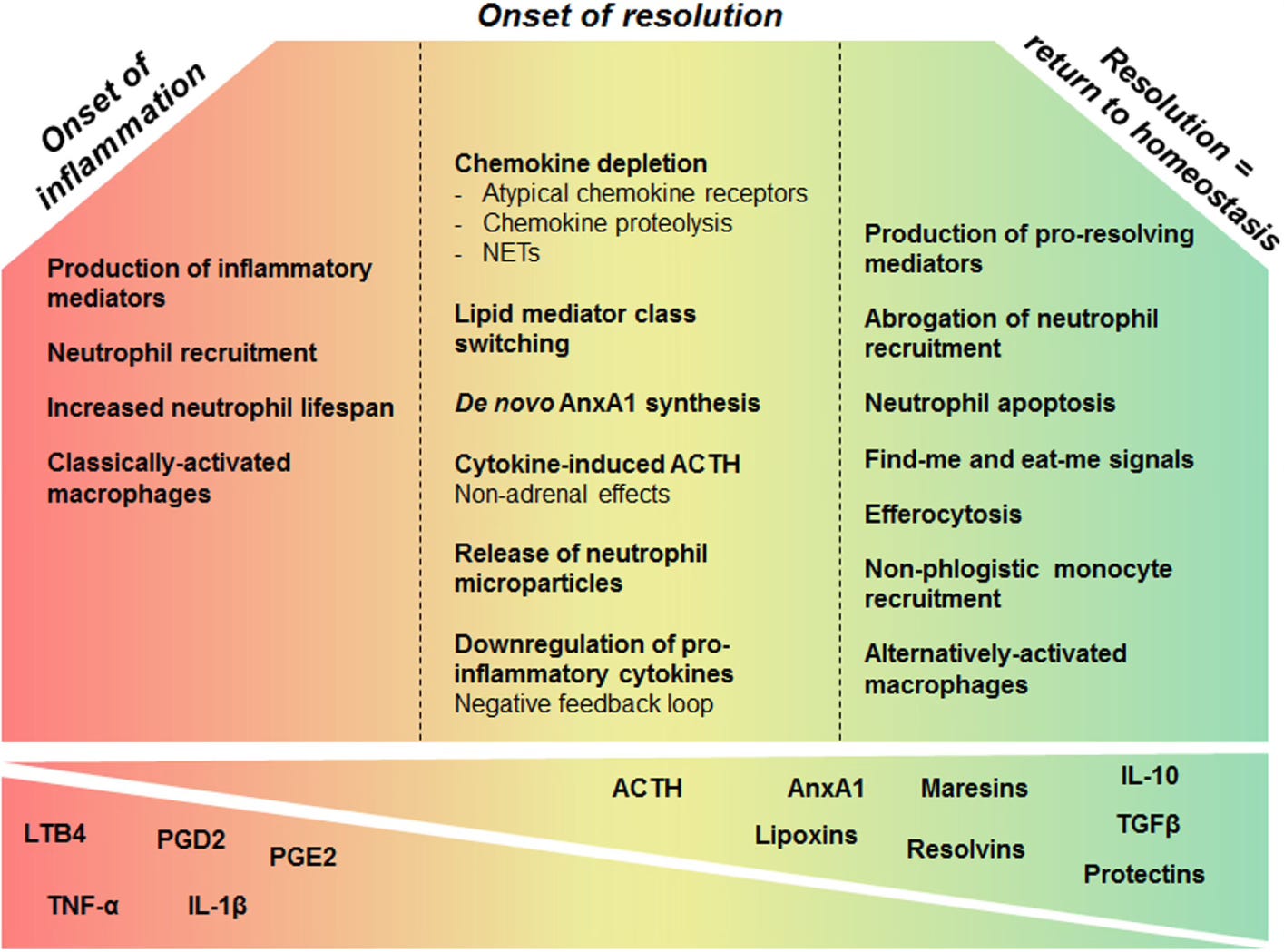
Resolution Phase
Everything so far describes inflammation working the way it’s supposed to. It fires, it does its job, it shuts down. Sometimes, though, that last step doesn’t happen, and the response that was meant to be temporary just keeps running. This is chronic inflammation, and it sits behind a huge number of long-term diseases.
Think back to that cleanup crew for a second, because a lot of chronic inflammation traces to the cleanup going wrong. When the macrophages can’t clear the dead cells fast enough, or the clearing process breaks down, those dead neutrophils pile up and rupture instead of getting cleared away (Ge et al., 2022). All the toxic contents they were holding spill straight into the tissue. To your body, that spill reads as fresh damage: new DAMPs, new alarm signals, another round of recruitment. The very thing that was supposed to end the response ends up feeding it.
Without that cleanup, the macrophages never get the message to change. That soothing M2 program, the one that produces IL-10 and starts calming everything down, only comes online when the macrophage receives the right signal, and eating those dead cells is the signal. No cleanup, no signal, so the M2 genes never get transcribed. The macrophages stay in their aggressive, inflammatory M1 mode, and the site stays hot (Ge et al., 2022). Now damage and repair are both running at once, indefinitely, with neither one ever finishing.
When tissue is caught in that loop of constant injury and constant attempted repair, the repair signals keep telling your fibroblasts to lay down collagen into a site that never heals. Fibroblasts are the connective-tissue cells that produce collagen and the other structural proteins making up the extracellular matrix, the scaffolding your tissues are built on. Here they just keep building, with no signal telling them to stop. Over time all that excess collagen accumulates as scar tissue, a process called fibrosis, and it slowly stiffens and reshapes the tissue until the organ can no longer work the way it used to (Chimenti et al., 2022).
So the story of inflammation comes down to balance. You want a response strong enough to protect you but smart enough to know when to quit, firing hard when a threat appears and then standing down once the work is done. Your body is good at the firing part. Staying well depends just as much on the shutting down, on that whole resolution program running the way it should and pulling the response to a close.
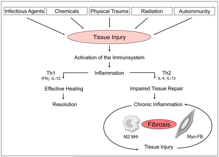
Pathogenesis of fibrosis.
We started with a single cut on a can lid, and from there your body ran an entire operation: resident cells spotting the intruders, mast cells throwing open the vessel walls, a crew of neutrophils swarming in to fight, then a careful, deliberate shutdown that cleared the mess and handed the site over for repair.
That’s initiation, amplification, and resolution, each phase flowing into the next.
So much of whether this protects you or harms you comes down to the ending. All that firepower stays safe only because the body has an equally strong system for standing down, so when that standing-down fails, the same response that defended you begins wearing at your own tissue, which is why the stopping carries just as much of the work as the starting ever did.
If this piece helped inflammation click for you, the biggest way to support the work is to consider a paid subscription.
I’m keeping it free because this foundation should be available to anyone, and paid members are what make that possible, so I can keep publishing work like this without putting the important pieces behind a paywall.
Every subscription helps keep this accessible for everyone.
If you found this useful or helpful, please like, comment, restack, or share it with someone who’d enjoy it! It really means a lot.
Until next time,
— Agy | The Buffalo Herbalist
Comments
Nothing yet. Say the first thing.
Sign in to join the conversation.