
Hello,
This is the 1st in a new series of 3 posts on metabolic emergencies. Today's post will cover two distinct causes of hypoglycemia with different laboratory findings and treatment options.
We were also happy to announce that registration for the ABGC Board Review Bootcamp is now open! The bootcamp will take place from June 11th - July 23rd, 2026 and is for genetic counselors preparing for the August 2026 ABGC board exam. Our course includes 30 hours of live instruction that cover all 5 domains on the ABGC content outline, plus access to a 500-question qbank for additional practice. Early-bird pricing of $399 is available through June 1st ($499 regular price thereafter). You can sign up and learn more about the bootcamp at our website: www.studyrare.com/abgc-bootcamp

Please feel free to reach out (daniel@studyrare.com) with any questions about the course or this post. I hope you have a great week!
-Daniel
A 9-month-old boy presents to the emergency department with a seizure after a febrile illness. He has had decreased oral intake over the past 2 days. Physical exam reveals a lethargic infant with hepatomegaly. Laboratory studies show a glucose of 28 mg/dL (low), elevated venous lactate, elevated triglycerides, and elevated uric acid. Glucose levels do not respond to glucagon. Which of the following enzymes is MOST likely deficient in this patient?
A 14-month-old girl presents to the emergency department with lethargy after a 24-hour febrile illness with poor oral intake. Laboratory studies show a glucose of 22 mg/dL (low), and serum beta-hydroxybutyrate is also low. An acylcarnitine profile shows elevated medium-chain acylcarnitine species. Newborn screening was declined at birth. Which of the following BEST describes the primary mechanism of hypoglycemia in this patient?
Question 104: B) Glucose-6-phosphatase
Question 105: C) Impaired fatty acid oxidation
Hypoglycemia is one of the most common metabolic emergencies. It is dangerous because the brain runs almost entirely on glucose (with ketones as its only backup fuel), and prolonged hypoglycemia causes seizures, coma, and permanent neurologic injury if untreated. While many cases in children have multifactorial causes, recurrent or unusually severe hypoglycemia should prompt a workup for an inborn error of metabolism. This post walks through several pathways that help maintain blood glucose between meals, a decision tree used to narrow the differential diagnosis, and two classic IEM causes of pediatric hypoglycemia: GSD Ia and MCAD deficiency.
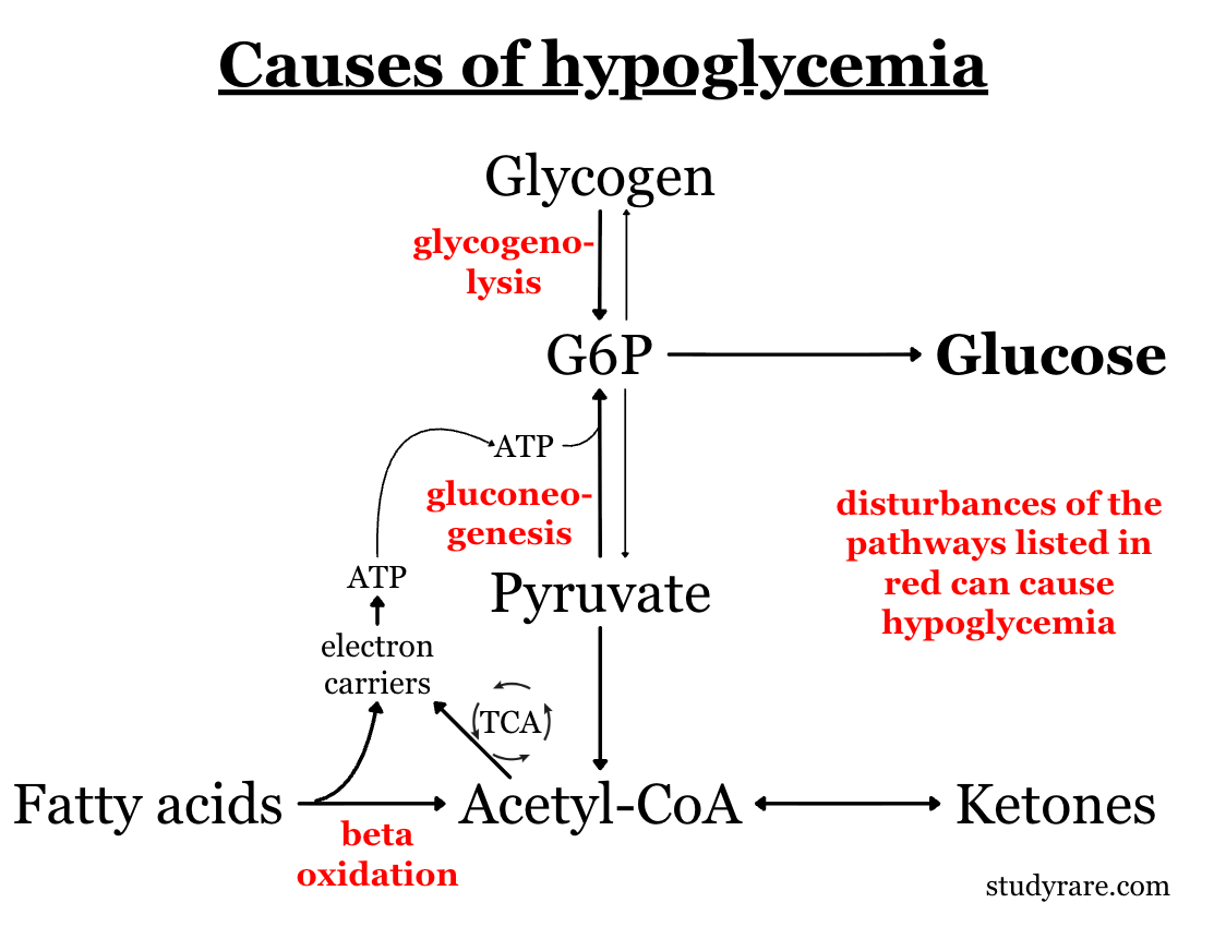
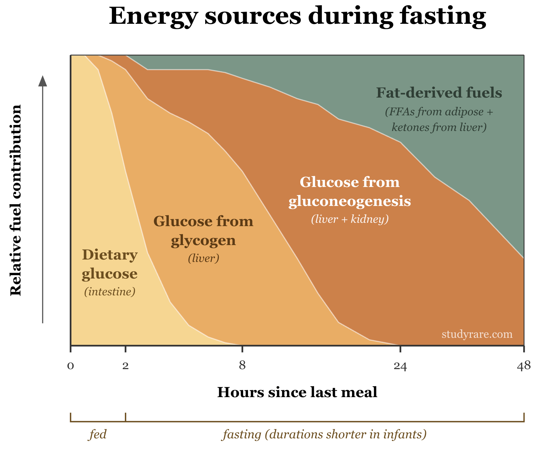
Three pathways keep blood glucose stable between meals. Disruption of any one of them can cause hypoglycemia. These pathways function at different times after a meal (see “Bonus” section at the end of this post for more details). The two diseases covered in this post each affect different pathways:
Glycogenolysis - the liver breaks down glycogen to release glucose. First line of defense while fasting, lasts roughly 12 hours in older children (much shorter in infants).
Gluconeogenesis - the liver makes new glucose from lactate, glycerol, and amino acids. Starts to increase as glycogen runs low.
Fatty acid beta-oxidation (FAO) - once glycogen is depleted, the body oxidizes fatty acids. FAO does not make glucose directly, but it produces the ATP, NADH, and acetyl-CoA* that power gluconeogenesis. It also generates ketone bodies (e.g., beta-hydroxybutyrate) that serve as an alternative fuel for the brain.
*Acetyl-CoA activates pyruvate carboxylase, the enzyme that catalyzes the 1st step of gluconeogenesis.
Insulin shuts down energy production
Insulin is the master off-switch for all three pathways. When you eat, insulin rises and shuts down glycogenolysis, gluconeogenesis, and lipolysis (the mobilization of fat that feeds FAO), and also promotes glycogenesis (production of glycogen). It also promotes the uptake of glucose from the bloodstream into cells. As a result, glucose levels in blood drop. During fasting, insulin falls and these pathways turn back on. This is why inappropriately elevated insulin in a patient with hypoglycemia is so dangerous: all three pathways are blocked simultaneously, and the body cannot even make ketones as a backup fuel.
💡 Remember: insulin drives glucose into cells, lowering glucose in the bloodstream.
When you see a hypoglycemic child, one useful lab test to consider sending is ketones (e.g., beta-hydroxybutyrate). While fasting, there is less glucose available, and so the body instead mobilizes fat and makes ketones for energy. So during hypoglycemia, ketones should be high. Low ketones in the setting of hypoglycemia is pathologic and helps narrow the differential, as seen in the diagram below:
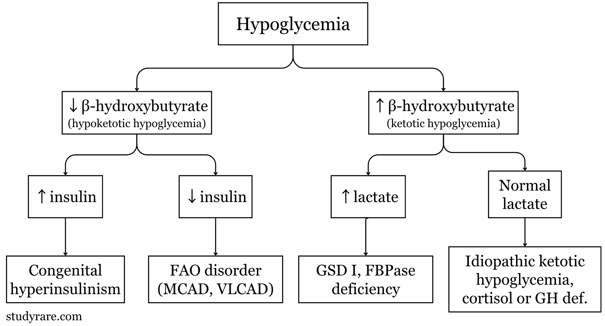
Low ketones (hypoketotic) → 2 possibilities:
Hyperinsulinism - insulin is suppressing lipolysis (the mobilization of fat), so the body cannot make ketones. Insulin is inappropriately elevated (should be low because glucose is low).
FAO disorder - the body tries to make ketones (via FAO) but the pathway is broken. Insulin is appropriately low.
High ketones (ketotic) → body responded correctly, but glucose production still failed:
Lactate high - points to GSD I or a gluconeogenesis defect (glucose-6-phosphate is backed up or cannot be formed).
Lactate normal - points to idiopathic ketotic hypoglycemia, hormone deficiency (growth hormone or cortisol), or the milder GSDs (types 0, III, VI, IX).
The two cases in this post sit on opposite sides of the diagnostic algorithm. Question 104 presents a case of ketotic hypoglycemia (right half), while question 105 presents a case of hypoketotic hypoglycemia (left half).
The patient in Question 104 has glycogen storage disease type Ia (GSD Ia), caused by deficiency of glucose-6-phosphatase (G6Pase) (Choice B). This enzyme catalyzes the final step of both glycogenolysis and gluconeogenesis, which is the conversion of glucose-6-phosphate (G6P) to free glucose. Without G6Pase, glucose cannot be released into the bloodstream from either pathway, and patients become hypoglycemic once feeds space out or when they cannot eat during illness.
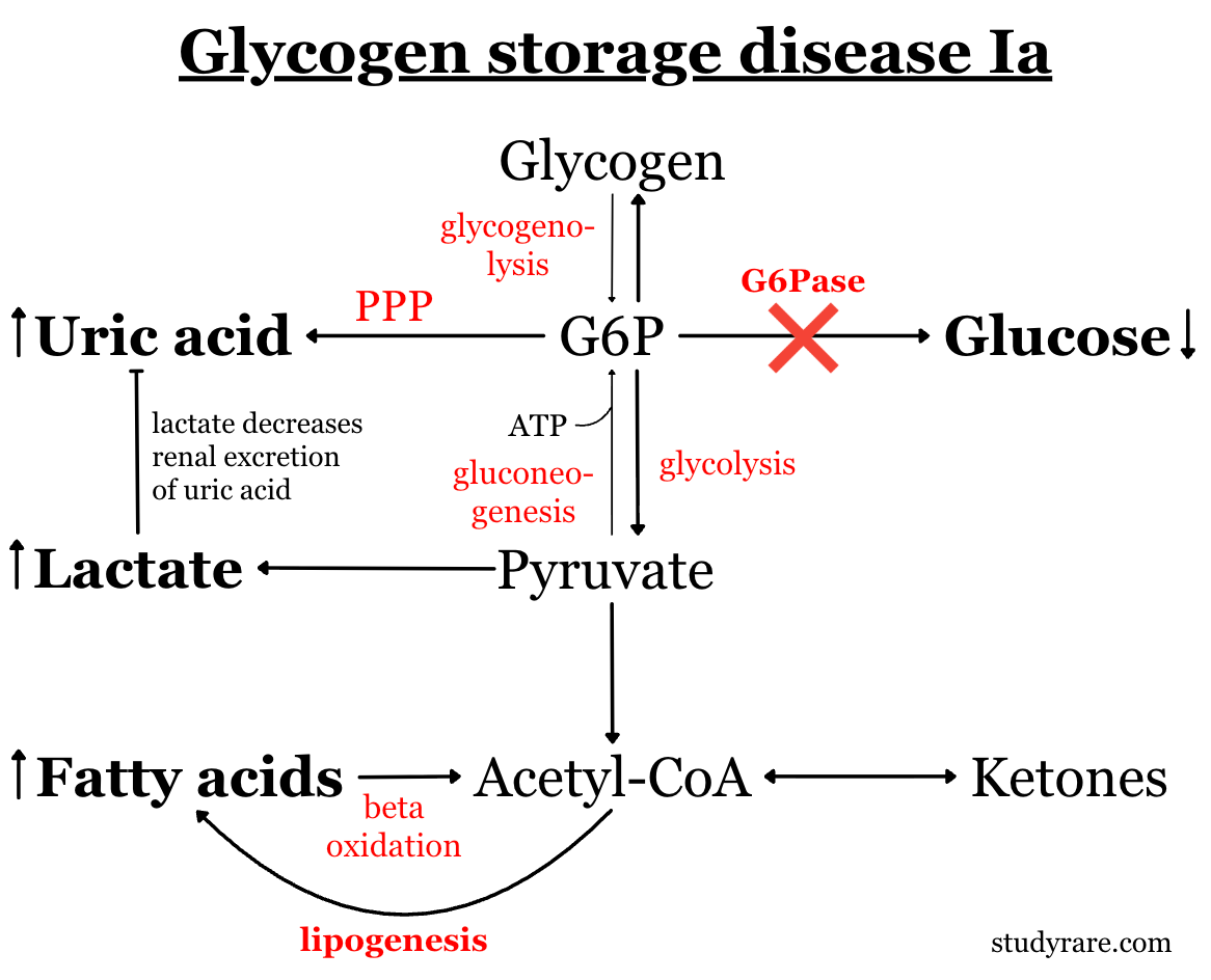
The combination of fasting hypoglycemia, lactic acidosis, hypertriglyceridemia, and hyperuricemia in a patient with hepatomegaly should raise concern for GSD Ia.
As seen in Question 104, patients with GSD Ia do not respond to glucagon (a medication given to hypoglycemic patients to raise blood glucose). But why? Glucagon is a hormone with the opposite action to insulin and stimulates glycogenolysis (glycogen breakdown), which generates more G6P. But without G6Pase, G6P has nowhere to go and instead shunts into lactate. The absent glucose response to glucagon (often with a paradoxical rise in lactate) is a classic finding in GSD Ia and places the patient on the ketotic, high-lactate branch of the decision tree.
Management of GSD Ia includes frequent feeds and uncooked cornstarch (a slow-release glucose polymer that provides 4-6 hours of glucose). Patients should avoid fructose and galactose, as both are converted inside cells to G6P, which cannot be released and instead worsens the lactate and triglyceride load. Hepatic imaging for adenomas (which carry a risk of malignant transformation) is indicated, and liver transplant is an option for patients with severe complications.
The patient in Question 105 has medium-chain acyl-CoA dehydrogenase (MCAD) deficiency, the most common fatty acid oxidation disorder. The classic presentation of MCAD is a previously well infant or toddler who decompensates during an illness (e.g. viral gastroenteritis) with poor oral intake. Between episodes, patients are asymptomatic.
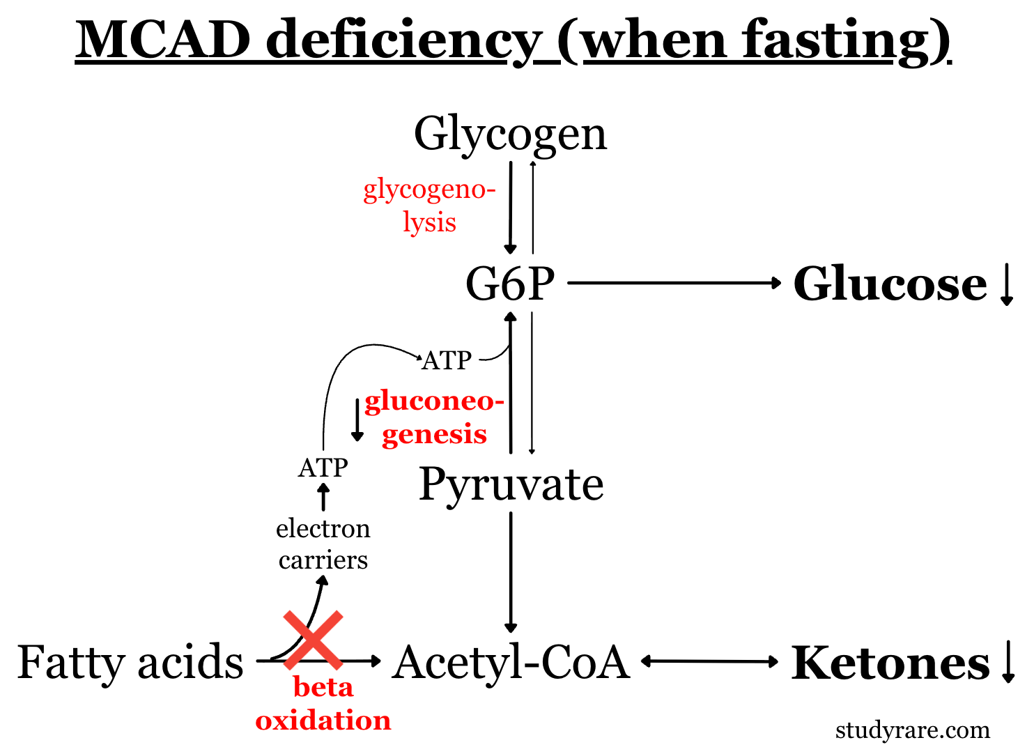
MCAD catalyzes the first step of beta-oxidation for medium-chain fatty acyl-CoAs (C6–C12). During prolonged fasting, fatty acid oxidation becomes an important fuel source. This process generates cellular energy directly and produces ketones that serve as an alternative fuel for the brain. MCAD deficiency blocks this process, so patients present with hypoketotic hypoglycemia, which is low blood sugar without the expected compensatory rise in ketones (mechanism described in figure below). It is the hallmark of FAO disorders and the reason MCAD sits on the hypoketotic branch of the decision tree.
A plasma acylcarnitine profile is a useful biochemical test when diagnosing a suspected FAO disorder. In MCAD deficiency, medium-chain species (particularly C8, octanoylcarnitine) are elevated. Newborn screening uses a similar test and catches most cases before a clinical crisis. Scenarios like the one in Question 105 can arise when comprehensive NBS is declined or otherwise not available.
Management includes avoiding fasting (exact duration varies by age) and ensuring patients follow an emergency protocol (dextrose-containing fluids orally or IV) during illness. No dietary fat restriction is needed for patients with MCAD, unlike long-chain FAO disorders (VLCAD, LCHAD, CPT I/II), which typically do require fat restriction.
Question 104
(Choice A) Deficiency of acid alpha-glucosidase causes Pompe disease (GSD II), which is a lysosomal and glycogen storage disorder. Pompe presents with severe hypotonia and hypertrophic cardiomyopathy in the infantile form, not fasting hypoglycemia with lactic acidosis.
(Choice C) MCAD deficiency does cause fasting hypoglycemia, but the metabolic profile here (lactic acidosis, hyperuricemia, hypertriglyceridemia) points more specifically to GSD I. In MCAD deficiency, lactate and triglycerides are normal, and the hallmark is hypoketotic hypoglycemia.
(Choice D) Deficiency of muscle glycogen phosphorylase causes McArdle disease (GSD V), which presents with exercise intolerance and rhabdomyolysis in adolescents and adults. It does not cause fasting hypoglycemia or hepatomegaly. The defect is confined to skeletal muscle, which has a different glycogen phosphorylase isoform than the liver.
Question 105
(Choice A) Congenital hyperinsulinism (increased insulin secretion) can cause hypoglycemia with suppressed ketones, since insulin inhibits lipolysis. However, hyperinsulinism does not cause elevated medium-chain acylcarnitines. The acylcarnitine profile in this patient points specifically to a block in fatty acid beta-oxidation.
(Choice B) Defects in gluconeogenesis (e.g., fructose-1,6-bisphosphatase deficiency) cause fasting hypoglycemia with lactic acidosis. Fatty acid oxidation is intact in these disorders, and the body can therefore still produce ketones. We would expect elevated, not suppressed, beta-hydroxybutyrate.
(Choice D) Impaired glycogenolysis is the mechanism for most glycogen storage disorders (GSD). Patients with GSD do not present with hypoketotic hypoglycemia, as the breakdown of fatty acids into ketone bodies is not impaired.
GSD Ia and MCAD deficiency both present with severe fasting hypoglycemia in infancy but result from defects in different metabolic pathways. The ketone level at the time of hypoglycemia helps narrow the differential diagnosis in an undiagnosed patient. GSD Ia presents with ketotic hypoglycemia and lactic acidosis (G6Pase block shunts G6P into lactate, triglycerides, and urate), while MCAD presents with hypoketotic hypoglycemia (the FAO pathway is broken, so the body cannot make ketones or sustain gluconeogenesis). GSD Ia is managed with frequent feeds and cornstarch, while MCAD is managed by avoiding fasting, especially during illness.
2025 ABMGG General Exam Blueprint | V. Single gene inheritance > d) Single gene disorders > Metabolic disorders (glycogen storage diseases, fatty acid oxidation disorders); IX. Population screening > Newborn screening
2023 ABGC Exam Content Outline | Domain 1C. Genetic Conditions. (Both MCAD and GSD are conditions listed on the ABGC self-study guide)
Additional resources
1. Glycogen storage disease type I (GeneReviews)
2. MCAD deficiency (GeneReviews)
3. Recognition, assessment and management of hypoglycaemia in childhood (BMJ)
Consider reviewing our earlier post (Question 3), which covers the related GSD Ib subtype and its treatment with empagliflozin.
The pathways discussed in this post do not all contribute equally at every point during a fast. Dietary glucose covers the first 1-2 hours. Hepatic glycogen then takes over for the next several hours, with gluconeogenesis becoming more and more dominant as glycogen stores are depleted. With prolonged fasting, fat-derived fuels (free fatty acids from adipose plus ketones from the liver) eventually become the largest contributor. Note that these durations are all shorter in infants, which is part of why a healthy 9-month-old cannot tolerate fasting nearly as long as an adult, who can fast for days without decompensating.
Try mapping the IEMs discussed in this post onto this chart. GSD Ia shrinks the glycogen and gluconeogenesis bands, so patients become symptomatic relatively quickly once dietary glucose runs out (hence the need for near-continuous glucose sources like cornstarch). In contrast, MCAD shrinks the fat-derived fuels band, so patients become symptomatic later in the fast, once FAO should have taken over. This video walks through this framing in more detail (min ~5:30-10:30).
Comments
Nothing yet. Say the first thing.
Sign in to join the conversation.