
The last post clearly showed how the hydrogel rubber clot could be “hidden" in the currant jelly clot if clotting was allowed to occur for 30 min prior to centrifugation. Due to being hidden with the old method, it could be possible to attribute a negative expression of hydrogel to a physical property or even a therapeutic effect but this would be false. I have my doubts on any test that allowed 30 min (or enough time to have currant jelly clot) prior to the centrifuge step. It may be nearly impossible to sort out a therapeutic effect in the future. I will discuss this later.
The PRF method for demonstration of hydrogel rubber clot requires collection of blood into glass (plastic vacutainer tubes with a silica pro coagulant is acceptable) and performing an immediate centrifugation for 8 min at a reduced force of about 1,500 rpm. The tube may require an additional 5 min to form the fibrin rich clot after the initial spin. I feel the plain red top plastic vacutainer tubes without pro coagulant can be used but may require additional time to form the fibrin rich clot after the spin. You can tell if the fibrin clot has formed by noting a gel in the serum.
I took two 5 ml glass red top tubes and obtained my blood via venipuncture. I placed both tubes in the centrifuge and spun for 8 min at 1,500 rpm. Note that it not necessary to invert or agitate the tubes prior to spin since the idea is to get the fibrin away from the red cells prior to clotting ( but you will need clotting to demonstrate hydrogel). Following the spin an additional 5 min or so may be required for a fibrin clot to form as confirmed by a gel.
Here is a tube after the spin and given 5 min to form the hydrogel. Let me just say that the fibrin hydrogel is absolutely a normal finding in any living person but its change to a calamari state I will discuss later.
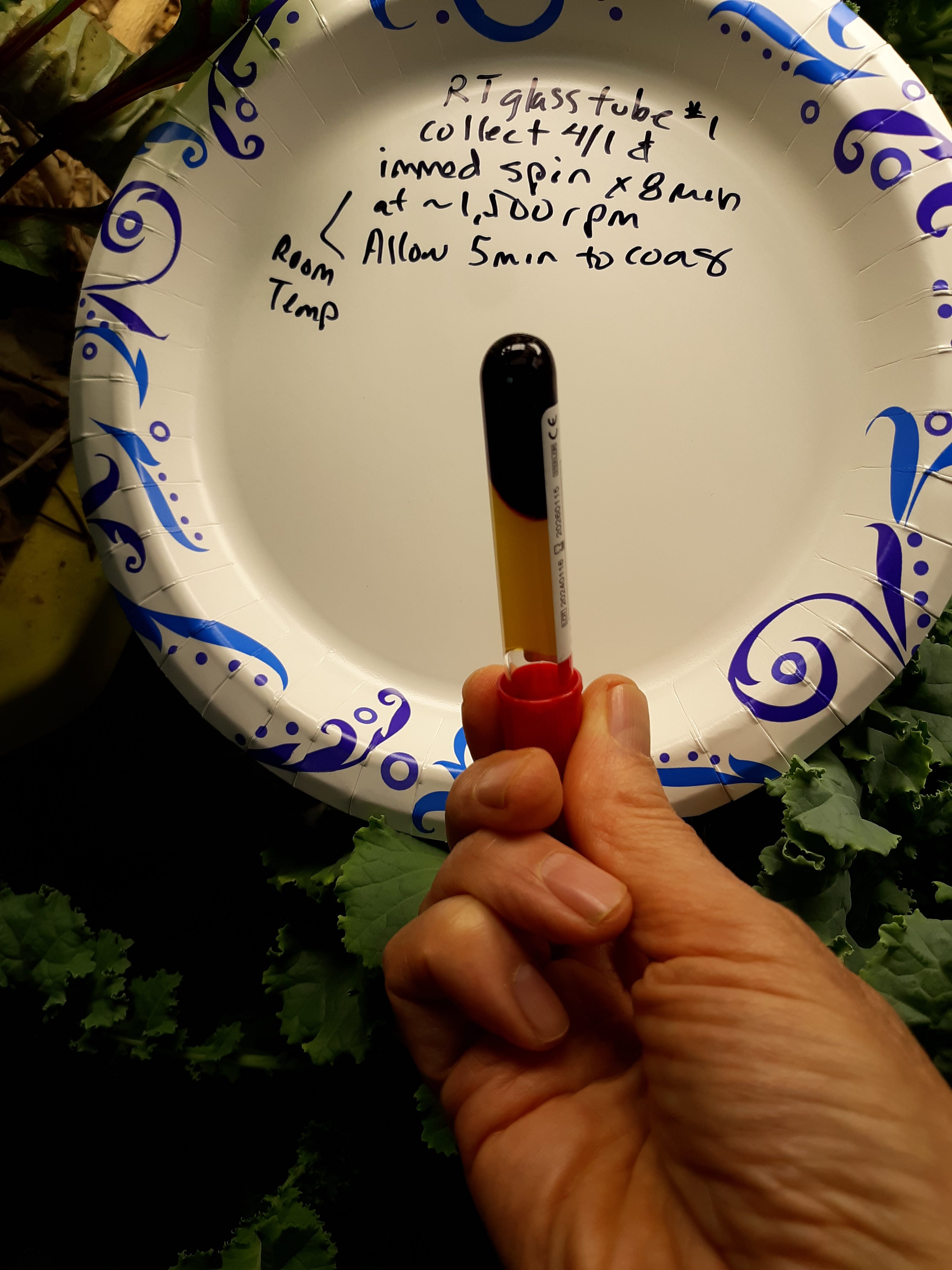
I can immediately examine the PRF by emptying the contents onto a paper plate after I am sure the fibrin hydrogel has formed. The first sample was a gel after the 5 min after the spin. I forgot to get a pic on the plate so here is the 2nd sample that is examined about 20 min after the spin.
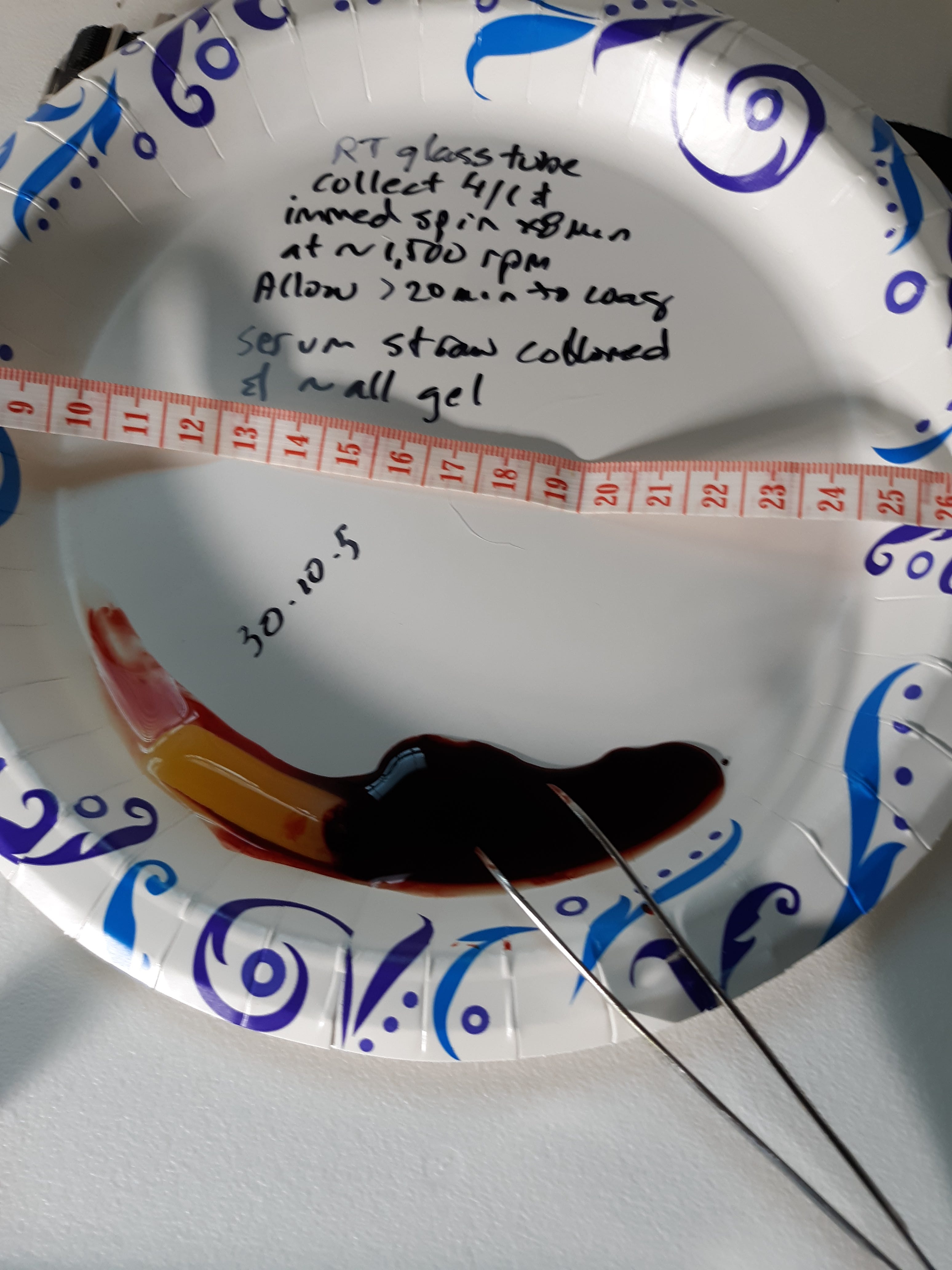
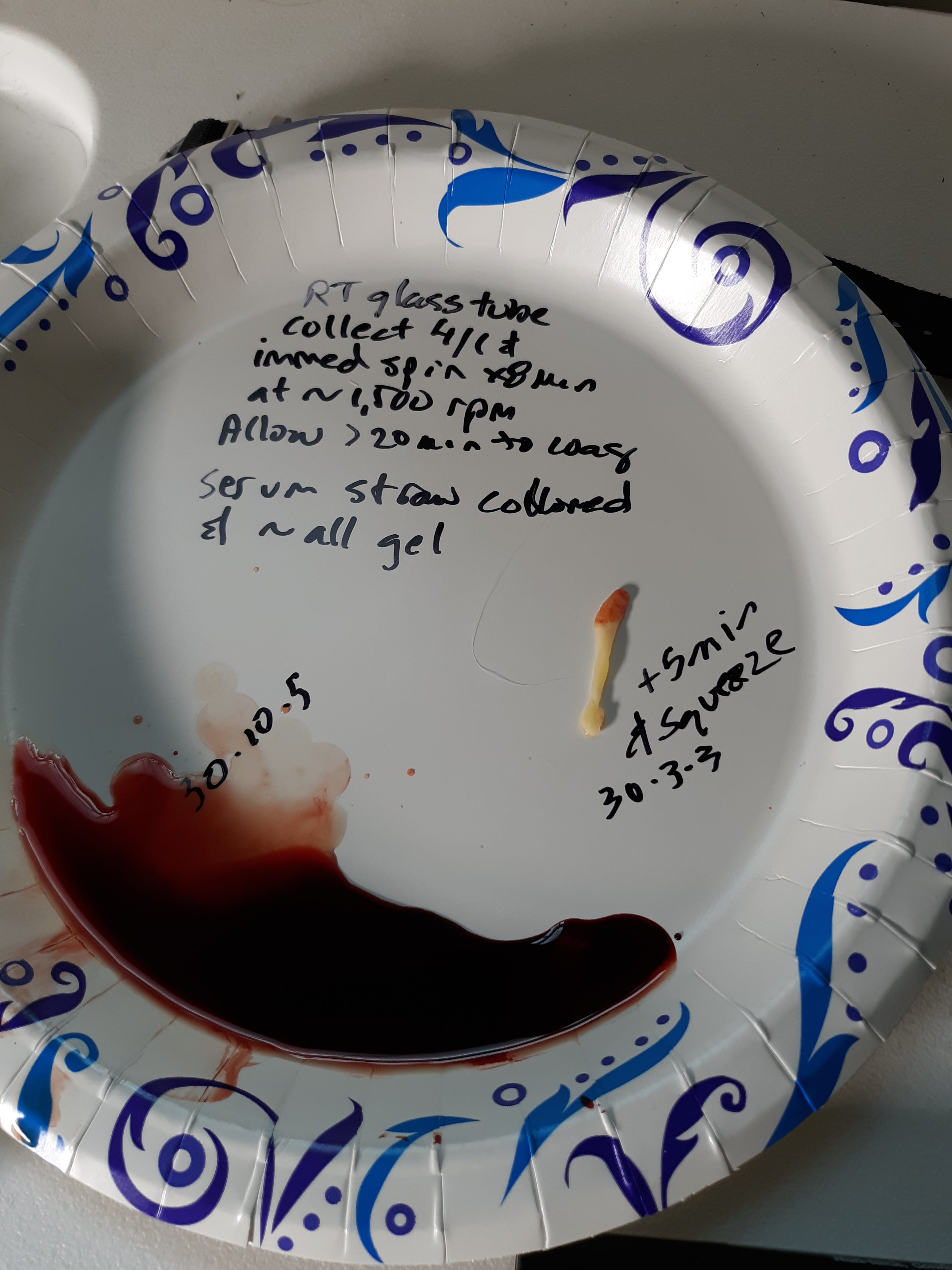
As in the past the gel will shrink and become whiter when the serum is squeezed out after a few minutes. The so called calamari state that is very rubber like.
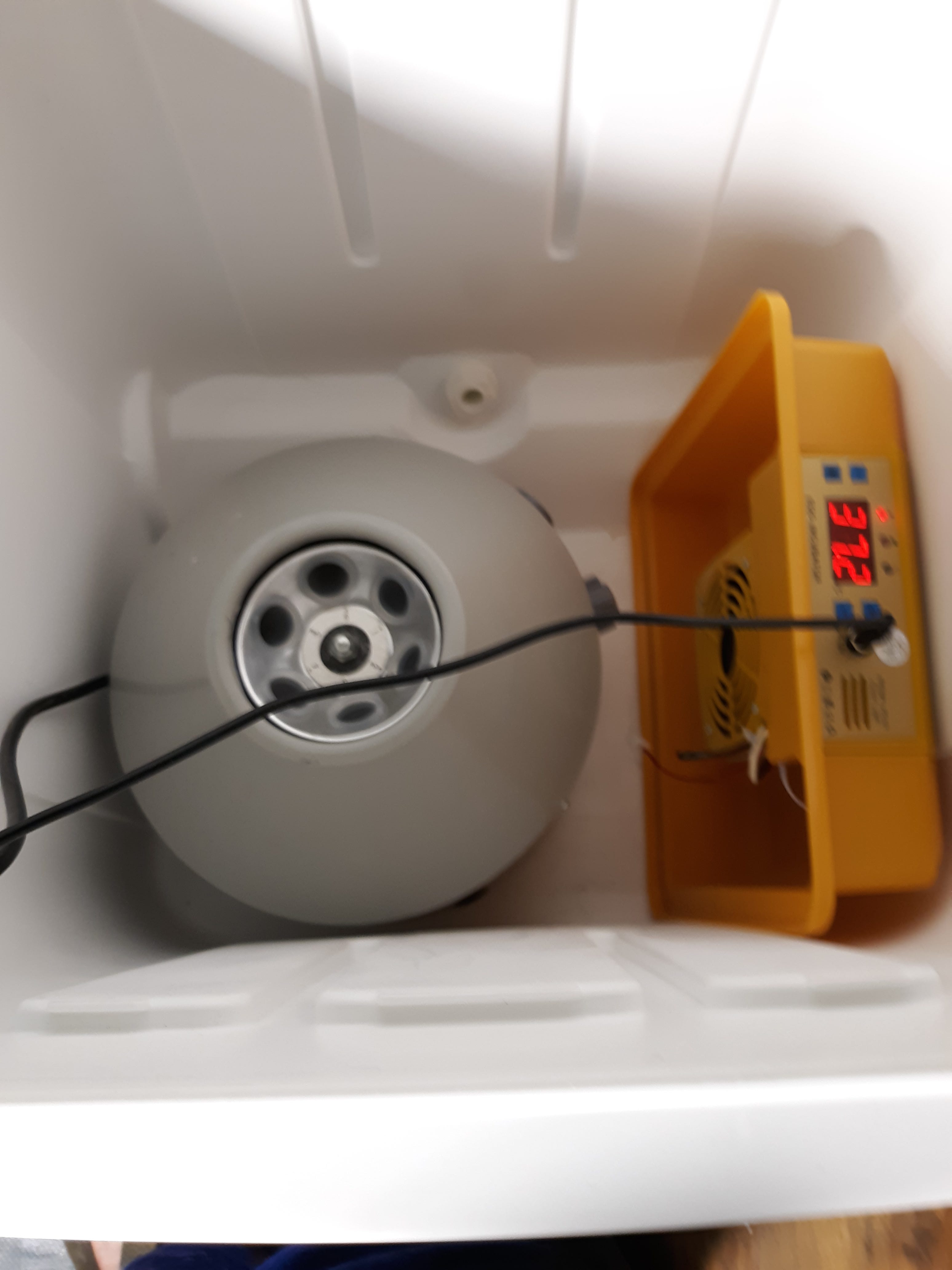
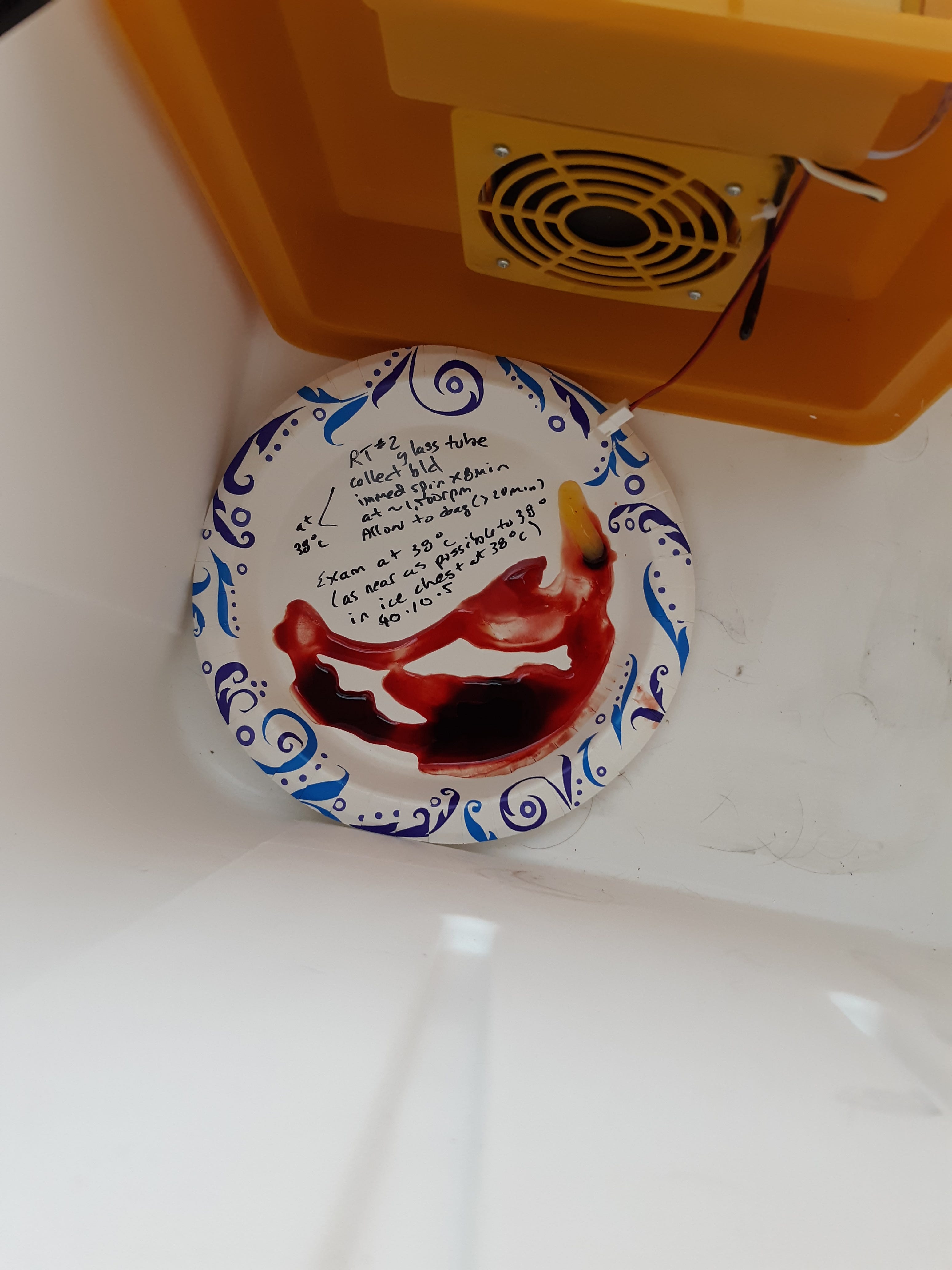
I wished to see if performing the PRF method at 38°C (100°F) had any effect. I collected blood into two 5 ml glass red top plain tubes and immediately centrifuged them at 38°C within the insulated ice chest for 8 min at 1,500 rpm. I allowed 5 min after the spin for fibrin clotting at 38 °C. The set up is pictured.
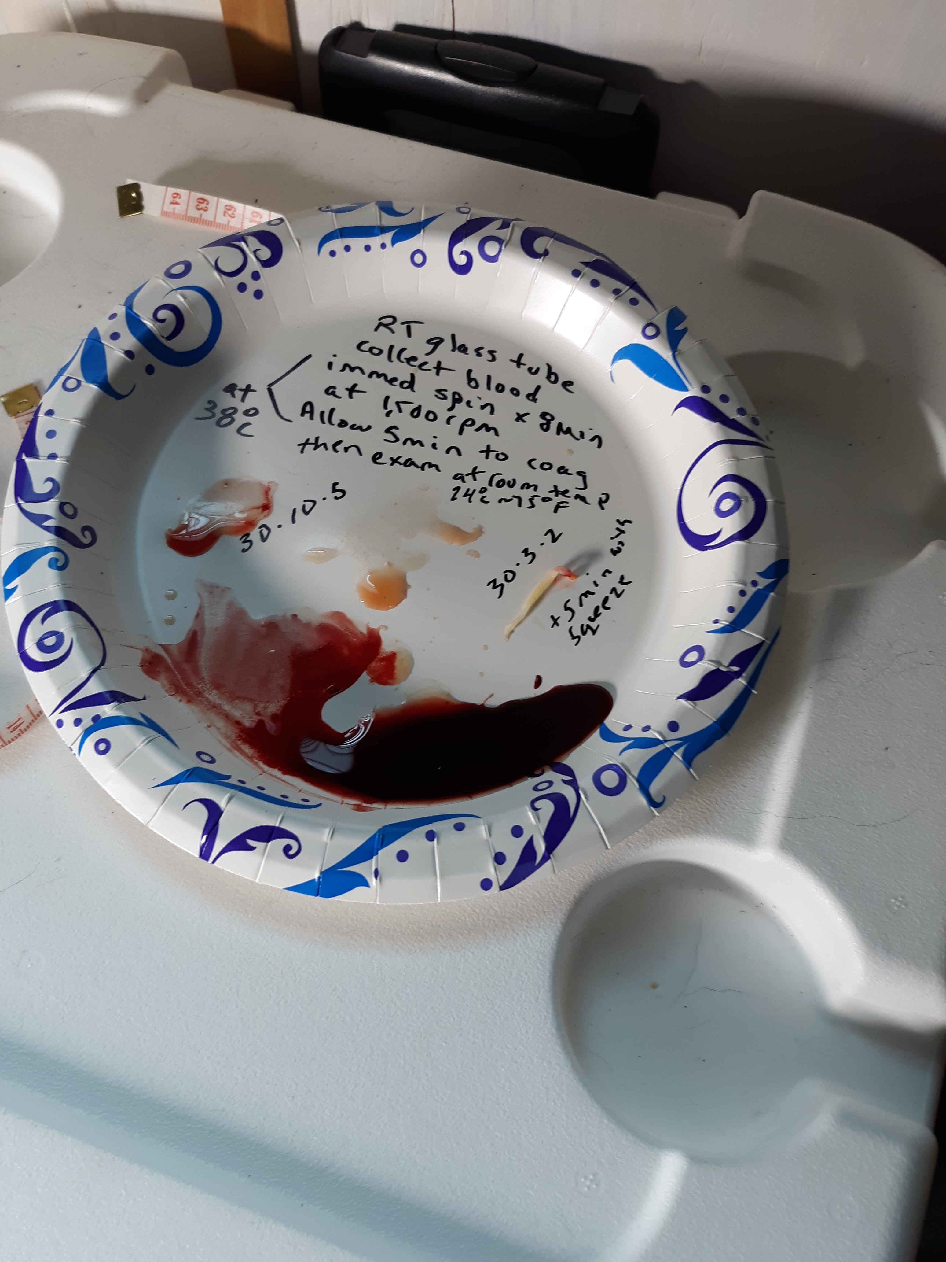
The following pic shows the exam of tube contents at room temp of about 24°C (75 °F).
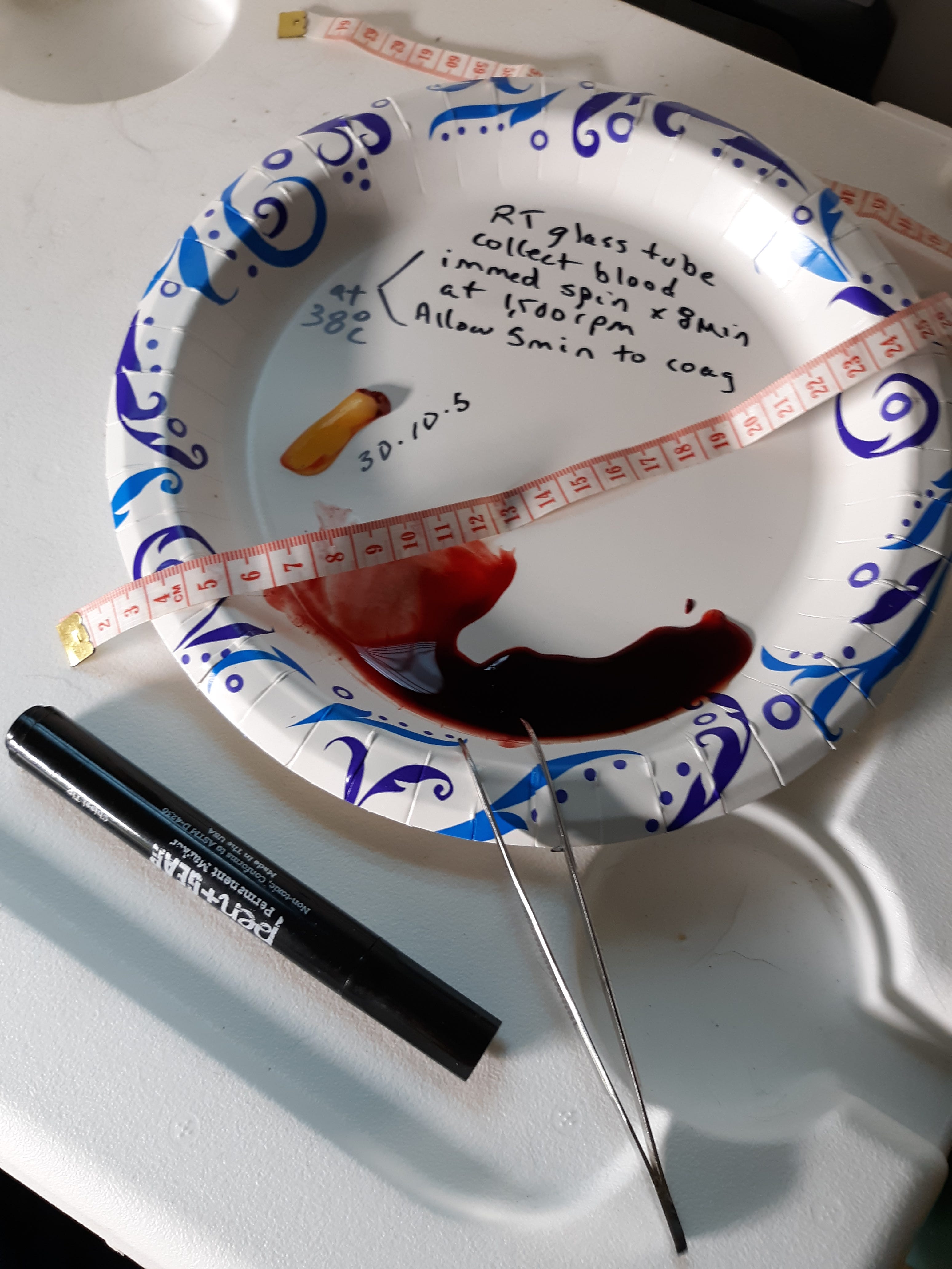
The following pic shows the effect of squeezing, shrinkage, and about 5 min. The specimen is rubbery with calamari effect.
I felt the above result showed that performing PRF method at body temperature made no difference from doing the same at room temperature. I tried to extend the body temp exposure to the exam of tube contents at 38°C with the 2nd tube I had collected.
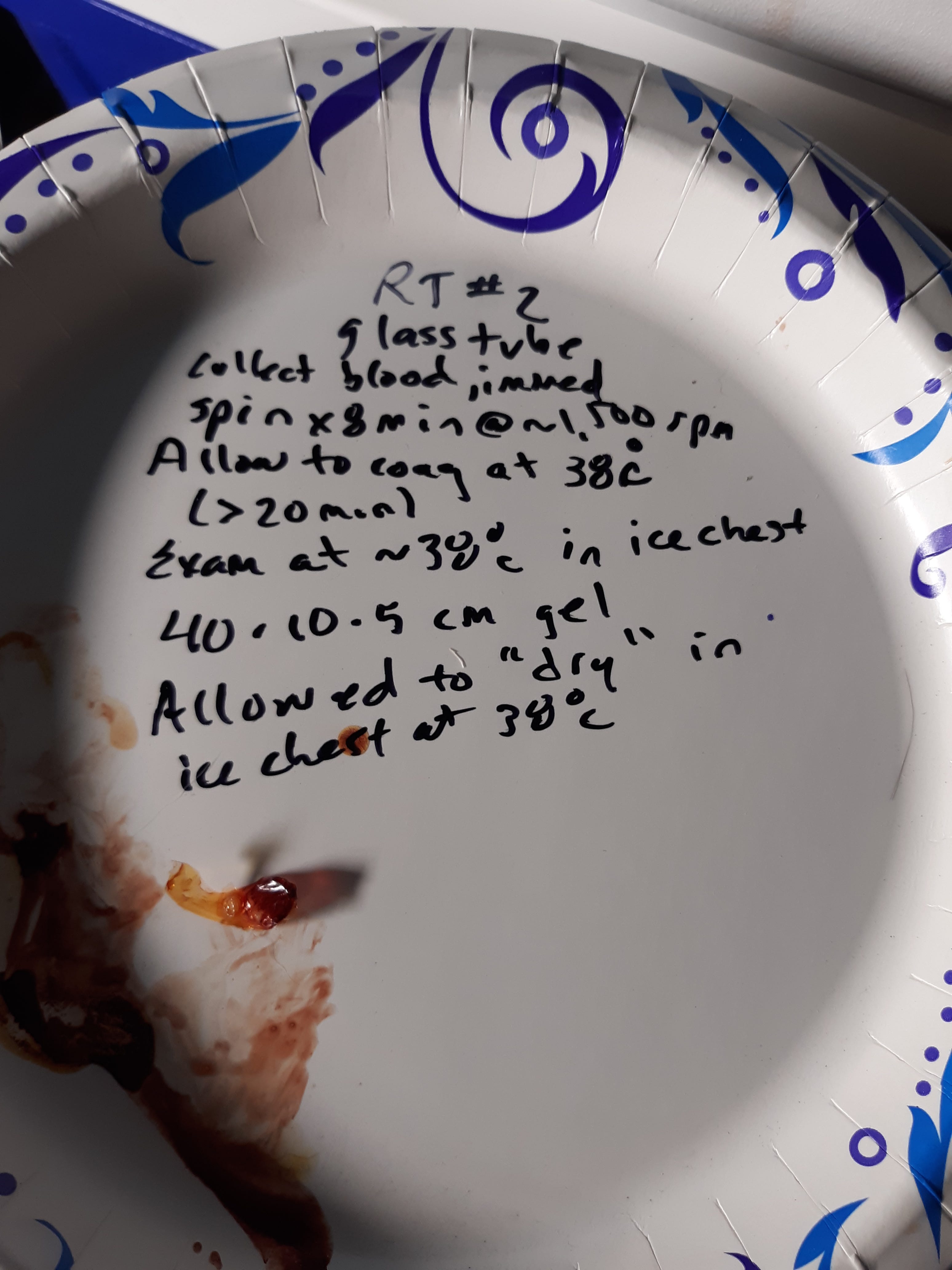
I left the specimen to dry and shrink in the heated ice chest. The specimen appeared to over dry in the ice chest over the next few hours.
Summary and Discussion. PRF method is a more reliable way of demonstrating the hydrogel rubber clot. PRF collection has been used mainly in dentistry and been described and used since 2001. PRF is collected in that field for its healing and rejuvenation properties. A common use was having it placed into the defect after wisdom tooth extraction to avoid “dry socket” condition. If the collection of PRF is done correctly every living person should show a hydrogel with the test. The act of fibrinogen being converted to fibrin is when soluble fibrinogen (dissolved) is converted to a solid (hydrogel). So some definitions are needed. Fibrinogen is an essential blood clotting factor made by the liver and is normally dissolved in the circulating blood. Fibrinogen is converted to fibrin during normal blood clotting and becomes a hydrogel. A hydrogel is when monomers of say fibrinogen are acted upon by other clotting factors to become fibrin fibers that can form a 3D net that holds serum and other components like platelets and red cells if in the vicinity.
Is the fibrin hydrogel that is formed with PRF an abnormal variety? We can say that it can carry out its main function still and that is to form clots and prevent hemorrhage. The issue is it abnormal with an amyloid component? The amyloid component would cause it to break down less readily and thus cause issues when perfusion (flow) is hindered more than it should. If you have not seen this recent video from Vejon Health on YT make sure you watch it. https://www.youtube.com/live/ySMqoy0Nc00?si=lW33Al8HVMwbcdY1
Dr Douglas Kell presents evidence that spike (or your favorite nano particle) can act as a prion that attaches to fibrinogen and when it converts to fibrin it is changed to have an amyloid character (fibrinaloid). It is as above in that is more resistant to breakdown, but still it is possible and the fibrin proteases may help (nattokinase, lumbrokinase, serrapeptase, bromelain). Dr Kell's publications page is very interesting. http://dbkgroup.org/longcovid/
The following is from his publications page: addition of small amounts of purified (virus-free) SARS-CoV-2 (‘alpha’) spike protein, and that this amyloidogenesis was also true for the plasma of individuals with Post-Acute Sequelae of COVID-19 (PASC, i.e. Long COVID). A more recent paper expands on this. Thus the SARS-CoV-2 spike protein alone was sufficient to induce this anomalous microclotting that was more resistant to the normal means of removal (‘fibrinolysis’, mainly catalysed by the enzyme plasmin).
Dr Kell in the video above seems to imply that the fibrinaloid micro clots could grow into the large new clots the coroners are finding. What is needed is characterization of the fibrin hydrogel clots as to having an amyloid component. I suspect they do since the hydrogel rubber clots are a relatively new phenomenon.
What was shown in the above testing is that there is no evidence of a temperature sensitive hydrogel rubber clot with PRF testing. The old way of waiting 30 min for blood to clot in the test tube could give erroneous results since the fibrin hydrogel could be “hidden” in the current jelly clot.
An unanswered question for me is that prior to the pandemic did PRF demonstrate a hydrogel that would not convert to a calamari like rubber clot? It would be nice to show the rubber clots to an endodontist familiar with PRF from 20 years ago and see if they are different. Still that would not be definitive since a rubbery nature only reflects the entanglement of the fibers and normal fibrin may be able to do that. In the meantime the clots need to be analyzed for amyloid content for a definitive answer. Dr Kell suggests that labs out there do amyloid testing with the thioflavin t dye!
A merry heart does good, like medicine, But a broken spirit drys the bones. Proverbs 17:22
No posts
Comments
Nothing yet. Say the first thing.
Sign in to join the conversation.