
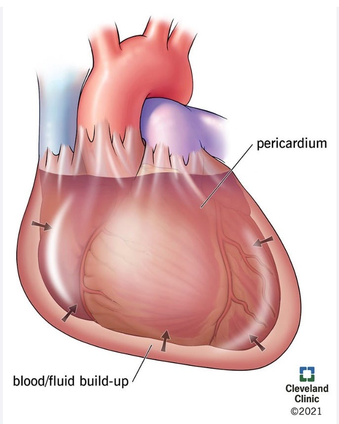
Pericardial disease has always been tough to diagnose especially before portable echocardiography and cardiac MR were readily available.
One of my Rules from those days is “If They Have Peripheral Edema, Have Seen Three Cardiologists, and No One Knows What It Is, It’s Constrictive Pericarditis.” I will write about that later, but my recent week of service coverage brought up another rule that can be related to pericardial disease. The Rule is:
Don’t Make or Dismiss a Diagnosis Based on One Parameter –
We were asked to follow a young patient with an almost 2 cm pericardial effusion. She had had a prolonged hospital stay because of infection and renal failure requiring persistent dialysis. We were told she was not in pericardial tamponade because her echo did not show any right ventricular compromise. But the echo did show “stranding” consistent with the development of fibrous bands between the parietal and epicardial pericardium. I could not do a formal pulsus paradoxus measurement because we could not find a manual sphygmomanometer, but I thought she had pulse diminution with inspiration by palpation. Dr. E. William Hancock, a pericardial disease expert at Stanford when I was a fellow there, once told me that clinically important pulsus paradoxus could usually be felt by femoral pulse palpation. Dr. Hancock was worth listening to on pericardial disease because he was the first to define effusive constrictive pericarditis in 1971. (1) Effusive-constrictive pericarditis is rare and occurs when there is both visceral pericardial constriction and a tense fluid effusion. The key finding is that that the high right heart pressures fail to normalize after draining the fluid.
Because of Dr. Hancock’s comments on the peripheral pulse in tamponade, I was concerned our patient had pericardial tamponade because of her pulse decrease. I wanted to tap the effusion to exclude infection and because I thought others would also be concerned about tamponade later. The invasive cardiology team inquired if I was sure I wanted to do it because she was not in tamponade by echo. I told them my concerns and they agreed to proceed. I always request that the invasive cardiologists measure the pericardial pressure at the start and end of the tap, another Hancock teaching. The pericardial pressure was 22 mmHg at the start and decreased to 2 to 4 mmHg at the end of the case so she had been in tamponade.
Whew! I am glad I requested the tap. I am not presenting this as “Boy, was I smart,” but as “Boy, was I lucky.” My patient could have gotten seriously sick without that drainage.
I present this case because it emphasizes the importance of not making or excluding a diagnosis on the basis of one parameter. I encouraged the tap because she did not look “great” to me; she had a big effusion; and I thought I felt paradox on her peripheral pulse.
I do not know why her right ventricular chambers did not show collapse on the echo, as they should in tamponade, but it could be that the fibrous strands held the right-sided structures open. Dr. Sean McMahon, director of our cardiac echo lab, also mentioned that high right atrial and ventricular pressures can prevent diastolic collapse. Loculation of fluid can also cause restricted cardiac filling without right chamber collapse on echo.
I have one other pericardial case that I use when I tell trainees “not to make a decision on the basis of one parameter.”
I had just moved from Brown University to the University of Pittsburgh and was on the consult service. I was called emergently to the dialysis unit where a patient had had syncope and was in near arrest. Given that it was the dialysis unit, and renal failure can produce pericardial effusions and tamponade, I was pretty sure the patient was in tamponade, but he had absolutely no important pulsus paradox. I measured it myself because those were the days when manual sphygmomanometers were readily available. This was before widespread portable echocardiography so no echo was available. Also, I had been taught that tamponade was a clinical diagnosis based on a paradoxical pulse. I hesitated for a minute - the “new boy on the block” didn’t want to over-react and there was no paradoxical pulse, but the situation was desperate. So I asked for the pericardial needle and syringe and went ahead. I blindly (without echocardiography) used the old landmarks: slightly to the patient’s left of the xiphoid process, at a 30 degree angle, aiming for the left shoulder. When I popped through the pericardium, the bloody fluid was under such pressure that it pushed back the plunger in the syringe. The patient improved immediately.
I was shaken because I had almost not done the tap. I went downstairs and related my story to Jim Shaver, MD, the Chief of Cardiology at that time at Pitt. He proceeded to describe his observations that some patients with renal failure in tamponade did not have a paradoxical pulse.(2,3)
Here is how that works:
Inspiration increases blood return to the right ventricle. The right ventricle expands, shifts the septum leftward, and this compresses the left ventricle. This compression restricts left ventricle filling , which decreases left ventricular stroke volume. This reduces the systolic blood pressure (SBP). This is “pulsus paradoxus” or “paradoxical pulse” and it occurs to a slight degree (less than 10 mmHg), even in normal individuals without effusions. However in cardiac tamponade, the fluid in the tightly stretched pericardium compresses the ventricles. So, when inspiration fills the right ventricle, the leftward shift of the septum greatly reduces left ventricular filling and produces a large reduction in SBP (>10mm HG or >10% of SBP).
This septal shift cannot happen if the septum is thick and stiff from conditions such as long-standing hypertension, hypertrophic cardiomyopathy, or aortic stenosis. So, my dialysis patient with long-standing hypertension could not shift his septum to produce the paradox. I didn’t know that, but I am glad I did not forgo the tap just because the patient lacked a paradoxical pulse.
The Rule – Don’t Make or Dismiss a Diagnosis Based on One Parameter.
References:
1. Hancock EW. Subacute effusive-constrictive pericarditis. Circulation. 1971 Feb;43(2):183-92. PMID: 5540704
2. Shaver JA, Reddy PS, Curtiss EI, Ziady GM, Reddy SC. Noninvasive/invasive correlates of exaggerated ventricular interdependence in cardiac tamponade.J Cardiol. 2001;37 Suppl 1:71-6.PMID: 11433831
3. Reddy PS, Curtiss EI, O’Toole JD, Shaver JA. Cardiac tamponade: hemodynamic observations in man. Circulation. 1978 Aug;58(2):265-72. doi: 10.1161/01.cir.58.2.265.PMID: 668074
#cardiactamponade; #pericardialdisease; #pericardialeffusion: #jamesshavermd; #ErnestwHancock; #BillHancock; #JamesShaver; #effusiveconstrictivepericarditis
Sean McMahon, MD, reviewed this piece for me, but I am totally responsible for its final content.
No posts
Comments
Nothing yet. Say the first thing.
Sign in to join the conversation.