

Sometimes we forget how fast technology changes.
This is an image of one of the very earliest CT scans of the brain1.
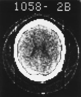
It took almost 5 minutes to do, and the results, while absolutely groundbreaking at the time, are pixelated and lacking any of the fine resolution of the scans we do today.
This all happened in the early 1970s.
Because scanning was so slow, the patient (And the organ of interest) had to remain perfectly still for minutes at a time.
This is why CT scans of the heart were such a late advancement.
The old ‘beating 60 times a minute’ presented quite the problem.
The heart just wouldn’t stay still long enough.
It would be the late 90s before CT scanning technology was good enough to scan the heart arteries with any reasonable accuracy.
Today’s scans are almost unrecognisable from the original CT scans.
The primary goal of a CT coronary angiogram is to answer two questions.
Is there plaque?
Where is it, and is it likely to be obstructing flow down the vessel?
Modern scanning technologies are exceptional at this task.
Below is an image of a photon-counting CT coronary angiogram2.
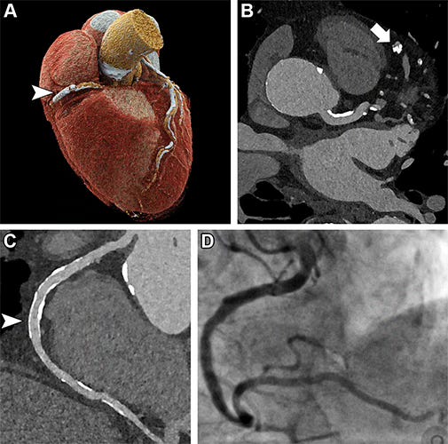
The bottom left image is the CT scan result. The bottom right image is an invasive angiogram.
The results are almost identical.
The CT scan is an entirely non-invasive test and comes with very few risks compared to an invasive angiogram.
We are living in the future.
The ability to acquire these types of images when I started my training as a doctor was something we could have only dreamed of.
While cardiac CT has been mostly used to identify if there are blockages in the coronary arteries that cause chest pain, we are increasingly using these scans to see IF a patient has coronary artery disease and what their future risk of a heart attack is.
This has mostly been the role of CT coronary artery calcification (CT-CAC) scans.
CT CAC scans quantify the amount of calcium in the coronary arteries and the risk of a future heart attack.
The more plaque/calcium, the greater the risk of a future heart attack.
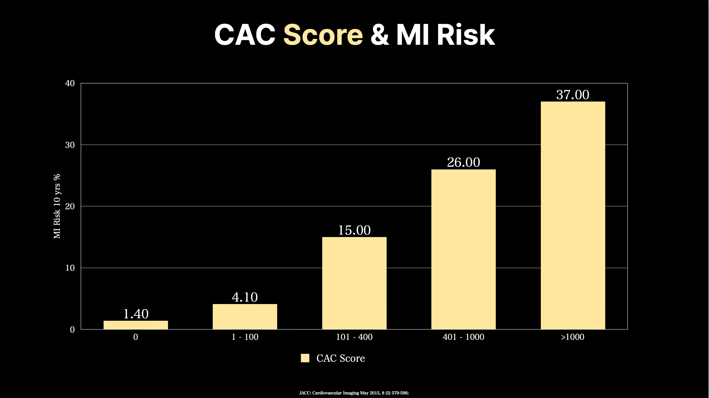
A calcium score of 0 suggests a very low risk of a heart attack in the next 10 years.
A calcium score of greater than 300 suggests the same risk of a future heart attack as someone who has had a heart attack before3.
So very high.
In simple terms, at least over the near term, you cannot die from a condition you do not have.
About 10% of patients with a CAC score of 0 DO have evidence of plaque on more advanced scanning, such as CT coronary angiography4.
So a CAC score of 0, while reassuring, will miss plaque (and risk) in quite a few people.
Secondly, and more importantly, heart disease (plaque in the arteries) usually doesn’t kill people.
Heart attacks do.
A heart attack occurs when plaque in the coronary arteries ruptures, causing a clot to form in the artery and stopping blood flow to the heart.
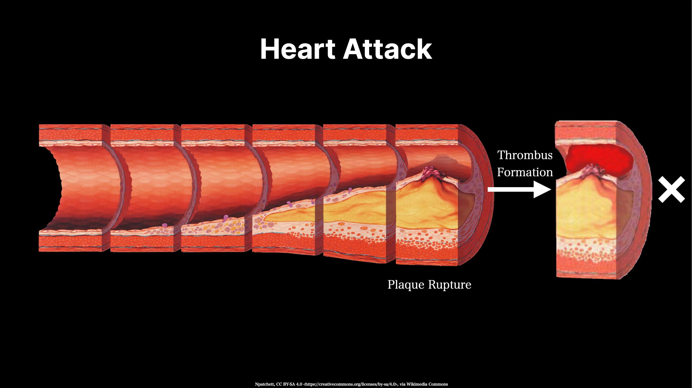
Plaque that ruptures is typically considered to be a vulnerable plaque.
Plaque that does not rupture (and cause a heart attack) is generally stable plaque5.
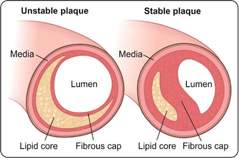
Vulnerable plaque usually has an actively inflamed cholesterol core with only a very thin wall separating it from the lumen of the artery.
Stable plaque is typically heavily calcified, has a small cholesterol core, minimal inflammation and has a thick fibrous wall separating the lipid core from the lumen of the artery.
Stable plaque is the type of plaque you die with, not from.
The type you want.
To date, cardiac CT scans have had limited ability to identify whether a plaque is stable or not.
We had very limited ways of identifying if there was active inflammation.
Until Now.
One of the key factors that separates stable from vulnerable plaque is the amount of inflammation in and around the plaque.
This can be measured using existing CT coronary angiograms with added software that assesses the amount of inflammation called a perivascular fat attenuation index (FAI)6.
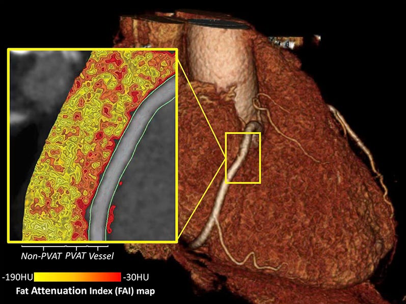
Think of this like a ‘Heat Map’ scan of the coronary artery.
The question is, how predictive are these scans at detecting the future risk of a heart attack or dying from heart disease?
The answer is very predictive.
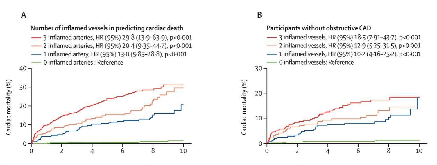
Compared to having no inflammation identified in any coronary arteries, having inflammation in just one artery increases the risk of dying from heart disease by 13-fold 7.
Having inflammation in all three main coronary arteries increases the risk by almost 30 fold.
This is a huge difference.
This is what separates patients who will die WITH rather than FROM coronary artery disease.
While perivascular inflammation is a process occurring around the vessel, Euclid and other software tools can assess the amount of plaque within the vessel that is more likely to rupture.
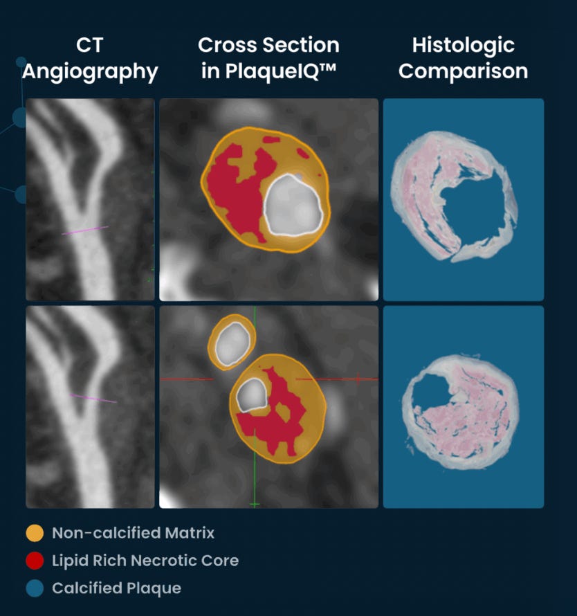
These tools look directly for non-calcified, vulnerable/soft plaque.
Again, looking to make a distinction between plaque you will die from rather than with.
Combined with the rapid change, the CT scanners themselves deliver the technology platforms that now assess and interpret this data have also raced ahead.
There are many other examples, including Heart Flow and Cleerly, which can assist with plaque composition and also assessing whether plaque is obstructive.
There is no doubt that what we are seeing here will soon be the standard of care for all CT scans in the future.
We have come a very long way from the grainy images first acquired by the early CT scans over 50 years ago.
But even still, there are more exciting changes yet to come.

https://radiologykey.com/computed-tomography-8/
Accuracy of Ultrahigh-Resolution Photon-counting CT for Detecting Coronary Artery Disease in a High-Risk Population. Radiology. 2023 Jun;307(5):e223305.
Budoff M, Kinninger A, Gransar H, et al. When Does a Calcium Score Equates to Secondary Prevention?. J Am Coll Cardiol Img. May 2023
Pedersen ER, Hovland S, Karaji I, et al Coronary calcium score in the initial evaluation of suspected coronary artery disease Heart 2023;109:695-701.
https://www.jaypeedigital.com/eReader/chapter/9789351524144/ch1
Perivascular Fat Attenuation Index by Computed Tomography as a Metric of Coronary Inflammation. J Am Coll Cardiol. 2018 Jun 12;71(23):2708-2709.
ORFAN Consortium. Inflammatory risk and cardiovascular events in patients without obstructive coronary artery disease: the ORFAN multicentre, longitudinal cohort study. Lancet. 2024 Jun 15;403(10444):2606-2618.
No posts
Comments
Nothing yet. Say the first thing.
Sign in to join the conversation.