
Your heart pumps blood downward — to your feet, your calves, your lower legs.
But blood has to return upward.
Against gravity.
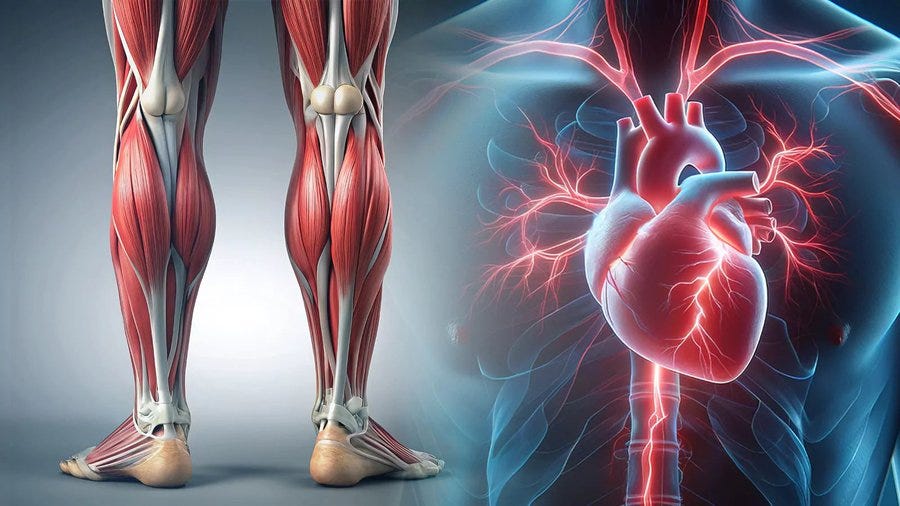
Through vessels that have no muscular wall of their own.
The question most people have never been asked: how does it get back?
The answer is your calves.
Your calf muscles — specifically the soleus and gastrocnemius — contract with every step you take and squeeze the venous blood vessels running through them, physically pushing blood back up toward the heart.
This is called the skeletal muscle pump.
And it is not a backup system.
It is the primary mechanism by which blood returns from your lower body to your heart.
Without it: blood pools in the lower legs. Venous pressure builds. Circulation stagnates. Every system that depends on blood flow — your brain, your lymphatic system, your nervous system — begins to suffer the downstream consequences.
Most people are sitting on this mechanism for 8–10 hours every day.
And there is a specific reason it fails even when people do move.
The calf contains two muscles that operate as a coordinated pump:
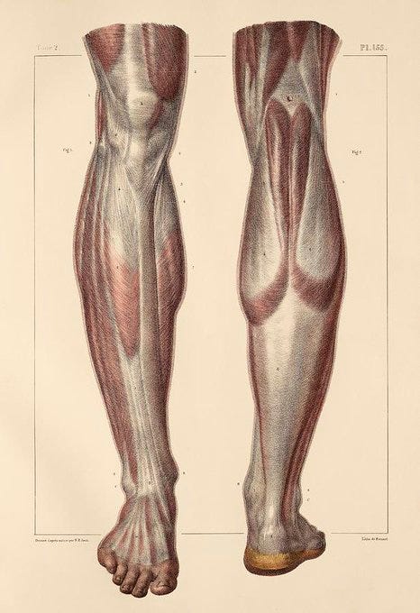
The soleus — the deeper, flatter muscle that runs from below the knee to the heel. It is the primary venous pump — contracting during walking, standing, and weight-bearing to compress the deep veins of the lower leg and push blood upward.
The gastrocnemius — the more superficial muscle that forms the visible calf shape. It contributes to venous return during dynamic movement — running, jumping, climbing.
Around and between these muscles sits fascia — the connective tissue matrix that surrounds, separates, and connects every muscle, nerve, and vessel in the body.
When fascia is hydrated and mobile, it slides freely — allowing the calf muscles to contract fully, compress the veins maximally, and pump blood upward with each step.
When fascia is dehydrated, sedentary, or chronically tensioned — from prolonged sitting, stress, or insufficient movement — it becomes dense and adhesive. It restricts the calf muscles’ range of motion. The pump weakens. Blood return from the lower body becomes progressively less efficient.
This is not a dramatic event.
It is a gradual mechanical failure — accumulating silently across years of desk work, sedentary commuting, and chronic stress — until the downstream consequences appear as symptoms that seem completely unrelated to the calf:
Cold feet. Brain fog. Chronic fatigue. Stiff knees. Bloating. Anxiety that won’t resolve. Sleep that never feels deep enough.
All of them trace, in part, to the same upstream mechanical failure in the lower leg.
The term “second heart” for the calf muscle is not metaphorical.
It appears in cardiovascular medicine and vascular surgery literature as a functional description of the soleus muscle’s role in venous return.
The scale of what the calf pump manages: at rest, approximately 70% of the body’s blood volume is in the venous system below the heart. A significant portion of that volume must be actively returned against a gravitational column of pressure that reaches approximately 80–100 mmHg at the ankle — higher than the arterial pressure at that level.
The calf muscle pump generates enough pressure during contraction to overcome this gravitational column and push blood upward through the venous valves that prevent backflow.
When the calf pump is compromised:
→ Venous pressure at the ankle and lower leg increases — producing the heaviness and aching that most people attribute to “standing all day”
→ Venous stasis — blood pooling in the lower leg — creates the conditions for deep vein thrombosis in high-risk individuals
→ Reduced venous return reduces cardiac preload — the volume of blood the heart receives per beat — reducing cardiac output and oxygen delivery to every organ simultaneously
Research published in the Journal of Vascular Surgery confirmed that calf muscle pump function is a significant independent predictor of venous disease severity — and that calf muscle pump failure produces measurable reductions in systemic circulation that extend well beyond the lower leg.
The calf is not a leg muscle that happens to help circulation.
It is a circulatory organ that happens to be located in the leg.
→ Calf muscle pump and venous return — Journal of Vascular Surgery
Exercise activates the calf pump.
But exercise does not release locked fascia.
This is the distinction most fitness and health content misses entirely — and it explains why people who walk regularly, exercise consistently, and maintain active lifestyles still experience the downstream symptoms of calf pump failure.
Fascia is not muscle.
It does not respond to contraction and relaxation cycles the way muscle tissue does.
Fascia responds to sustained pressure, hydration, and temperature — the three mechanical inputs that change its viscosity and mobility.
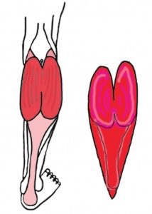
When calf fascia becomes dense through chronic sedentary posture, stress-induced tension, or dehydration:
→ It restricts the full range of muscle contraction — meaning the calf pump never reaches its full compression capacity even during active movement
→ It compresses the lymphatic vessels running through the calf — which sit immediately beneath the fascia and depend on fascial mobility for their own pumping action
→ It maintains a state of mechanical tension that the nervous system reads as a threat signal — keeping the sympathetic nervous system partially activated regardless of what the rest of the body is doing
This last point is the most clinically significant.
Research on fascial mechanoreceptors — the sensory receptors embedded throughout the fascial network — confirms that dense, restricted fascia generates continuous low-level afferent nerve signals that the central nervous system processes as background threat.
That background threat signal keeps cortisol slightly elevated.
Keeps the sympathetic nervous system slightly activated.
Makes complete parasympathetic recovery — deep sleep, genuine relaxation, full digestive function — impossible to achieve regardless of what other interventions are applied.
The fascial tension in the calf is not just a local mechanical problem.
It is a systemic nervous system problem that originates in the lower leg.
→ Fascial mechanoreceptors and nervous system signalling — PMC/NIH
The popliteal fossa — the diamond-shaped hollow at the back of the knee — is one of the most neurologically dense anatomical spaces in the lower body.
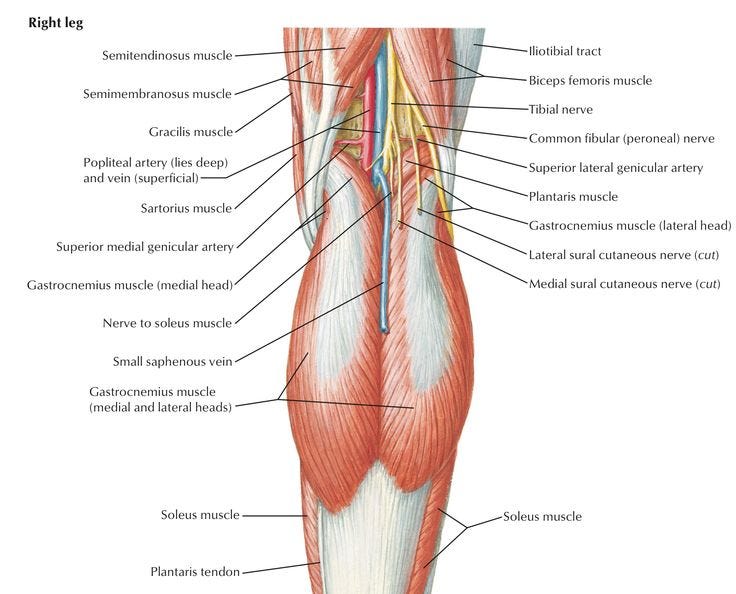
Running through it:
→ The popliteal artery and vein — the primary vascular supply to the lower leg
→ The popliteal lymph node cluster — the primary lymphatic drainage station for the entire lower leg and foot
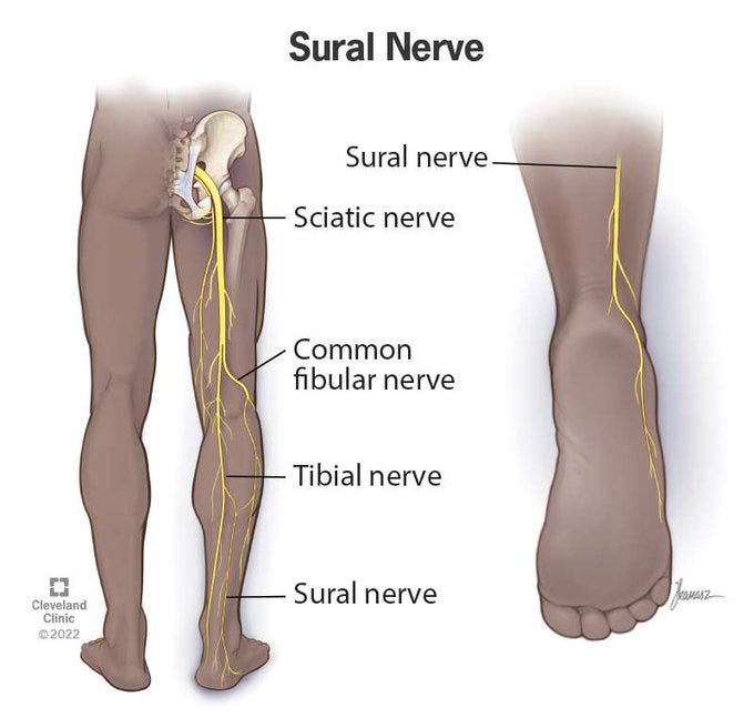
→ The tibial nerve and common peroneal nerve — branches of the sciatic nerve responsible for sensation and motor control throughout the lower leg
→ Fascial continuity connecting the hamstring fascia above to the calf fascia below
Sustained pressure applied to the popliteal fossa — through a ball or rolled towel in the kneeling position — simultaneously addresses all four of these structures.
The neurological mechanism most specifically studied: the tibial nerve runs immediately adjacent to the tibial artery in the popliteal fossa. Sustained gentle pressure on the tibial nerve at this location has been studied for its effect on the autonomic nervous system through a mechanism called neural tension release.
Research on neural tension release confirms that sustained pressure on peripheral nerve pathways produces measurable shifts in autonomic balance — specifically increasing parasympathetic tone through a mechanism that involves the afferent fibres of the tibial nerve transmitting a sustained non-threatening signal that inhibits sympathetic activation.
This is why the kneeling pressure technique produces the sensation of the nervous system “switching off” — not as metaphor but as documented neurophysiological event.
The pressure is not relaxing a muscle.
It is producing a specific neurological signal that the autonomic nervous system interprets as safety — and responds to by downregulating the sympathetic activation it had been maintaining.
→ Neural tension release and autonomic nervous system — PMC/NIH
The popliteal lymph nodes — sitting in the popliteal fossa at the back of the knee — are the primary lymphatic drainage point for the entire lower leg and foot.
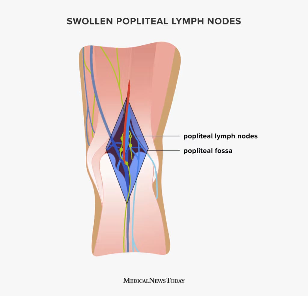
All lymph from the foot, ankle, and lower calf must pass through these nodes before continuing upward toward the inguinal nodes in the groin and eventually the thoracic duct.
When the popliteal nodes are congested — from sedentary posture, fascial restriction, or insufficient lower leg movement — the entire lymphatic drainage pathway for the lower body backs up.
The result: the swelling, puffiness, and heaviness in the lower leg that millions of people experience daily is not primarily a venous problem — it is a lymphatic problem that originates at a congested popliteal node cluster.
Sustained pressure on the popliteal fossa in the kneeling position directly decompresses the popliteal lymph node cluster — creating the mechanical conditions for lymph drainage to resume upward.
This connects to the lymphatic system post covered recently:
The legs-against-wall position drains lower body lymph toward the inguinal nodes using gravity.
The popliteal pressure technique clears the intermediate drainage station — the popliteal nodes — that legs-against-wall alone cannot reach.
The two techniques are not alternatives.
They are sequential: popliteal pressure first to clear the intermediate node, legs-against-wall second to drain the cleared lymph toward the inguinal nodes and into the thoracic duct.
→ Popliteal lymph nodes and lower limb drainage — PMC/NIH
The body stores unresolved stress not as a psychological phenomenon but as a physical one.
Fascia is the primary storage medium.
Research in the field of somatic neuroscience — particularly the work of Dr. Peter Levine and the documented phenomenon of trauma resolution through body-based approaches — confirms that unresolved stress activates the body’s defensive motor responses and that these responses become encoded in the fascial tension of the muscle groups most involved in the stress response.
The calves are specifically implicated.
The defensive response to threat — the impulse to run, to flee — is encoded in the lower leg musculature. When threat is perceived but escape is impossible (chronic workplace stress, emotional conflict, financial pressure), the motor impulse that was generated but never completed remains encoded in the calf and lower leg fascia as chronic tension.
This is the mechanism behind the observation that many people experience unexpected emotional responses — tears, shaking, sudden calm — when sustained pressure is applied to locked calf fascia.
The pressure is not producing the emotion.
It is releasing the incomplete motor response that has been stored in the tissue — allowing the nervous system to complete what it began and return to baseline.
Research published in the Journal of Bodywork and Movement Therapies found that sustained fascial pressure techniques produced measurable reductions in cortisol markers alongside the physical release — confirming that the emotional component of fascial release is physiological rather than purely psychological.
This is why the technique works for anxiety, for sleep difficulty, and for the feeling of being emotionally stuck — not because it is a wellness ritual but because it is completing a physiological process that chronic stress interrupted.
→ Fascial release and cortisol reduction — Journal of Bodywork and Movement Therapies
The placement determines everything.
One centimetre too high — above the popliteal fossa — and the pressure lands on the hamstring tendon insertion rather than the lymph node cluster and tibial nerve pathway.
One centimetre too low — into the upper calf belly — and the pressure compresses the gastrocnemius muscle rather than the fascial junction where the five mechanisms above converge.
The exact placement — defined by three anatomical landmarks you can identify without any medical training — and the specific breathing sequence that research shows doubles the vagal activation during the hold — are below.
Where does your body hold tension most consistently?
The calves and lower legs that feel heavy by evening.
The knees that feel stiff every morning.
The anxiety that arrives without obvious cause.
The sleep that never feels complete despite the hours.
Exact placement — the three landmarks:
Sit on the floor with your legs extended in front of you.
Find the crease at the back of your knee — the horizontal fold that appears when you bend the knee slightly. This is the base of the popliteal fossa.
Move your fingers upward from the crease by approximately 2–3 centimetres — you are now at the centre of the popliteal fossa, between the two heads of the gastrocnemius muscle.
The ball or rolled towel should be placed at this exact point — not at the crease, not in the upper calf belly, but at the soft centre of the popliteal fossa where the skin is most hollow and the tissue is deepest.
Comments
Nothing yet. Say the first thing.
Sign in to join the conversation.