
1: A connectome is a static snapshot of the microstructure of the brain. And yet, many people expect that it may eventually be sufficient for simulating the brain with a sufficient degree of accuracy to recapitulate many interesting cognitive and behavioral properties. How could this possibly be?
The canonical answer is separate “side experiments” that will allow us to learn about how the nervous system works first, which will allow us to run those simulations. But what might those side experiments consist of?
A nice example comes from a new study using Patch-seq on close to 1500 human neurons expressing the neurotransmitter glutamate (i.e. glutamatergic neurons) across all six cortical layers. This method allowed the authors to measure the morphology, electrophysiology, and transcriptome from each individual neuron.
If we want to simulate a connectome, we need to know not just which neurons connect to which, but how those neurons function. Their use of Patch-seq allows us to build sort of a lookup table, so that given a neuron’s transcriptomic “type” (which we can infer from its morphology or biomolecular staining, and could be multidimensional), we can predict its electrophysiology.
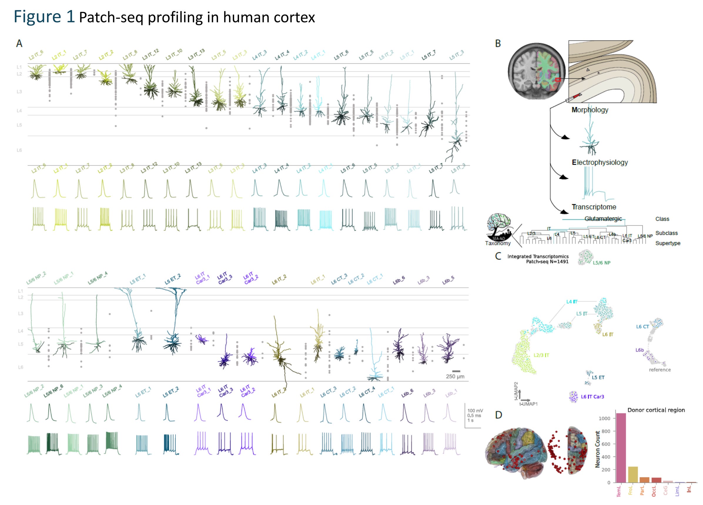
Consider the action potential, which is the “spike” that neurons use to communicate (and the namesake of these monthly update emails!). There are a ton of aspects of the action potential that differ between neurons.
For example, when you inject current into different neuron types, some fire readily while others are more resistant. L4 IT (Layer 4 Intra-Telencephalic) neurons, which are the main recipients of thalamic input, need very little current to start firing. L2/3 IT and L5 ET neurons require around two to three times as much. This property, called the rheobase, was the single most distinguishing electrophysiological feature across neuron subclasses.
As another example, once a neuron starts firing, how does it respond as you push the current harder? That is captured by the F-I slope, which measures the rate at which firing frequency increases with more injected current. L4 IT and L5/6 NP (Near-Projecting) neurons have the highest F-I slopes, thus allowing them to relay graded input strengths. On the other hand, L6 IT neurons have nearly flat F-I slopes, meaning that they fire a burst at stimulus onset and then barely respond to further increases. This was the third most distinguishing electrophysiological feature between neurons.
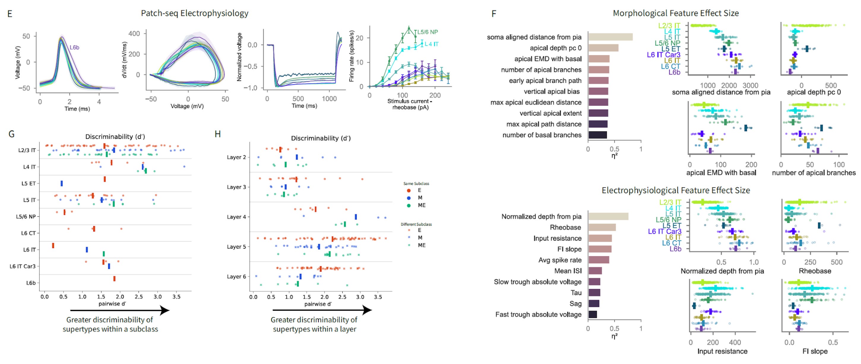
This cell type-electrophysiology lookup table approach is very helpful. But part of the problem with it is that there may still be variation within cell types that also matters for their function.
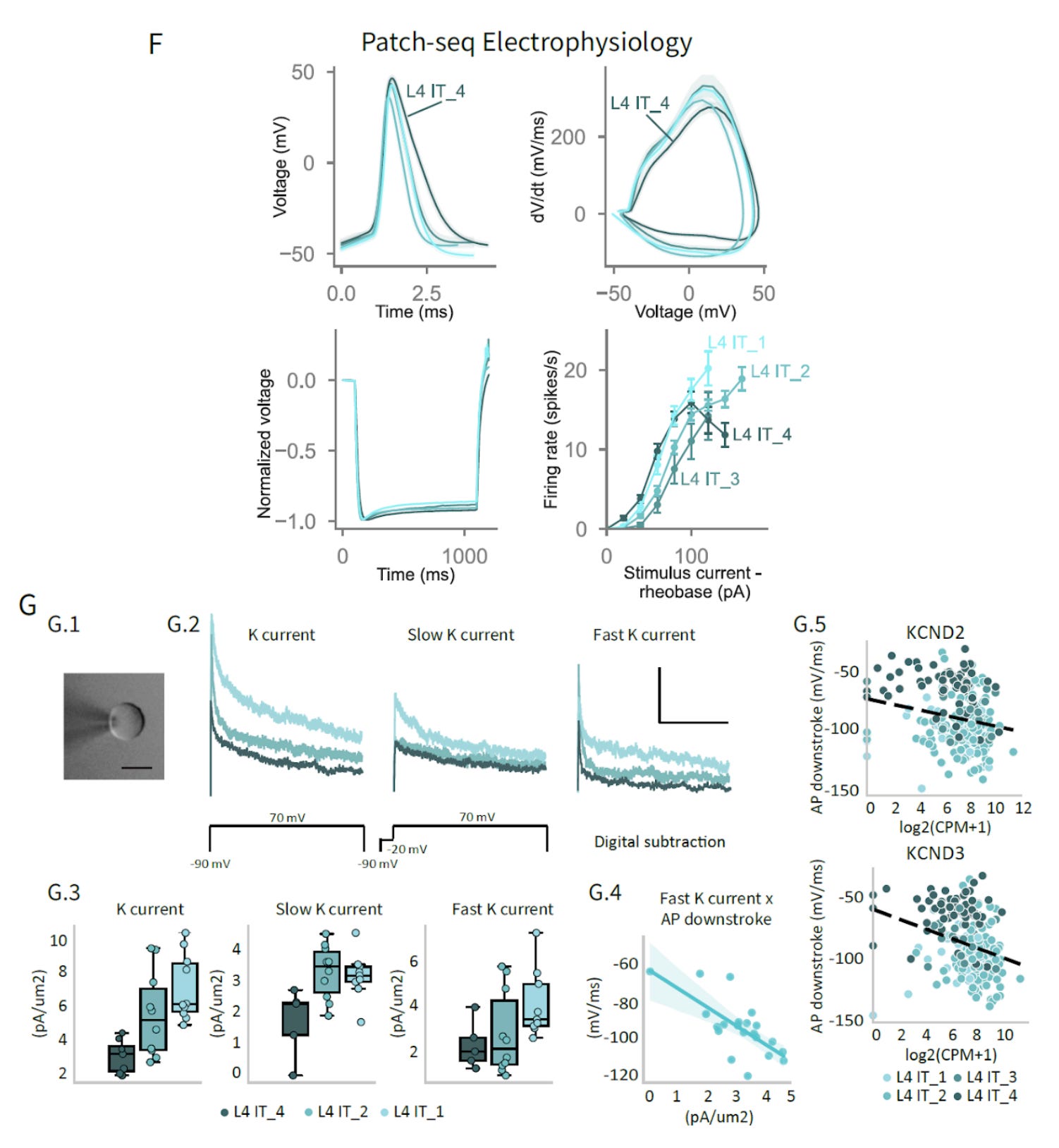
The paper presents a nice example of this by zooming in on L4 IT neurons. Even within this single subclass, the action potential shape varies. In particular, there is a subtype called L4 IT_4 neurons, which have broader, slower spikes than the other L4 IT subtypes.
What explains this? They used a technique called nucleated patch recording, which involves pulling off part of the cell body, and which allows them to measure potassium currents directly. They found that L4 IT_4 cells have the least fast-inactivating potassium current (called A-type), while L4 IT_1 cells have the most. A-type potassium channels drive fast repolarization of the spike, so less of them means a slower downstroke and a broader action potential.
They found that the RNA levels of KCND2 and KCND3, which are the actual genes encoding the Kv4.2 and Kv4.3 channels that carry A-type current, predict the downstroke speed across the L4 IT population. But this expression varies continuously within the cell type, not discretely between types. So the transcriptome-derived cell type gets you pretty far, but there is still a spectrum of ion channel expression within each category that further shapes how individual neurons behave.
In this data set, it seems like the connectome plus morphologically-derived cell type classifications would probably allow you to predict a lot of the electrophysiological variance between cells. The remaining variance may require measuring, or somehow inferring, further molecular details such as the potassium channel expression. What degree of precision would be required for accurately simulating different aspects of cognition and behavior is still very much an open question.
2: A study uses artificial hibernation in mice as a new paradigm to study what structures are required to maintain long-term memories. Basically, they chemogenetically activate a subset of neurons in the hypothalamus to induce a hypothermic and hypometabolic torpor-like state for around 48 hours.
Because the subset of neurons activated are called Q-neurons, this is called Q-neuron-induced hypometabolism and hypothermia, or QIH for short. (It seems this is probably not possible in humans because we don’t have a clear analog of the Q-neuron population.)
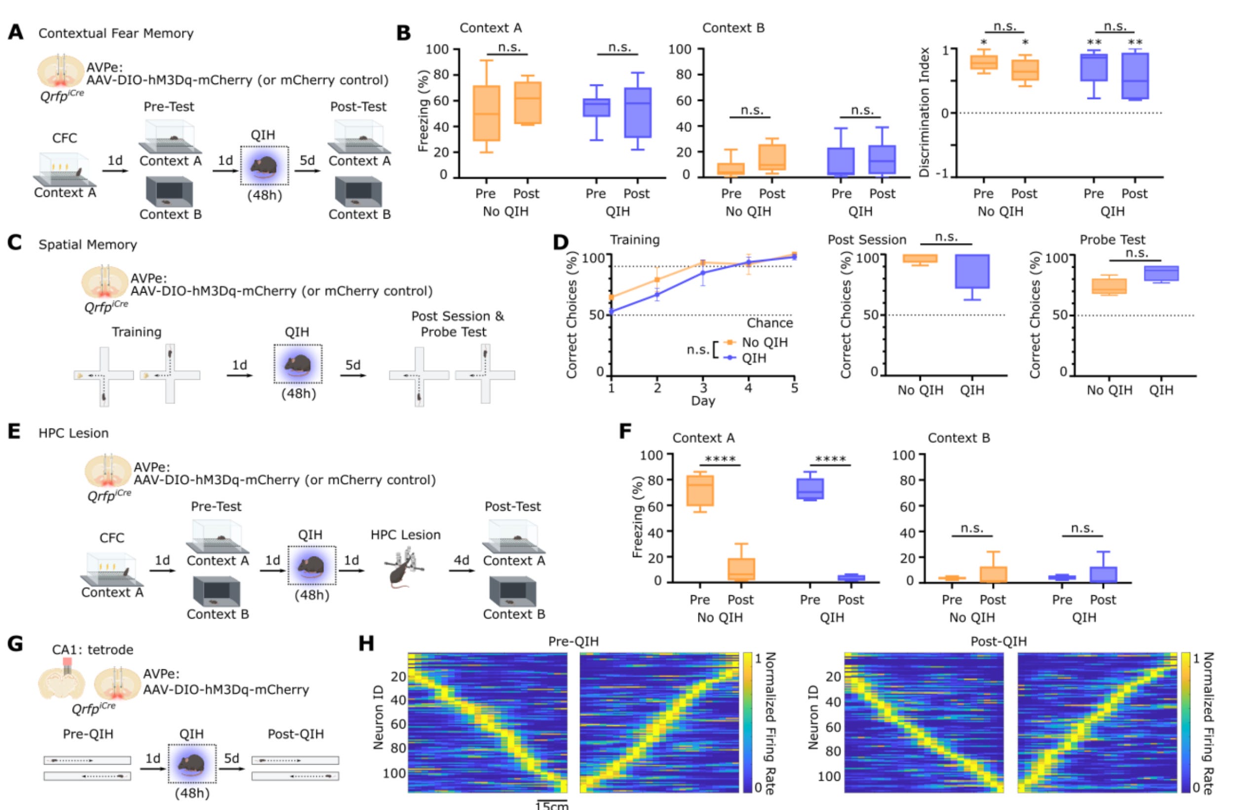
During this artificial hibernation, the hippocampal firing rate drops by around 70% and the synapse density falls by more than 50%.
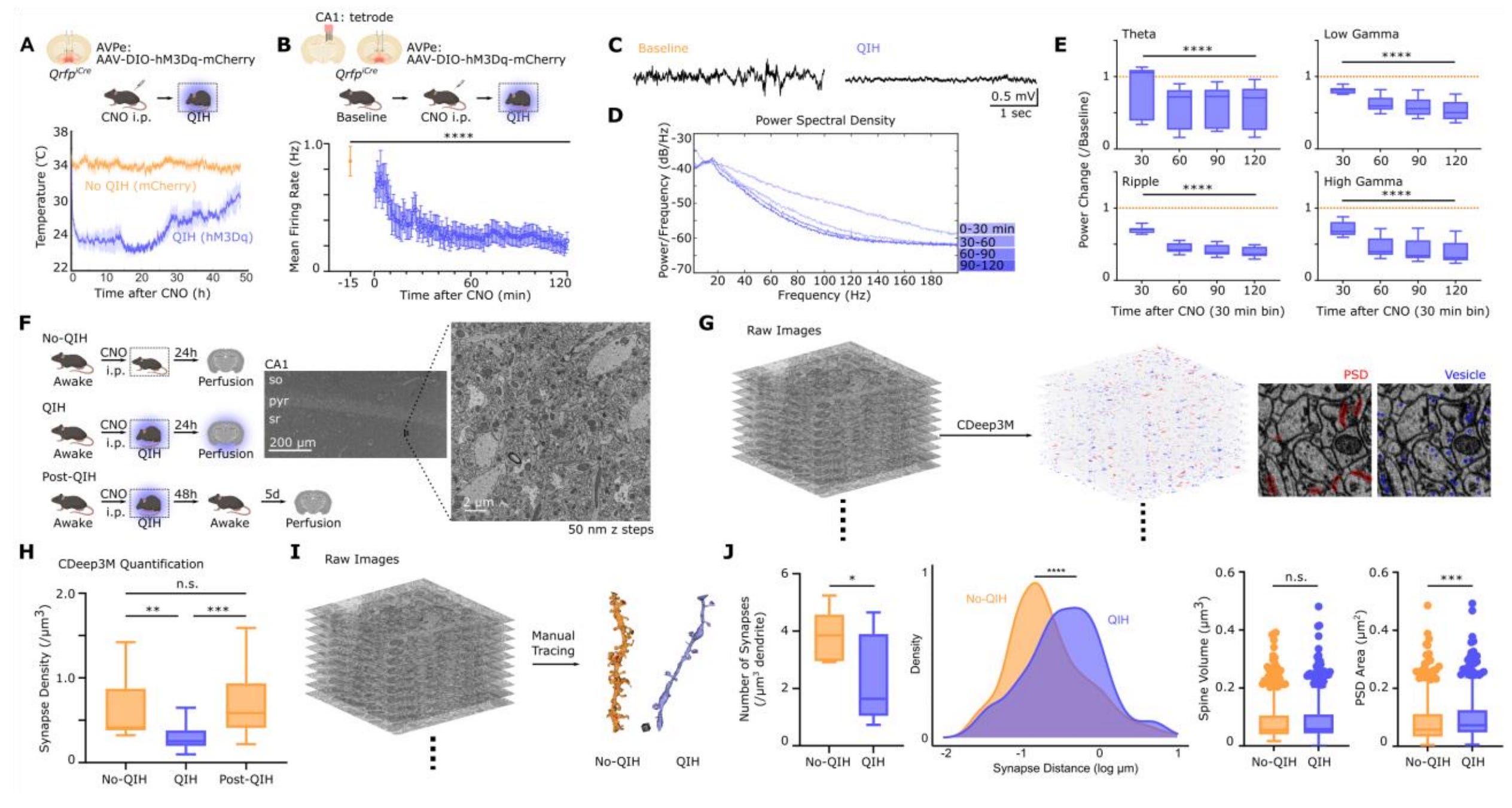
They next tested whether this loss of synapses led to a loss of long-term memories that were created prior to artificial hibernation. They found that these memories were not lost. The mice woke up with contextual fear memories and spatial navigation intact, and place cell representations maintained the same.
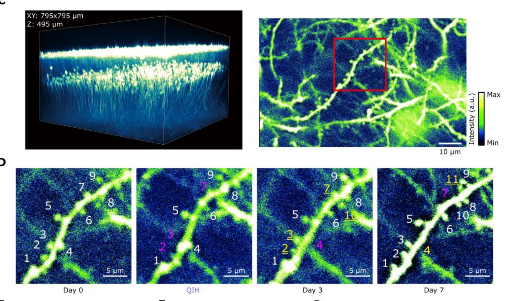
They then used two photon imaging to track individual dendritic spines through hibernation. They found that the dendritic spines that persisted maintained their original volumes.
And interestingly, the found that dendritic spines which were eliminated tended to regrow at the same dendritic locations within 24 hours. Specifically, they found 82.1% site-specific regrowth. This suggests that there might be some sort of structural scaffold at those dendritic locations that persists through the elimination and guides regrowth.
They next found that while overall engram synapse density drops during hibernation, spatially clustered engram synapses tended to be selectively protected. The non-clustered engram synapses tend to get pruned, while the clusters tend to survive. And these clusters tend to occur at multi-synaptic boutons (MSBs), which are axonal varicosities that synapse onto multiple postsynaptic partners.
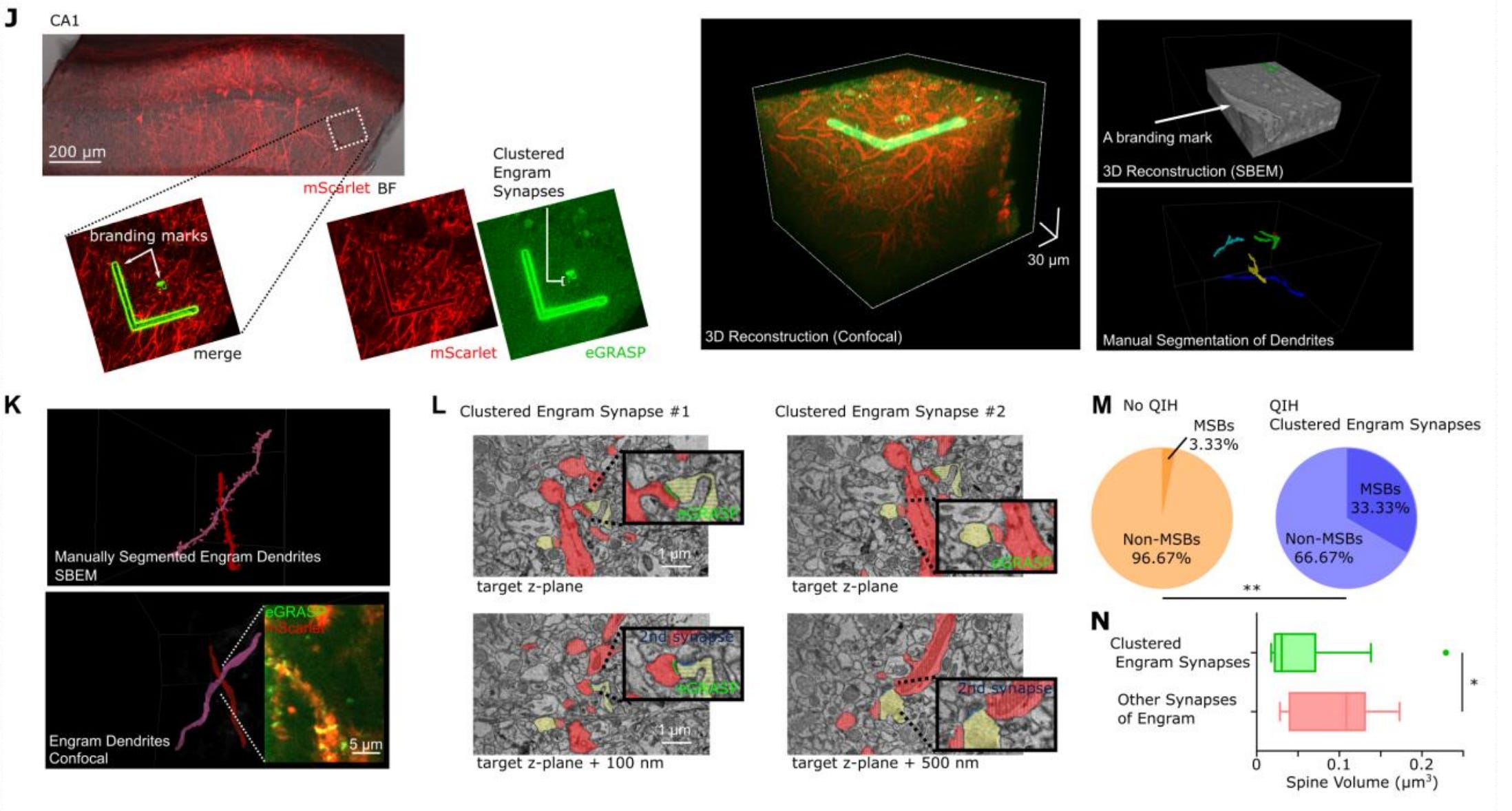
There are a couple of possible interpretations of this study. The authors propose that a sparse “core memory trace,” which is defined by spatial clustering and multi-synaptic bouton connectivity, may be the critical aspect of long-term memory retention. In this case, non-clustered synapses would be dispensable. Alternatively, something about the hibernation state could enable network reconstruction upon arousal, with these clusters serving as seeds.
The finding that consolidated long-term memories can survive hypometabolism, hypothermia, and more than 50% synapse loss, as long as certain clustered synapses are selectively protected, adds to the existing evidence that long-term memory depends on key structural relationships rather than labile molecular states or ongoing electrophysiological activity.
3: Some of the wisdom of neuroscientist Adam Marblestone is captured in his recent podcast interview with Dwarkesh, which is mostly about how current AI models are similar and different to the brain. Here is my interview with Adam from 2015.
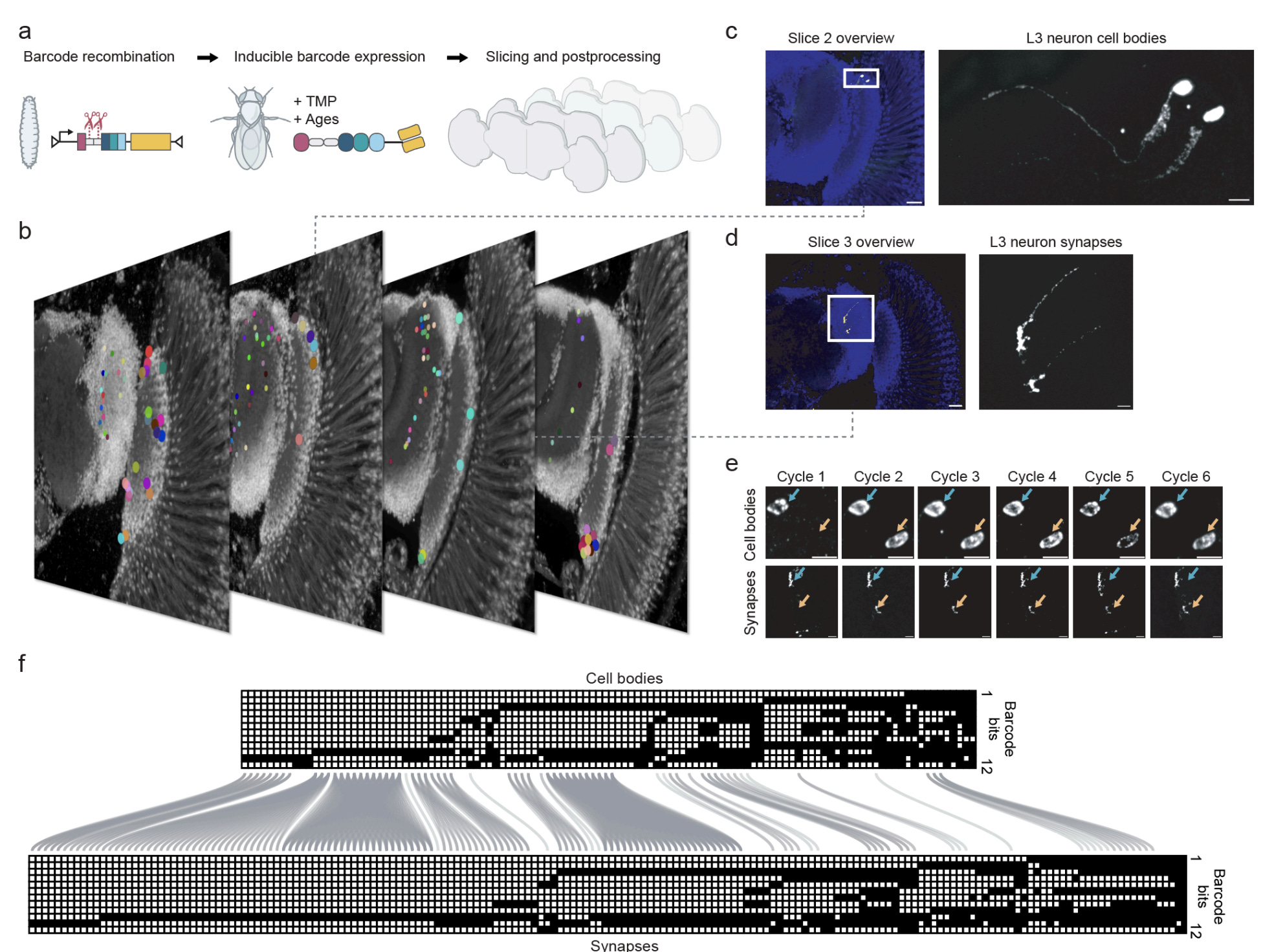
4: A new genetic method allows mapping of synapses to cell bodies using protein barcoding and light microscopy, which could be much more easily scaled than methods using electron microscopy. It is currently limited by a small number of epitopes (12) incorporated into the barcodes.
5: An argument that neuroscientists should study animals without neurons more often. This is because data has shown that multicellular organisms like Trichoplax adhaerens can still respond to neuropeptides. For example, they respond to endomorphin-like peptides in ways that suggest changes in their feeding behavior.
Neuropeptides operate via slower changes than electrophysiological ones, working primarily due to signaling via G-protein-coupled receptors. And yet, they alone are capable of mediating certain behavioral differences between animals.
To me this reminds me of the difference between a state and a trait. One might argue that the moment-to-moment variability in states between humans, such as whether one is hungry or thirsty, are not as essential for personhood as variability in traits such as long-term memories or personality. (More precisely I should say that at least that rings true for me, but I also think others should be free to choose what they value for themselves.)
6: A punchy argument by Nassir Ghaemi that mixed states (i.e. where manic and depressive symptoms are both present in the same patient) are crucial for understanding mood disorders. He argues that the DSM’s separation of bipolar disorder from major depressive disorder is wrong, that mixed states are highly under-recognized, and that this has clinical consequences.
He thinks this is especially the case in people with depression who also have baseline hyperthymic temperment, which manifests as constant high energy and a high degree of optimism. He notes that he thinks one of his friends had this condition, had it worsened with serotonin reuptake inhibitors (like Prozac or Lexapro), and then died of suicide.
As a result of this belief that mixed states are underrated, he now treats most of his patients with low-dose lithium (300-600 mg/d) rather than serotonin reuptake inhibitors.
Slightly too “me vs the world” for my taste, but it’s certainly a provocative essay and he might be onto something important.
7: An argument by Haim Belmaker, one of the pioneers of lithium treatment and research, that lithium should not be considered the gold standard treatment for bipolar disorder.
He notes that the superiority of lithium over alternatives has not been clearly demonstrated in meta-analyses. As alternatives to lithium, he sometimes uses valproate, carbamazepine, or combinations of the three. He says that he often has good results instead with low doses of second generation antipsychotics such as olanzapine 2.5 mg or risperidone 1-3 mg.
He also argues that despite decades of research, we do not really know the molecular mechanism of lithium. He notes that its proposed mechanism has conveniently shifted with academic fads in each era, from monoamines in the 1970s, second messengers in the 1980s, third messengers in the 1990s, and finally to neuroprotection in the 2000s. These findings were usually not replicated before being abandoned for the next fashionable framework.
I’m not sure I agree with him that the side effect burden of lithium is not much different than the alternatives. While this is a highly individual consideration, it seems to me to be meaningfully lower than some of the alternatives. However, because one of the major side effects of certain second generation antipsychotics like olanzapine is weight gain, this calculus has been changing rapidly in recent years with the new anti-obesity medicines.
8: An article about Mitul Desai, who after decades of helping manage his brother’s schizophrenia while building a successful career, quit to launch a company that works with insurance to provide coaching and peer support to caregivers dealing with mental illness in loved ones.
It’s a good summary of how caregiving for people with mental health problems is so different from other types of caregiving, due to there being unpredictable crises, isolation driven by stigma, and the impossibility of forcing treatment on adults.
9: A study of 85 participants ages 20-84 finds that chronological age is associated with lower reported baseline cognitive fatigue levels, while the “brain age difference” (how relatively old the brain appears relative to the chronological age based on MRI morphology) predicts how quickly cognitive fatigue accumulates during mental tasks.
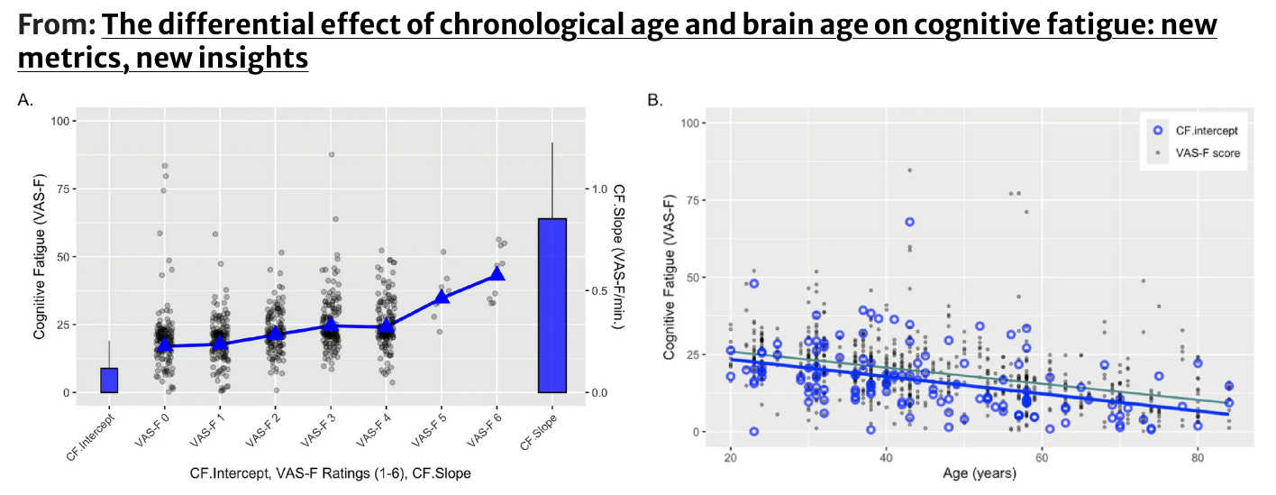
A paper I was a part of previously found that histologically, brain age is most strongly linked to vascular structure. So to take a leap on my jumping-to-conclusions mat, I would hypothesize that the accumulation of cognition fatigue during tasks might be especially related to cerebrovascular function.
10: Merge labs, a new company planning to make brain computer interfaces that avoid implants and reach deep into the brain, has raised $252 million in funding. It seems they are primarily focused on using ultrasound devices.
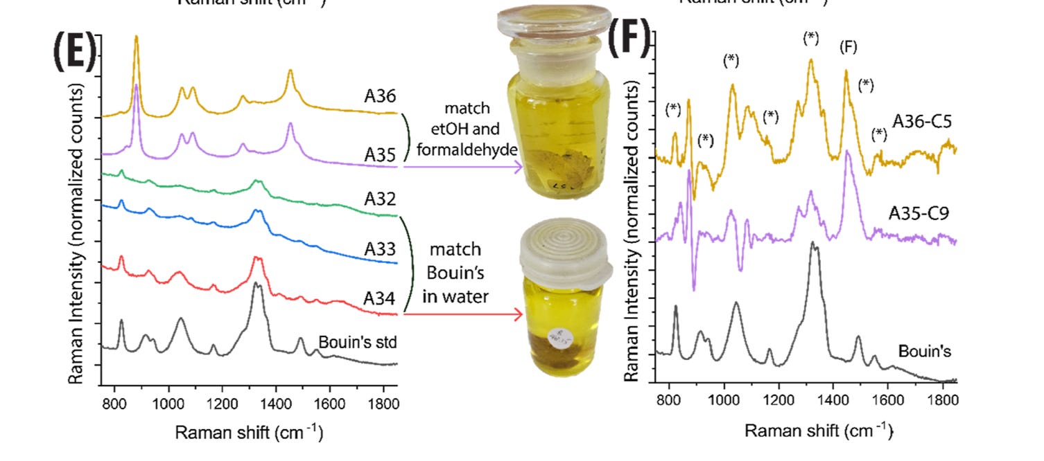
11: A new study uses a portable device that does Raman spectroscopy on samples preserved in natural history museums to determine what type of fluid preservative was used:
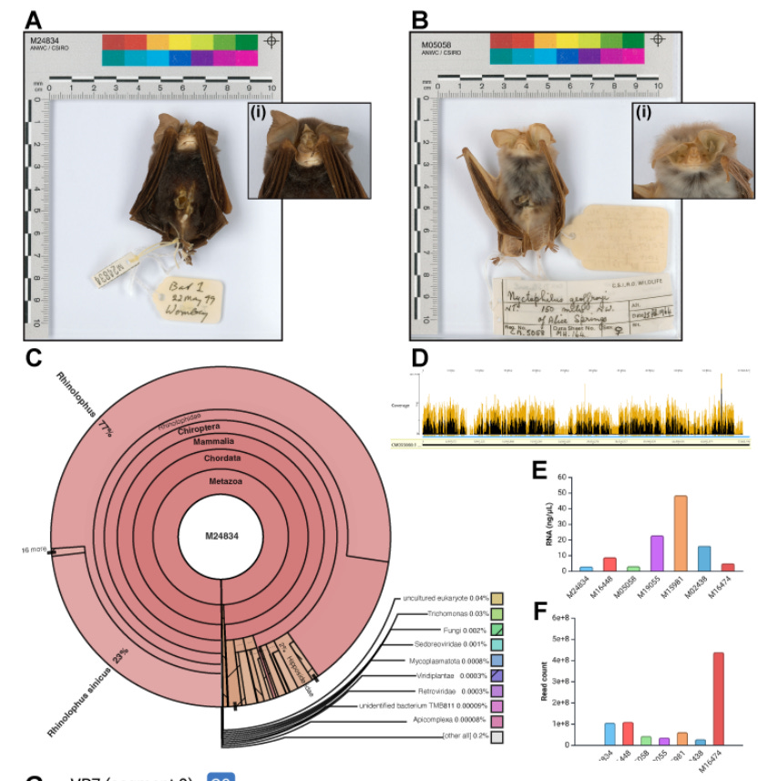
12: A study finds that bats preserved in the fluid state for decades can still have the RNA content in their bodies analyzed precisely:
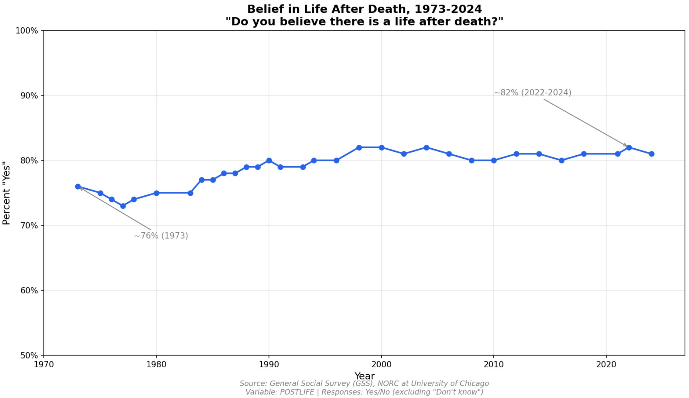
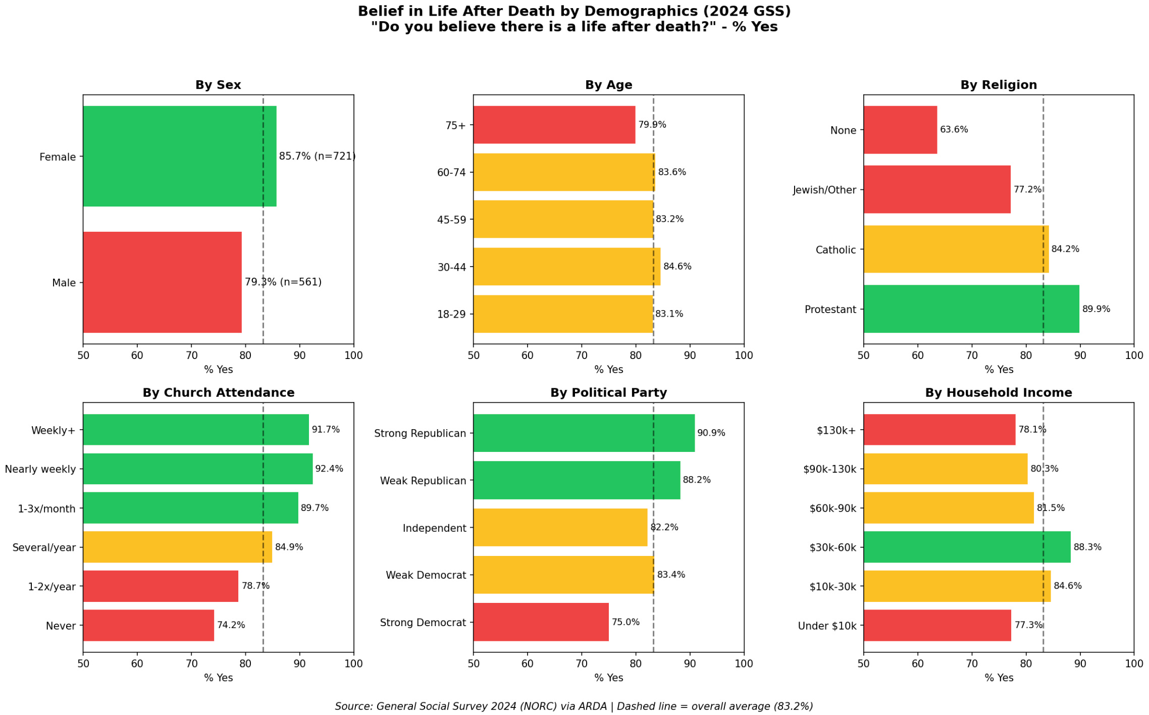
13: New-to-me data from 2025 shows that while religiosity in the US is down, belief in an afterlife is either fairly steady or actually seems to be slightly increasing. Here’s a plot of the data:
Belief in an afterlife is strong in all demographic groups, but is especially strong in women, Republicans, those who attend church frequently, and has an inverted u-shaped curve with household income:
It’s interesting to me that the dominant cultural narrative about death has not budged in the past 50 years, despite so many other changes in the world. If anything, it has gotten stronger.
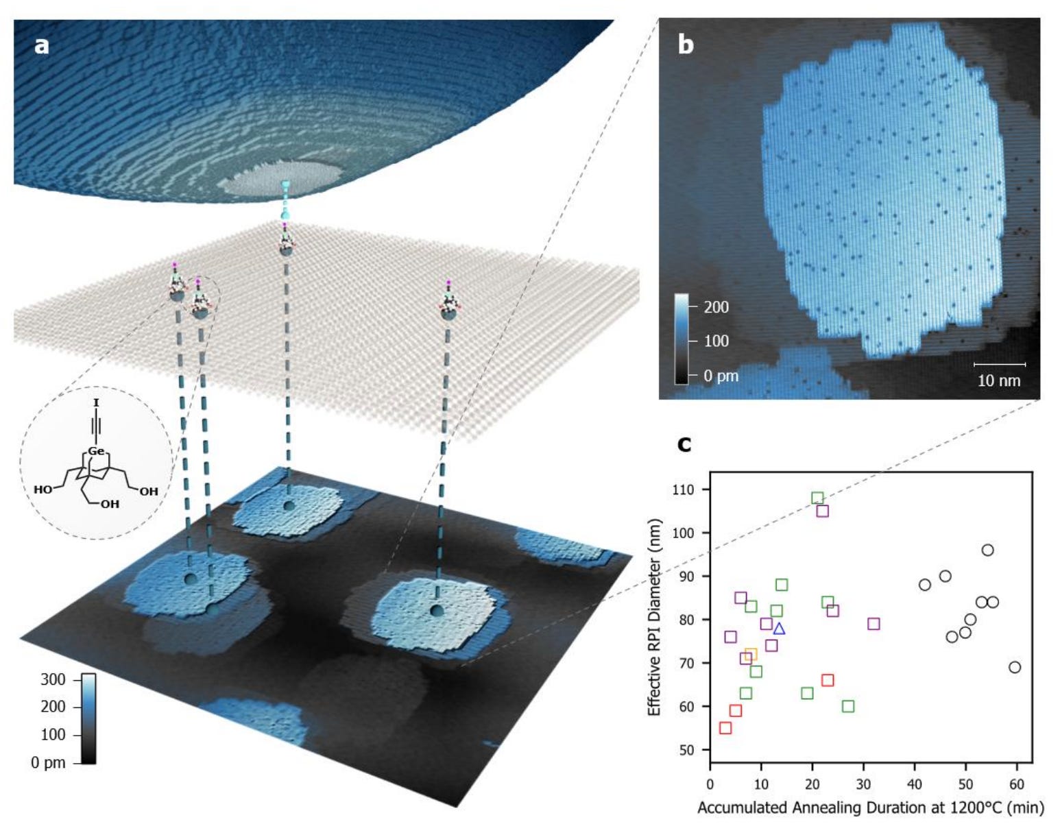
14: An advance in scanning tunneling microscopy (STM), as a new paper introduces “inverted-mode STM.” Normally in STM, the probe tip manipulates or images atoms on a sample surface.
Here they flip it, so that specially designed molecules stand upright on a silicon surface, and as the probe scans over them, each molecule acts as a tiny imaging tip that maps the probe apex from below. This lets them verify the atomic structure of the probe tip while also enabling controlled chemical reactions at the junction.
They demonstrate the technique by reproducibly removing single hydrogen atoms from the probe’s silicon surface (27/28 success rate). The way this works is that a molecule with a reactive radical terminus, when positioned close enough, forms a stronger bond with the hydrogen than the silicon does, so the atom transfers to the molecule.
This is progress toward “mechanosynthesis”, i.e. using precise mechanical positioning, rather than applied voltage or heat, to drive chemical reactions and build structures atom-by-atom.
This is not my field, but as far as I can tell, this paper is a big deal. I’m surprised it has not gotten more interest. Robert Freitas and Ralph Merkle are co-authors.
15: Neuroscientist Peter Rupprecht’s annual “intuition about the brain” post includes a section on connectomics and its limitations for whole brain emulation. He distinguishes between current technical barriers (such as imaging scale and data analysis bottlenecks) and more fundamental questions about whether pure electron microscopy-based connectomics alone could ever suffice, even given future technological advances.
In the comments, he clarifies that he’s skeptical that connectomics without molecular annotation will ever be sufficient for reviving a person in silico, since neurons have variable distributions of ion channels and other proteins that electron microscopy alone cannot measure. But he notes that expansion microscopy with biomolecular labeling might eventually resolve these limitations. He concludes: “So, from the perspective of a brain preservation person, there is definitely room for hope for such future technology to reconstruct and revive brains in 100-200 years (although one can argue whether this preservation effort is justified).”
16: I was honored to see so many interesting comments on my post about the memory decoding award (see also on Reddit here and here, and Ken’s opening remarks on the award ceremony here). I wanted to signal boost and follow-up on part of Nicholas Kircher’s interesting comment here:
I have been playing around over the last year or so with the connectomics datasets from flywire, thinking of ways I might be able to simulate some structure or other, though my main limitation is that I don’t have data on what the expected result should be from a real specimen in terms of the neural firing patterns.
Ay, there’s the rub! That’s the exact problem. Nobody has great ways of figuring out what the expected neural firing patterns should be, and our ground truth data is thus far pretty sparse. For those who are interested in this area, here are a few avenues to consider:
A: Randal Koene has thought about this problem very hard, possibly harder than anyone else. He has created the Brain Emulation Challenge, which has synthetic electrophysiology data that modelers can try to predict.
B: So far the largest real-world data linking electrophysiology to connectomics I know of is in the MICrONS data set, which is from the mouse visual cortex. However, as far as I know, nobody has yet attempted to analyze this data to try to predict function from structure (as opposed to the other way around).
It would be interesting to develop benchmarks in this structure-to-function prediction, because we would expect our ability to do so to get better over time, as we learn more about the brain and can therefore infer more about the expected electrophysiology from the electron microscopy images.
C: People at Janelia, Harvard, and Google are working on a data set that will have combined calcium imaging and connectomics data from a larval zebrafish, called ZAPBench. This will probably be the best data set for benchmarking neural circuit models once it is released.
17: Aschwin de Wolf has started a substack, here is his first post.
18: Michael Cerullo has also started a substack. Here are his thoughts on Sebastian Seung’s recent podcast. In that podcast, when asked directly about the possibility of mind uploading, Seung deflected by framing it as a religious belief. Cerullo notes that this form of psychoanalysis is inaccurate, and it is more like a guilt by association argument. I agree.
19: BRLS is a non-profit that supplies $8-9 million yearly to cryonics research and has long been the major funding mechanism of the field. One of the best ways to figure out what the funded companies are up to is through Ben Best’s yearly updates, since he is the Director of Research Oversight at BRLS. Here is this year’s update.
One of the most exciting updates is the work at 21st Century Medicine (21CM) led by Ralph Spindler. He reports that they have done electron microscopy on rabbit brains, by opening the blood-brain barrier with detergent so that the cryoprotectant M22 can get into the brain. Work in this area from 21CM is reportedly being submitted to PLOS One.
It would be fantastic if another brain preservation protocol is able to achieve high quality structural preservation, especially if it can be done reliably in realistic conditions. More competition is good for the consumer.
I’m not sure what the methods were, and the available images aren’t high resolution enough for me to actually evaluate their data. I look forward to seeing the actual paper so that I can actually do so.
20: My talk at the December Sparks Brain Preservation open house is now online. The talk was about historical perspectives people have had on the topic of revival following long-term preservation, including those of John Hunter, Ben Franklin, Evan Cooper, Robert Ettinger, and Robert Prehoda.
No posts
Comments
Nothing yet. Say the first thing.
Sign in to join the conversation.