
There is a particular kind of silence that settles over a room when someone reads their knee MRI report for the first time.
They came in for one thing, the ACL, and what came back was a paragraph that reads like the guest list for a wedding nobody wanted to attend. Complete ACL rupture. Extensive bone marrow oedema of the lateral femoral condyle and posterolateral tibial plateau. Impaction fracture. Meniscocapsular junction tear. Deep MCL injury. And then the question, usually delivered very calmly, which somehow makes it worse: “So is all of that bad?”
The honest answer is that it is normal, it is important, and it really should change how we plan the next six months.
If you take a large group of people with fresh ACL tears and scan them properly, the ACL turns out to be travelling with a fairly substantial entourage. Cristiani et al. (2024) did exactly that with more than two hundred and fifty patients aged fifteen to forty, all imaged within a few weeks of injury, with every scan read independently by both an orthopaedic knee surgeon and a musculoskeletal radiologist.
Medial meniscus injuries turned up in well over half of the group, with ramp lesions the most common variety. Injuries to the medial collateral ligament complex appeared in around four in ten. Lateral meniscus injuries in roughly a quarter. Cartilage injuries, Segond fractures and lateral collateral ligament injuries all made appearances too, in smaller numbers. It is a long and slightly humbling list.
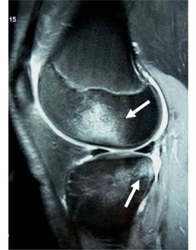
But the finding that should stop you in your tracks is that bone bruising was present in more than nine out of ten knees, and in most of those the pattern followed the classic pivot shift footprint, with oedema sitting in the posterolateral corner of the lateral tibial plateau and the central part of the lateral femoral condyle (Cristiani et al., 2024). The tibia slides forward, the femur crashes into it, and the bone quietly keeps the receipt.
So when a patient asks whether it is normal to have all that other stuff on the report, the answer is genuinely yes. The unusual scan is the clean one.
This is the piece of language I would most like to retire, because the word “bruise” does an enormous amount of damage to how seriously everyone takes it. People hear bruise and picture the purple thing on their shin that they poke for a week and then forget about, and clinicians can drift into the same casual reading without meaning to.
What has actually happened is a compression injury to bone. The internal scaffolding has been crushed, there is bleeding and swelling inside a rigid structure that has nowhere to expand into, and Galloway et al. (2023) found that the volume involved can be surprisingly large, sitting mostly in the lateral compartment across the tibial plateau and the femoral condyle. Their average total volume across the joint came in at around seven millilitres of bruised, bleeding bone marrow, which is roughly a teaspoon and a half, all of it inside a joint you would quite like your patient to bend today.
Once you sit with that image for a moment, the swollen, stiff, load averse knee in front of you in week two makes a great deal more sense. It is not deconditioning and it is not poor motivation. It is a bone injury, just one that does not get a cast or any sympathy.
This, for me, is the whole point. A bone bruise is a sign that the joint has been dealt a major blow and has absorbed a serious amount of trauma, and that is something to respect rather than something to note and move past.
The report will tell you that bone marrow oedema is present. What it will not tell you, at least not in any way you can act on, is how much.
That difference matters enormously in the first few months, which is why I will always ask to look at the actual images rather than working from the written summary alone. When I have the film up, I am trying to build a feel for a few things. How much swelling is this knee actually holding, and is that consistent with what I am seeing and feeling in the clinic. How much stress has landed in the lateral femoral condyle and in the tibial plateau, and how far does it extend. Is the joint surface above it intact, or is there a depression sitting underneath it.
And then the question that actually shapes the plan. Is this a knee we can work with carefully and sensibly, respecting it while still making progress, or is this genuinely stressed bone that needs a period of real protection.
That protection question is broader than most people assume, because the conversation usually leaps straight to running and jumping. Bone does not particularly care whether load arrives through a hop test or through a heavy leg press, and it does not distinguish between a training session and a long day on your feet. So if the film tells me the lateral compartment has taken a serious hit, then gym loading, weight bearing and general daily volume all come into the conversation alongside impact, and we shape the programme around what the bone can tolerate rather than around what the calendar says we should be doing by now.
None of that is visible in a one line report. You have to go and look.
There is one more distinction worth carrying with you, and Mostowy et al. (2023) draw it neatly. In a bone bruise, the shape of the underlying bone is unchanged. In an impaction fracture, the compression has been forceful enough to leave a visible depression in the lateral femoral condyle, which shows up on imaging as the lateral femoral notch sign.
These are not rare, having been reported in up to about half of ACL injuries depending on the imaging used, and Cristiani et al. (2024) found them in close to half of their own group. So a genuine dent in the femur is a very common travelling companion.
Dents also behave differently from bruises. Across the studies Mostowy et al. (2023) gathered, the great majority linked impaction fractures with cartilage injury and progressive degeneration, they are associated with lateral meniscus injury, and in longer term follow up both the depression and the cartilage changes sitting inside it were still there many years later. Bruises tend to resolve. Dents largely do not, which is another good reason to know which one you are dealing with before you start writing loading progressions.
Here is the thing that ties it all together. The injury is the first trauma and the reconstruction is the second one, and if those two events land close together then the bone has not had much of a chance in between. Tunnels are drilled, grafts are harvested, menisci are repaired, and the whole joint is re-inflamed on top of a lateral compartment that is still quietly recovering from the original impact.
Which changes what I think the pre-operative phase is actually for.
If someone has twelve weeks between their injury and their surgical date, that can feel like a generous amount of runway, and there is a real temptation to fill it. Get them running again. Sneak a bit of hopping in. Turn up to theatre in the best shape of their life.
I understand the appeal, and I very rarely do it, and the bone is the reason why.
Impact work before a planned operation adds a genuine dose of joint stress in exchange for a return you are about to hand straight back on the operating table. When you know a second insult is already booked into the calendar, spending your risk budget on pre-op jumping is not a trade I am often willing to make.
So the sequence I use instead, which I have written about at more length in the pre-op ACL guide, is deliberately unglamorous at the start. The early weeks are proper physio based work aimed squarely at calming the knee down, so swelling control, pain control, and restoring symmetrical range of movement, and I would much rather spend an extra week here than rush it. From there we move into roughly a month of bilateral loading, building real strength and confidence through both legs working together. That flows into roughly a month of unilateral loading, where the operated side has to start carrying its own weight rather than borrowing from its neighbour.
And only then, if there is genuinely time left over and the knee has earned it, would I start layering in hopping, jumping and running with a known bone bruise sitting in that lateral compartment.
The order matters far more than the ambition here. If the surgical date moves closer and the timeline shortens, impact is the first thing that comes out of the plan rather than the last, because nothing in that final block is worth arriving at surgery with an angry joint.
The moment all of this tends to resurface is around three months after surgery, when we first ask the bone to accept impact again through jumping, landing and change of direction. Range of motion is fine, strength is tracking nicely, swelling has settled, and yet hopping feels wrong in a way the patient struggles to put into words.
It is worth being straight about where the evidence sits. Galloway et al. (2023) looked specifically at whether the size of the bone bruise predicted how people did, and found that it did not appear to influence when they returned to sport or how they rated their knees at two years, with the large majority of their group getting back to cutting and pivoting sport. That is genuinely reassuring, and patients deserve to hear it plainly, because a big bone bruise does not appear to decide your long term outcome.
What that study cannot tell us much about is the middle of the journey, and the authors say as much themselves, noting that their outcome measures were collected long after the bruising had settled and that the more interesting relationships may well sit in the earlier phases of recovery. So the honest position is that bone bruising probably does not change where you end up, while still having quite a lot to say about how comfortable month three feels along the way. And month three is exactly where a lot of rehab plans quietly lose the patient’s trust.
For clinicians early in their careers, it comes down to a handful of habits. Read the whole report rather than the first line, because the associated injuries are the plan. Ask for the images, because the difference between a knee that is a bit sore and a knee whose lateral compartment has taken a genuine beating is not something a written summary can convey. Change the language you use, because calling it a compression injury to bone rather than a bruise turns patient frustration into understanding almost instantly. And when you are building load back in, whether that is before surgery or after it, think about all of it together, impact and gym and daily life, rather than just the running.
For anyone reading this with a knee of their own, your report is not a horror story. It is a description of a normal, messy, high energy injury, and the other names on that list are the reason your rehab looks the way it does. If your physio seems to be holding you back from running before your operation, that is not caution for its own sake. And if landing still feels rubbish at three months while everything else is tracking along nicely, that is very often the bone having its say rather than a sign that something has gone wrong.
Grumpy is not the same as broken, and your knee is allowed a slow reply.
If this is the kind of reasoning you want more of, a wider ACL library lives at www.learn.physio. Fifty plus hours of masterclasses, practicals, lectures and roundtables covering the whole pathway, from the first week after injury right through to return to sport.
No pressure either way. But if you have been finding these useful and you want the long form version, that is where to find it.
Thanks for reading, and thanks for the work you do getting people back to the things they love.
Mick The ACL Hub
Cristiani, R., van de Bunt, F., Kvist, J., & Stålman, A. (2024). High prevalence of associated injuries in anterior cruciate ligament tears: A detailed magnetic resonance imaging analysis of 254 patients. Skeletal Radiology, 53, 2417–2427. https://doi.org/10.1007/s00256-024-04665-9
Galloway, C., Ward, H., Higbie, S., Kleihege, J., Kumaravel, M., Lowe, W. R., & Bailey, L. (2023). Relationship between bone bruise volume and patient outcomes after ACL reconstruction. Orthopaedic Journal of Sports Medicine, 11(2), 23259671221146205. https://doi.org/10.1177/23259671221146205
Mostowy, M., Ruzik, K., Ebisz, M., LaPrade, R. F., & Malinowski, K. (2023). Impaction fractures of the lateral femoral condyle related to anterior cruciate ligament injury: A scoping review concerning diagnosis, prevalence, clinical importance, and management. Clinics in Orthopedic Surgery, 15(5), 781–792. https://doi.org/10.4055/cios22278
No posts
Comments
Nothing yet. Say the first thing.
Sign in to join the conversation.