
Exosomes, schmectasomes. It is time I talked about exosomes. I revisited the Onpattro (patisiran) data and was struck by how much research into Onpattro’s regulatory authorization helps us understand mRNA technology and mRNA platform pharmacology.
Skip to the Summary for the TL;DR version.
Note: I use exosomes and extracellular vesicles interchangeably. Sorry. Technically, exosomes are a subtype of extracellular vesicle defined by their endosomal origin. Since plasma-isolation methods cannot prove that origin, scientists generally prefer “EVs” or “small EVs.” But given the nature of transfection, these EVs are likely mostly endosomes.
Most conversations about mRNA vaccines focus on the spike protein. The spike protein is toxic/biologically active, so the antigen itself is the problem. On the other hand, the “lipid nanoparticle” camp says the delivery system is the problem. But in order to understand the mRNA platform itself, we need to look at the pharmacokinetic data at something that’s been hiding in plain sight in the pharmacokinetic literature since 2020.
So you know, we pharmacists read pharmacokinetic data the way a mechanic reads an engine. Well almost. So when I reread the Onpattro data, which showed it has a two-phase elimination profile with a secondary plasma peak and a 60-day terminal half-life, you don’t shrug and move on. You ask what’s producing that second peak. And when the manufacturer’s own paper tells you EXACTLY what’s producing it, you pay attention.
The answer changes everything about how we understand mRNA vaccine toxicity and the platform itself. I raised this early in my research (late 2021), but it didn’t land, so I didn’t pursue it. Helene Banoun wrote an article on exosomes back in 2022 and that was attacked mercilessly. She was right of course, just early. Now that we understand the platform better, I thought we could talk about the exosomes/schmectasomes.
Let me start with Onpattro (patisaran), the first approved drug/therapeutic to use LNPs. The FDA approved Onpattro in 2018.
In 2020, Alnylam Pharmaceuticals published the pharmacokinetic analysis of patisiran (Onpattro),1 their siRNA therapeutic for hereditary transthyretin amyloidosis. Patisiran uses lipid nanoparticles, but with a different ionizable lipid called DLin-MC3-DMA instead of the ALC-0315 or SM-102 used by Pfizer and Moderna.
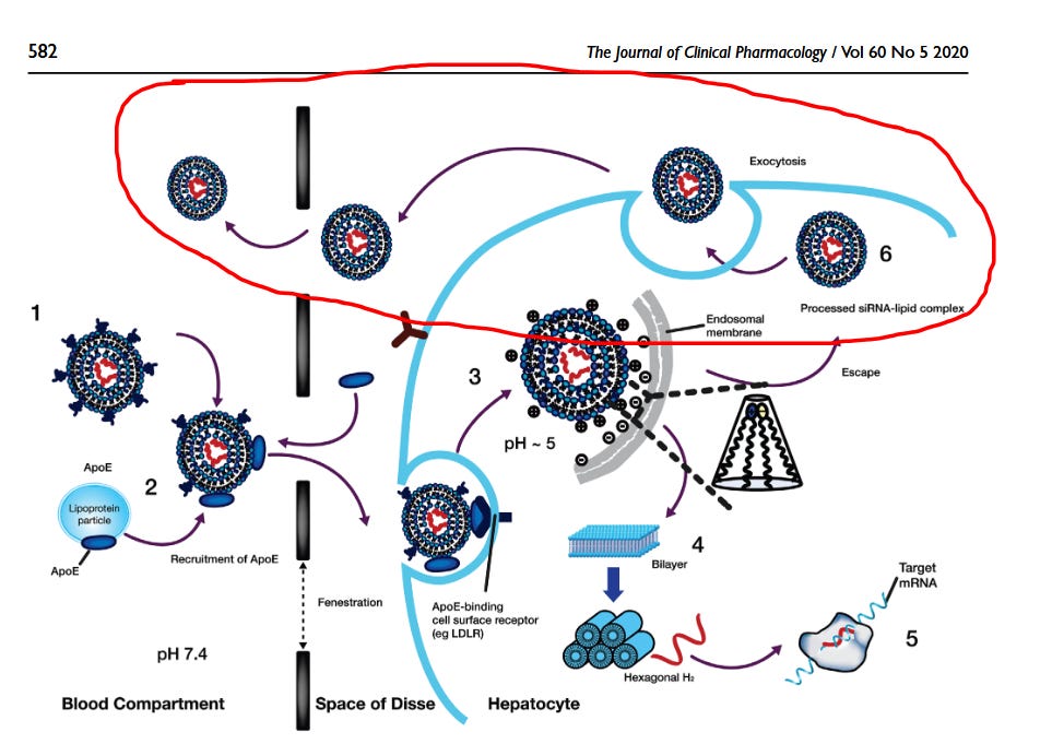
This paper established the intracellular fates of patisiran LNP–siRNA, including endosomal recycling and extracellular re-release.
The PK data showed something peculiar. After intravenous infusion, the siRNA plasma concentration didn’t decline smoothly. It dropped rapidly in the first few hours, that’s Phase 1, representing INTACT LNPs being cleared by the liver. But then it rose again. A secondary peak. Then it declined very slowly, with a terminal half-life of about 60 days for the ionizable lipid. To fully clear the lipids, it takes about 5 half-lives, or about 300 days.
The authors explained exactly what was happening:
“Imaging studies have shown that only a small fraction of the siRNA in the LNP is released into the cytosol and that about 70% of the siRNA that enters hepatocytes undergoes exocytosis through egress of LNPs from late endosomes/lysosomes back into the circulation.”
SEVENTY PERCENT! Most LNP cargo that enters cells is packaged into extracellular vehicles (EVs) and spit back into the bloodstream. This isn’t a minor pathway. It’s the dominant one.
And the exocytosed material isn’t just naked siRNA. It’s siRNA still complexed with ionizable lipids. The paper explicitly states that the second PK phase represents
“exocytosis of the siRNA-lipid complex from hepatocytes back into systemic circulation.”
The lipids travel with the cargo. They have a half-life of about 60 days, so they recirculate for about 10 months until they are all gone. Here is how I envision Onpattro LNP/exosome kinetics, based on the paper above.
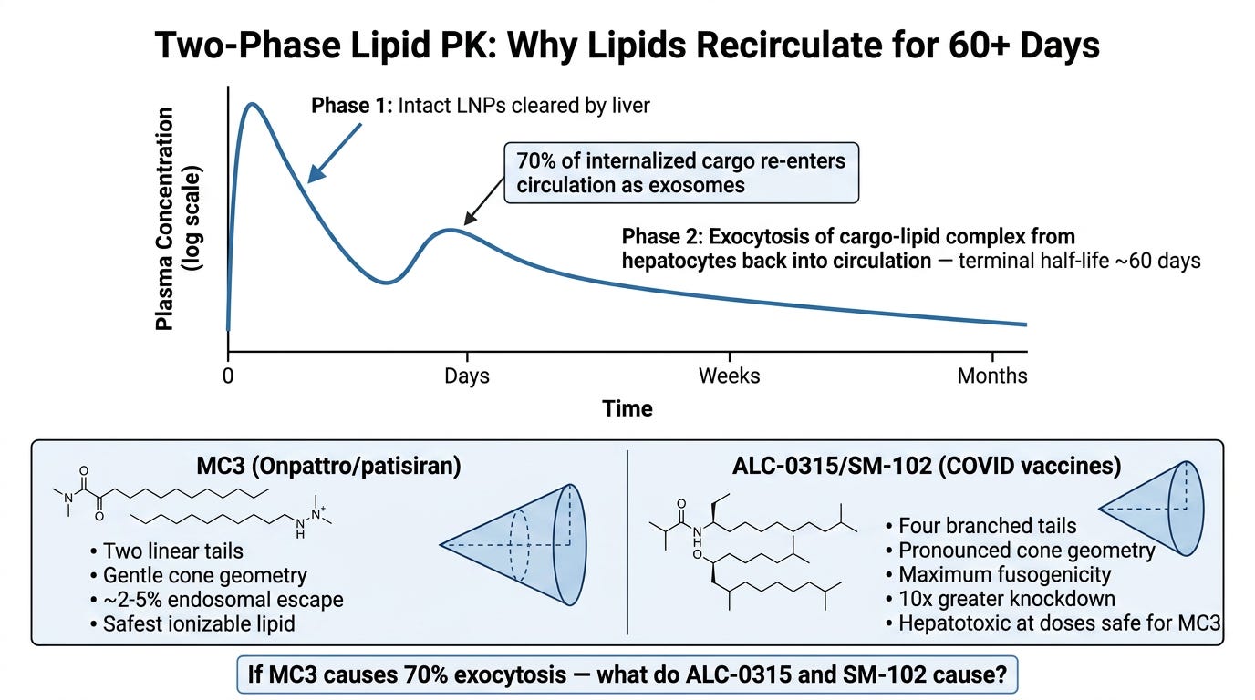
However, keep in mind Onpattro is given by IV infusion and is designed to go to the liver (about 90% does so). The vaccines are given IM, so these proportions regarding EVs may differ.
However, in the European assessment report for the Pfizer vaccine, they used the data on Onpattro (patisaran)2 to compare to ALC-0315 (pg 53-54).
Examination of the scaling of the comparable lipids (PEG2000-C-DMG, DLin-MC3-DMA) in patisiran indicates that the half-life of these lipids appears to scale with an exponent approaching the typically used half-life exponent (0.25). If this is the case for ALC-0315, we may expect a half-life approximating 20-30 days in humans for ALC-0315 and 4-5 months for 95% elimination of the lipid (Mahmood et al, 2010). Both lipids showed an essentially similar PK profile in clinic, with a strongly biphasic profile and long terminal half-lives.
Thus, it appears a similar two-phase distribution of the lipids occurs, and is most of that in exosomes? Even if lipid material remains detectable for months, it does not mean that intact, circulating, cargo-bearing exosomes remain for the same period. The lipid might reside in tissues, membranes, metabolites, lipoprotein complexes, recycled vesicles, or other compartments. We just can’t measure whether it is an intact supramolecular LNP, a hybrid LDL-LNP or something else during that terminal phase. Still, data from Bansal et al. and Maugheri (see below) suggest it is possible.
Before we continue, we need to establish the fundamental limitation on most LNP biodistribution studies. An LNP is not a single molecule. It is a supramolecular assembly containing RNA and four different lipids (plus contaminants, lol). But researchers usually track only one component, like a radiolabelled lipid such as cholesterol ONLY, the mRNA, or the expressed protein.
Whether researchers use a radiolabel or mass spectrometry, they generally follow a selected LNP component rather than the intact particle. Radiolabelled cholesterol, for example, could leave the original LNP and enter a cell membrane, extracellular vesicle, or LDL. Mass spectrometry may confirm that an ionizable lipid remains chemically intact, but it still cannot tell whether that lipid is in the original LNP or has been incorporated into a remodelled micelle, lipoprotein, extracellular vesicle, or cellular membrane. Similarly, PCR detects an RNA sequence but not necessarily the entire mRNA construct, while luciferase shows where protein expression occurred. None of these methods, by itself, demonstrates that an intact LNP was present.
Once inside the body, the original particle may disassemble and remodel. Its components can enter cell membranes, extracellular vesicles, LDL or HDL particles, micelles, metabolites, or new RNA–lipid complexes. The assay generally cannot tell which of these forms is carrying the measured component.
Thus, “LNP biodistribution” is often more accurately described as component biodistribution without particle identification. We imagine an LNP as a sealed parcel travelling from the injection site to different organs. But it may be more like a parcel that is opened at every depot, divided into pieces, repackaged in local containers, and given new address labels. Tracking one original component tells us where that component ended up, but not which parcel carried it there.
This helps explain why studies tracking different components can appear to produce different biodistribution results. They are measuring different pieces of a particle that has already come apart.
It also matters for the prolonged secondary plasma phase. Detection of RNA and lipid supports continuing association or parallel redistribution, but does not reveal whether the material is an intact LNP, a remodelled RNA–lipid complex, a lipoprotein hybrid, or cargo enclosed within an extracellular vesicle. The Onpattro study identified the returning material as an siRNA–lipid complex exocytosed from hepatocytes, but did not determine its physical structure or establish that it was enclosed within an exosome. Maugeri’s work (see below) did that, providing evidence that LNP-transfected cells can export functional mRNA within extracellular vesicles.
This matters when we hear from Pharma or regulators that the LNPs are cleared in 2 weeks. That is fake news.
Now, here’s where it gets dicey. MC3, which is the lipid in Onpattro was designed for gentle, controlled hepatic delivery. It has two linear tails producing a subtle cone shape. It achieves endosomal escape rates of roughly only 1-2%, 3 considered the “bottleneck” of mRNA therapy. It’s considered a less fusogenic, less hepatotoxic, more “gentle” ionizable lipid.
ALC-0315 and SM-102, the lipids in the Pfizer and Moderna vaccines, are completely different beasts. They have four branched tails producing a far more pronounced cone geometry. They were engineered for maximum fusogenicity, are faster, and produce larger “holes” in endosomes. 4
Ferraresso et al. paper5 demonstrated this directly: at identical siRNA doses, ALC-0315 LNPs caused 10-fold greater knockdown than MC3 LNPs, reached cell types MC3 couldn’t touch, and caused hepatotoxicity at a dose where MC3 showed no elevation in any liver enzyme.
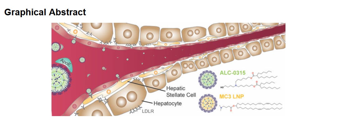
If approximately 70% of endocytosed MC3–siRNA material can be recycled extracellularly, what happens with the more fusogenic and membrane-disrupting ALC-0315 and SM-102 systems? Greater endosomal escape might reduce the amount available for recycling, while greater membrane perturbation could increase EV production or alter its cargo. So it is unknown and has not been measured, particularly after IM rather than IV administration.
With IM administration, LNPs stay in the muscles as a depot, then leak into the lymph and then the circulation. This is why Kent et al.,4 the best serial human blood kinetics data we have, showed Moderna’s SM-102 circulating in plasma for about 14-28 days, with the peak level ranging from 4 hrs to 2 days. That variability is in keeping with a slow leak from the muscle depot.
We actually have some data. Spike-carrying exosomes were detected in human plasma after COVID vaccination and before seroconversion. The Maugeri et al. study published in 2019 demonstrated that LNP-delivered mRNA can be packaged into cell-derived EV or exosomes, transferred to recipient cells, and remain sufficiently intact to produce protein in vivo. This isn’t hypothetical. It’s documented.
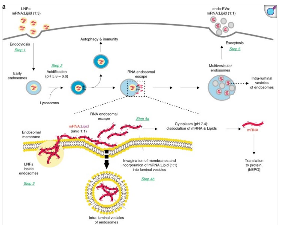
Maugeri concludes:
“Since LNPs with the same ionizable lipids used in this study are currently being utilized in clinical trials and endo-EVs contained hEPO mRNA acquired after the endocytosis of LNPs and delivered to other cells, we postulate that a similar scenario may occur in individuals administered with LNPs, suggesting that part of the mRNA delivery is achieved by such EVs.”
But the most direct evidence for EV-mediated spike distribution in vaccinated humans comes from Bansal et al. 2021,6 published in the Journal of Immunology as a “Cutting Edge” article, which deserves more attention than it gets.
Bansal isolated plasma exosomes from 8 healthy BNT162b2-vaccinated individuals before and after vaccination and at multiple post-dose timepoints. The findings:
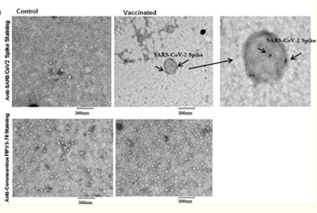
Circulating exosomes carrying SARS-CoV-2 spike protein (specifically the S2 subunit) were detectable at day 14 after dose 1. Antibodies were NOT yet detectable at that time point. Spike-positive exosomes appeared before the antibody response. Electon microscopy confirmed spike antigen on the exosome surface, not inside, where it is accessible to immune receptors. After the booster dose, both exosome-associated spike and antibody levels increased. Both peaked after dose 2 and declined together at 4 months.
This kinetic relationship is important: the exosome-spike curve mirrors the antibody curve. This suggests spike-carrying exosomes are part of the immune priming machinery, not an incidental byproduct (which we will discuss later with Marks et al.) Bansal confirmed this with a mouse experiment: immunizing mice with spike-positive exosomes from vaccinated humans produced anti-spike antibodies. Exosomes carrying spike are immunogenic in their own right. (Maugheri and Nawaz also confirmed this)
Here’s what they look like from Bansal. The spike protein is on the outside.

Nawaz et al. 20237 , Wang et al. 20258 and the review by Bader & Leroux 20249 have added further supporting evidence for EV-mediated mRNA redistribution.
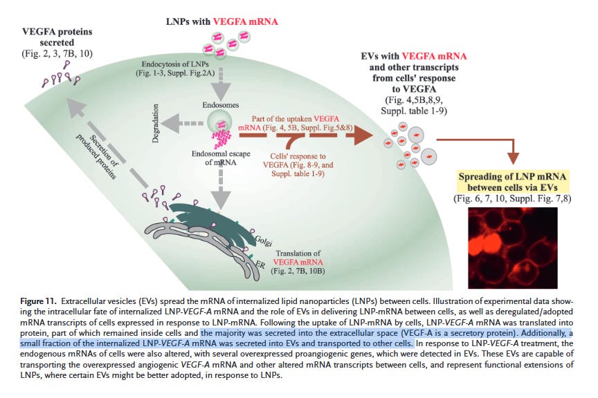
Here is Nawaz et al. 2023 showing that LNPs transform EVs into functional extensions that distribute active or functional therapeutic mRNA between cells, leading to protein expression in recipient cells (verifying Maugeri et al). This paper is worth the time to read, imho.
Wang et al. went one step further and fused LNPs with EVs to produce functional hybrid particles. This demonstrates that stable, biologically active LNP–EV chimeras are physically possible.
An interesting article by McCann et al.10 showed that lipid–RNA transfection complexes can be taken up, re-exocytosed and then co-purify with extracellular vesicles. They used RNAiMax, part of the Lipofectamine-family transfection reagents, optimized mainly for delivering siRNA and miRNA into cultured cells in cell and in vitro experiments. They found most transfected siRNA in their “EV” preparation separated at the density of RNAiMAX complexes, but not at the density of EV markers. They state:
If, transfection complexes contaminate sEV preparations, or are retained by cells and exocytosed in large quantities with sEVs, then the cited examples and many more in the literature could be due to contaminating transfection complexes.
The study used siRNA and is consistent with what the Onpattro (an siRNA) study reported above. We are not certain if the mRNA behaves similarly. If so, the particles observed by Maugeri, Bansal, and Nawaz could include bona fide EVs, or transfection complexes that could include recycled LNPs, remodelled lipid–RNA complexes, LNP–lipoprotein chimeras, or mixtures of these. So though we may not know the precise identity of the carrier, we CAN say vaccine-derived material can be repackaged or re-exported in various forms capable of secondary distribution.
And IF these secondary carriers are still SuPREX-like, i.e. RNA associated with membrane-active ionizable lipids, they could still perturb membranes and endolysosomal trafficking through the mechanisms as discussed in our L-DMD hypothesis. Their potency, tissue distribution and persistence, however, may differ from those of the original particle.
I think we have consistent data across multiple independent lines: EVs (or “transfection complexes”) are not a minor secondary pathway. They may be the primary mechanism by which vaccine antigen reaches the lymph node APCs that actually drive the immune response.
Another concept I have been considering is fractal spread. This is my speculation, and so far, there is little data. But if each cell transfected by an intact LNP produces exosomes, some of those exosomes transfect new cells. Can those new cells produce new exosomes? The vaccine signal doesn’t just disappear in a linear fashion from the injection site. Onpattro data showed secondary spread, and that may represent serial or branching redistribution through cell-to-cell transfers. This would require sufficient intact mRNA delivered to a secondary cell, translation in that cell, and repacking into new EVs and release for further spread. Or maybe it just passes intact mRNA from one exosome to another. Is that possible, and for how many generations can this cascade continue?
McCullough et al recently published an interesting case relevant to this hypothesis.
At 1,173 days post-vaccination, high-sensitivity ELISA detected free Wuhan spike protein in plasma (129.0 +- 4.1 fg/mL) and in circulating exosomes (11.6 +- 0.1 fg/mL). At 1,284 days, RT-PCR identified vaccine-derived spike mRNA within circulating exosomes, whereas PBMC RNA remained negative following DNase-treated extraction and amplicon-specific PCR targeting three spike ORF regions (S1-S3).
This is a long time. 3.5 YEARS. We don’t know whether this mRNA in the exosomes can be translated. Much of the mRNA in later EVs may consist of fragments or chemically altered molecules like adducts, which I discuss in this post.11 Now, the spike protein embedded in an EV membrane is another matter. It can remain immunologically active or otherwise biologically active, and potentially harmful without requiring new translation in the recipient cell. Exosomes can last a long time, but 3.5 years? Not sure.
The investigators did not test the exosomes for the ionizable lipid. That would be most interesting, since finding ALC-0315 or its metabolites in the same exosome as spike mRNA or protein could help determine whether multiple SuPREX components were repackaged rather than RNA or antigen persistence alone. I suspect that is the case.
This single case does not conclusively demonstrate fractal spread. You could also have a persistent source-cell reservoir that could explain the finding. Where? I don’t know. But this case raises the question of what process would continue to load or preserve vaccine-related material in circulating EVs years later? Fractal spread of exosomes could be one possibility.
Thoughts?
The debate about mRNA vaccine toxicity has largely focused on two questions: Is the spike protein toxic? Are the lipid nanoparticles toxic?
The spike protein camp argues that the immune response to the antigen is the central and unavoidable mechanism of harm. A recent paper which generated a lot of buzz was Marks et al in Nature Biotechnology12 earlier this year.
The Marks et al. paper proved this elegantly: even non-toxic GFP caused T cell infiltration and hepatocyte killing when delivered via LNPs. The influenza mRNA vaccine trials proved it clinically: the same flu antigens that are safe in conventional vaccines became highly reactogenic when delivered via the mRNA-LNP platform.
The LNP camp, and I count myself in this group, along with my co-author Falko Seger argues that the ionizable lipids cause direct membrane disruption, innate immune activation, and sustained signaling dysregulation independent of the antigen. Empty LNPs trigger ~9,500 gene expression changes13. They cause sickness behavior in mice reversible with a TLR4 inhibitor.14 The Ferraresso paper showed lipid-specific hepatotoxicity. Our L-DMD hypothesis15 documents ionizable lipid integration into cellular membranes and disruption of the phosphatidylinositol cycle, the cell’s master switch for membrane trafficking and inflammatory signalling. The lipids matter.
But the exosome data reveals something that hasn’t really been discussed: the platform doesn’t just deliver mRNA to cells. It turns those cells into a SECONDARY distribution route of spike protein, spike mRNA, and ionizable lipids throughout the body for weeks to months after injection. And maybe even years.
Bansal et al showed spike protein on the outside of exosomes
Maugheri et al showed mRNA INSIDE exosomes.
Onpattro kinetic data showed both siRNA (presumably similar to mRNA) AND LNP components inside the exosomes.
Nawaz et al. showed that the MAJORITY of the expressed protein was carried in EVs, along with small amounts of mRNA, which was shown to transfect OTHER cells.
So all bits of the mRNA-LNP complex, including its expressed protein, can be contained in exosomes, and these travel (in an exocrine manner, as elucidated by BioNTech founder Ugur Sahin himself in a paper he wrote in 2014) to other parts of the body.
How does this mechanism work, step by step? OK here is a brief overview.
LNPs are injected intramuscularly. Some stay in the muscle. Some drain through lymphatics (estimates is about 30-40% of the mRNA in the Covid vaccine dose “leaks”). Some leak into the bloodstream directly and reach the liver, spleen, ovaries, adrenals, and heart. This is what you see in the Pfizer biodistribution data.
Cells that take up LNPs face a choice. A small fraction of the cargo maybe no more than 15% for the COVID vaccine lipids escapes the endosome and reaches the cytoplasm, where the mRNA is translated into spike protein. The rest of the cargo stays trapped in endosomes.
Those endosomes don’t just sit there. They’re processed through the multivesicular body pathway. The trapped mRNA, spike protein, and possibly ionizable lipids are packaged into intraluminal vesicles. When these multivesicular bodies fuse with the plasma membrane, they release those vesicles as exosomes.
The exosomes circulate. They carry spike protein in its native membrane-embedded conformation. They carry residual mRNA that recipient cells can translate. And they carry the ionizable lipids, the same membrane-disrupting, TLR4-activating, PI-cycle-perturbing lipids that caused all the trouble in the first place.
These exosomes reach tissues the original LNPs never touched. They fuse with recipient cells. They deliver their cargo. The sheer volume of exosomes produced over weeks means cumulative antigen exposure can be substantial.
Since they have the cell’s own membrane encasing the mRNA and lipid bits, they may be trafficked differently than intact LNPs (like to the brain and bone marrow?). They are also less immunogenic because, based on the Onpattro data, most lipids are not in the original supramolecular LNP form, but in processed or remodelled lipid complexes.

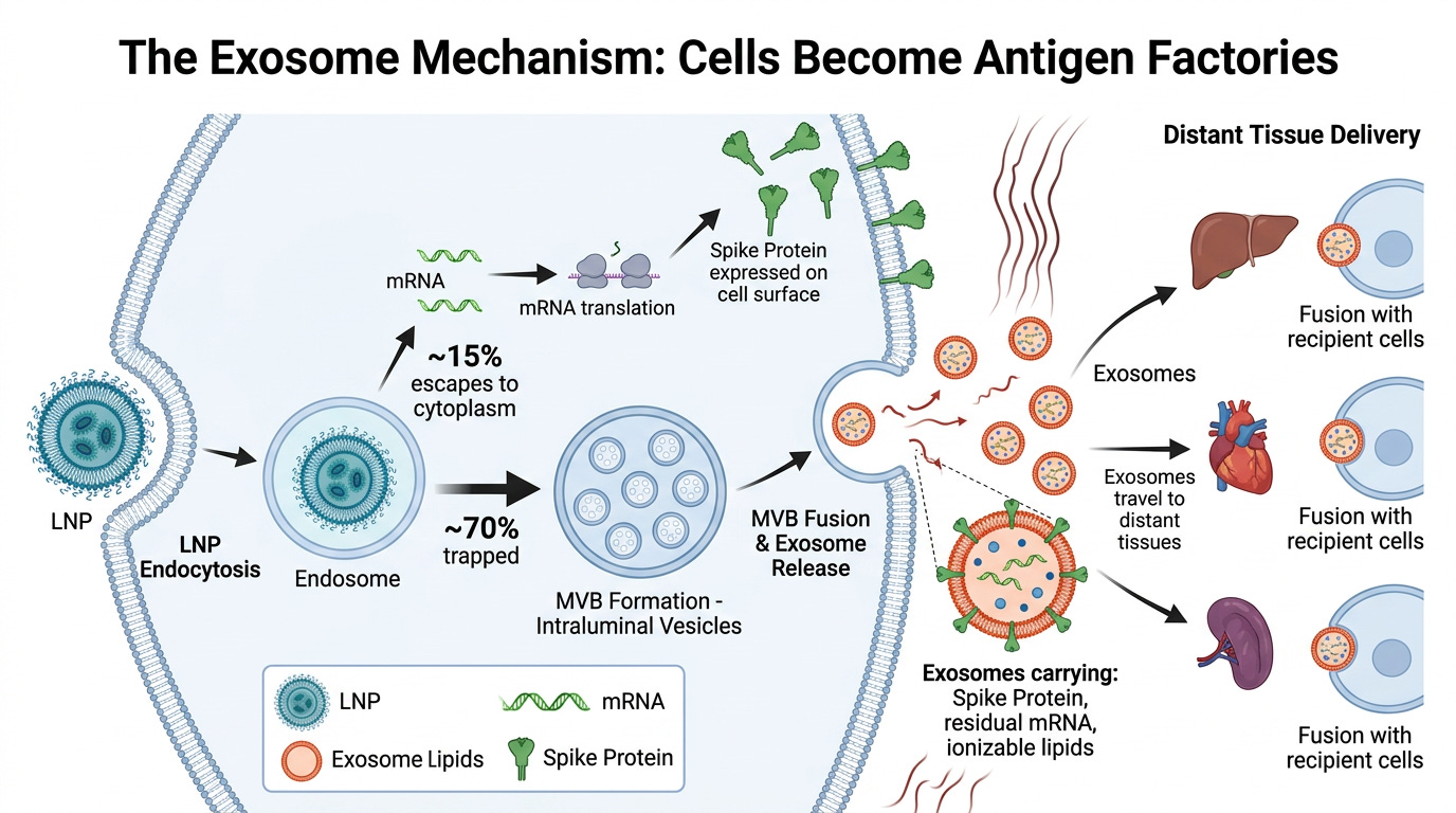
Should be cells become exosome factories but the AI (SciSpace in this case) does what it wants to do sometimes, lol.
This mechanism explains several findings that have puzzled researchers:
Why did Marks et al. find that dendritic cell expression of mRNA was dispensable for T cell priming? Because dendritic cells (DCs) don’t need direct transfection. They sample spike-carrying exosomes produced by transfected muscle fibers. Muscle produces the spike protein, and DCs just collect the product.
Why did silencing muscle expression reduce T cell responses by 30–50%? Because muscle fibers are the primary exosome factories after intramuscular injection. Shut down the factory, reduce the exosome supply, reduce the immune response.
Thus, the only mechanistically coherent explanation is cross-presentation via extracellular vesicles. Muscle cells package the spike protein into exosomes (as Bansal confirmed in humans). Exosomes travel to draining lymph nodes. Dendritic cells acquire exosome-displayed spike and present it on MHC-I to CD8⁺ T cells, without ever translating the mRNA themselves. Marks et al. did not directly identify how myocyte-derived antigen reached professional APCs. However, when their findings are considered alongside Maugeri’s demonstration of functional mRNA transfer through EVs and Bansal’s detection of spike-bearing EVs after vaccination, I believe EV-mediated transfer emerges as the most coherent, and I believe the best-supported mechanism.
Siguna Mueller, Ph.D., Ph.D. explains this possibility in her paper, which you can access here16. She also believes exosomes are vastly underappreciated, and, specifically during cross-presentation, may play a key role.
Why is spike protein detected in plasma for weeks or months after vaccination? The mRNA itself has a half-life measured in hours. Direct spike expression from the original transfection should peak within 48 hours. But exosome-mediated release continues as long as the transfected cells survive, and muscle fibers, in particular, are long-lived. But for how long? That is the question, and I don’t know that.
I’ve argued elsewhere that the mRNA-LNP platform, is what I call the SuPREX (Supramolecular Process-dependent RNA Expression system)
and it fails at two independent and irreducible levels.
The first is antigen-driven immune cytotoxicity, which many have documented. Any foreign protein expressed via this platform will be targeted by CD8+ T cells, and the expressing cells will be killed. This is inherent to the mechanism of action. You cannot have a vaccine without it.
The second is the lipid-driven membrane dysfunction that Falko, Dr Seneff and I have described in our L-DMD hypothesis. The ionizable lipids integrate into cellular membranes, disrupt phosphoinositide signaling, activate TLR4/NF-κB, suppress PPARγ, and trigger sustained inflammatory cascades that persist long after the antigen is cleared.
The exosome mechanism bridges these two failures and amplifies both. The lipids don’t just disrupt membranes locally at the injection site. They travel, via exosomes, to tissues throughout the body, activating innate immunity wherever they go. And the antigen doesn’t just get expressed in cells that received the original LNP dose. It gets expressed, or delivered pre-formed, to cells that received exosomes, thereby exposing cells to mRNA, spike or lipid that would ordinarily never be exposed (like the cornea, for example?)
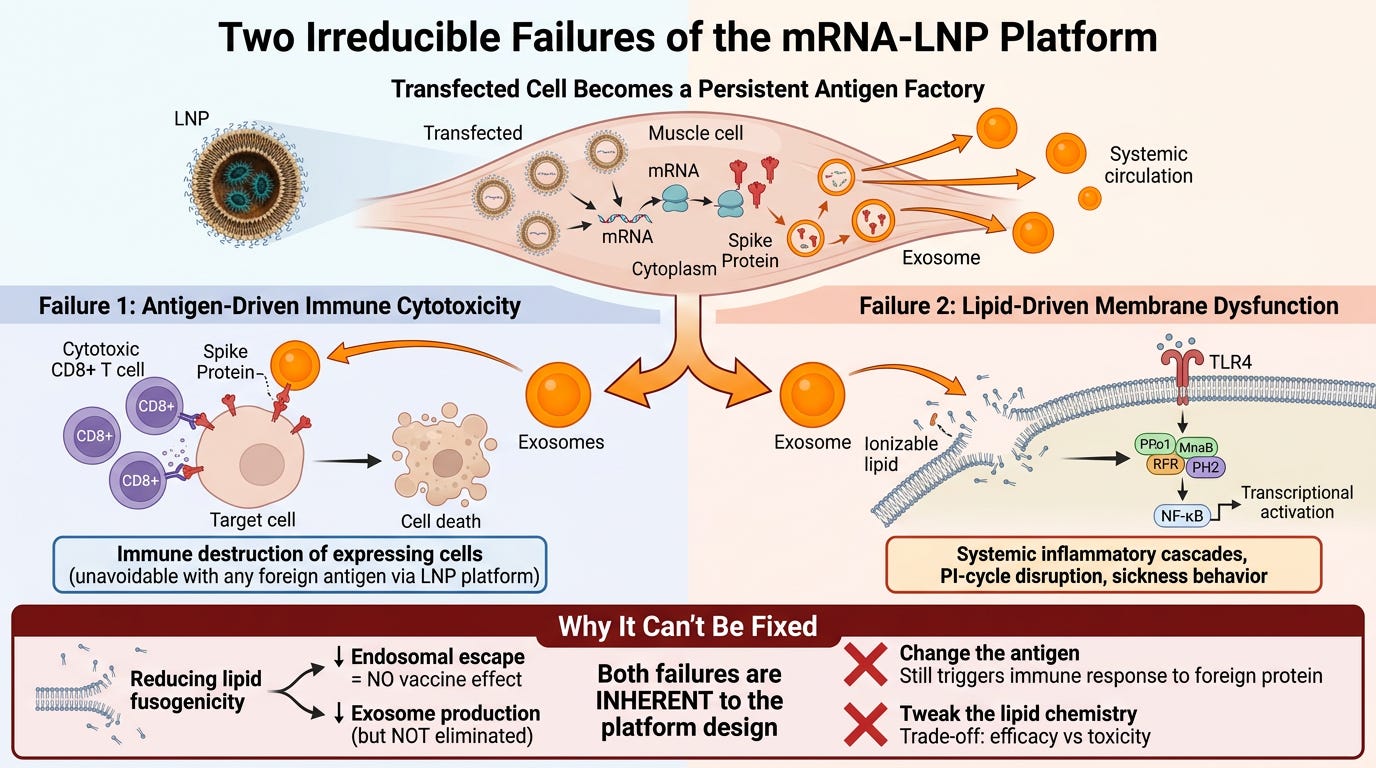
What I don’t know is if the LNP AS AN INTACT entity gets transported inside EVs. That is possible, but less likely. Other LNP-mRNA configurations are more likely, like micelles and the like. However, individual ionizable lipids, especially the amine heads, drive LNP immunogenicity by binding to Toll-like receptor 4 and CD1d and by promoting lipid-raft formation17. This is not good.
The booster schedule makes this worse in a specific and underappreciated way. The standard interval between dose one and dose two was 21 days for Pfizer and 28 days for Moderna. But the terminal half-life of the ionizable lipid, measured in the Onpattro data, is approximately 60 days. That means when dose two is administered, the body is not starting from a clean slate. Dose two was administered before complete elimination of lipid-related material was expected. Whether that residual material remained pharmacologically active, occupied EVs, or altered the response to the second dose was not examined. However, exosomes from dose one are likely still circulating at 3-4 weeks. Lipid-driven TLR4 signalling from dose one might still be occurring. The immune system has also already been primed: CD8+ T cells trained on spike protein during the first cycle are now present and ready to respond faster and harder to dose two. These mechanisms don’t reset between doses; they compound. Each subsequent dose is injected into a system that is likely executing both pathways simultaneously. It mechanistically explains why booster reactogenicity is consistently worse than the primary series, and why some individuals show cumulative injury patterns that don’t resolve between doses. And why these 3- or 4-week dosing intervals (prime and boost) for initial inoculation were, shall I say, very problematic.
What are the implications? If exosome-mediated redistribution is inherent to lipid-mediated transfection, and the Onpattro data proves it is, even with the least fusogenic lipid authorized, then I don’t believe there is any way to make the mRNA-LNP platform predictable or safer by changing the antigen or tweaking the lipid chemistry.
A less fusogenic lipid would reduce direct membrane damage but also reduce endosomal escape, affecting vaccine’s efficacy. A more rapidly degraded lipid might reduce long-term accumulation but would still trigger the initial wave of exosome production. Targeting LNPs to specific cell types might reduce the diversity of exosome-producing cells but wouldn’t eliminate exosome production from transfected cells. These issues will also affect LNP-based gene editing to some extent.
And the fundamental problem remains: you are injecting a system designed to turn human cells into exosome factories, and those exosomes don’t shut down when the original intact LNP dose has been cleared. They keep producing. They keep exporting. And they can circulate for a very long time. The exported material includes not just the antigen (spike protein), but possibly the very lipids that make the immune response to that antigen so destructive.
The mRNA-LNP platform is not a vaccine in the traditional sense. It’s a Supramolecular Process-dependent RNA Expression (SuPREX) system that transforms the recipient’s own cells into sustained sources of antigen production and systemic distribution. The spike protein is not the problem per se, it’s just the current payload. The lipids are not just delivery vehicles; they’re pharmacologically active agents that disrupt cellular signalling and travel with the cargo via exosomes to distant sites.
This is not a case where the mechanism was hidden or unknown. The Pfizer COVID-19 vaccine European Public Assessment Report (EPAR), explicitly cites Onpattro as the established model for LNP biodistribution. That literature includes the biphasic elimination profile. It includes the 60-day terminal half-life. The EPAR did not claim that ALC-0315 was exported in exosomes. But it explicitly used the patisiran (Onpattro) lipids as the clinical model for predicting ALC-0315’s prolonged human terminal disposition and described that terminal phase as “redistribution from tissues into which the LNP had been delivered.” As we saw in the Onpattro study earlier, the secondary plasma phase is attributed to cellular exocytosis of RNA–lipid complexes (ie in EVs). The obvious unanswered question is whether ALC-0315 undergoes an analogous cellular recycling process with EVs, but given the current evidence provided, I believe it is likely. And Moderna would be similar.
LNPs do not simply deliver their cargo and go pouf. After entering cells, much of their RNA–lipid material may be exocytosed back into circulation in extracellular vescicles (EVs) or exosomes. Transfected cells can also release the protein it encodes, usually surface bound.
These secondary particles are not necessarily intact original LNPs. Components may be repackaged into EVs, remodelled RNA–lipid complexes, lipoprotein hybrids, micelles, or cellular membranes, each potentially acquiring a new biological identity and, importantly, a new biodistribution pattern.
Most biodistribution studies track only one component of the supramolecular assembly, so they tell us where that component went, not what carried it there. We have been blind to the exosome mechanism.
Human studies have detected spike-bearing EVs after vaccination, and several experimental studies show that EVs can transfer mRNA between cells, which can transfect and make more protein.
Whether this can produce fractal spread remains unknown.
We need to know what is in these circulating exosomes, how long they remain biologically active, and whether they contain vaccine-derived ionizable lipid, mRNA, spike, or all three (and more, like plasmid DNA?).
There is one more thing, and I want to raise it carefully, because it’s the question that follows inevitably from everything above, and that is about “shedding.”
Shedding is a recognized phenomenon in gene therapy. When a viral-vector product is administered, regulators routinely require studies examining whether vector-derived material- that is, the vector itself (the virus), the transgene (DNA or RNA), or the expressed product (protein) enters urine, stool, saliva, tears, semen, or other secretions. Some of these products carry explicit precautions for household contacts, based on the possibility of transmission. This is standard practice. It’s uncontroversial. It’s expected.
So since we’ve established that the mRNA-LNP platform has the same characteristics (exosome factories, carrying the mRNA, expressed protein, and lipid components) circulating for weeks to months or more, you would think regulators would have asked whether vaccine-derived exosomes enter bodily secretions.
And if they do:
In what quantity, and in what form?
Are they biologically active and taken up by cells?
Can they produce any effect in another person?
I don’t know the answer. I don’t think anyone does. Because for mRNA-LNP products, this question appears never to have been asked of the regulators, let alone answered.
The mRNA-LNP platform produces exactly the kind of transmissible, vector-like particles that trigger mandatory shedding studies in every other gene-therapy context. And yet I can find no equivalent examination for the mRNA vaccines. The adenoviral-vector COVID vaccines, by the way (AstraZeneca/J&J), did include shedding language and precautions in their regulatory materials.
And given that the same regulators accepted the Onpattro data as the model for LNP biodistribution, including its 60-day half-life and its documented exosome production, the absence of a shedding question isn’t because the mechanism was unknown. It was known, cited, and then not followed to its logical next step. Why?
And that is yet another unanswered question in the regulatory assessment that has yet to be examined.
I appreciate my subscribers reading this technically dense stuff. Thank you. And as always, continue to pray the rosary and keep my new grandson in your prayers.
Thanks for reading Maria Gutschi! This post is public so feel free to share it.
* * *
https://pmc.ncbi.nlm.nih.gov/articles/PMC7187331/pdf/JCPH-60-573.pdf
https://www.ema.europa.eu/en/documents/assessment-report/comirnaty-epar-public-assessment-report_en.pdf
https://www.pnas.org/doi/10.1073/pnas.2307800120
https://www.sciencedirect.com/science/article/abs/pii/S0168365925006686?via%3Dihub
https://pmc.ncbi.nlm.nih.gov/articles/PMC9621687/pdf/nihms-1814479.pdf
https://pmc.ncbi.nlm.nih.gov/articles/PMC11073804/
https://advanced.onlinelibrary.wiley.com/doi/epdf/10.1002/advs.202206187
https://isevjournals.onlinelibrary.wiley.com/doi/full/10.1002/jev2.70201
https://www.sciencedirect.com/science/article/pii/S0169409X24002837?via%3Dihub
https://pmc.ncbi.nlm.nih.gov/articles/PMC9549735/
https://substack.com/@mariagutschi/p-181270476
https://www.nature.com/articles/s41587-026-03099-z
https://pmc.ncbi.nlm.nih.gov/articles/PMC8604799/pdf/main.pdf
https://pubs.acs.org/ancac3/article-abstract/18/36/24842/233323/Lipid-Nanoparticles-Elicit-Reactogenicity-and?redirectedFrom=fulltext
https://www.sciencedirect.com/science/article/pii/S2211383526004235
https://www.mdpi.com/2075-1729/15/10/1575
https://pmc.ncbi.nlm.nih.gov/articles/PMC11863198/pdf/nihms-2048549.pdf
No posts
Comments
Nothing yet. Say the first thing.
Sign in to join the conversation.