
There is something about GLP-1 agonists that almost nobody talks about.
Semaglutide, tirzepatide, and retatrutide are usually presented as variations of the same idea: drugs that activate the GLP-1 receptor.
That description is correct. But it is also incomplete.
Before we go any further, an important clarification: A major source of the physiological differences between these drugs is that they do not share the same receptor profile. Semaglutide selectively activates GLP-1R, tirzepatide activates GLP-1R and GIPR, and retatrutide activates GLP-1R, GIPR, and the glucagon receptor.
I am fully aware of that, but that is not what this article is about.
This article focuses on the one receptor they all have in common.
Although all three activate GLP-1R, they do not interact with it in the same way. They differ in receptor affinity and in how their molecular structures engage the receptor.
For some of these agonists, particularly tirzepatide, functional studies also show differences in intracellular signaling and receptor trafficking, which can substantially alter how the cell responds to the signal.
They also remain in the circulation for days, creating a form of receptor exposure that differs fundamentally from the short-lived signal produced by endogenous GLP-1.
This means that the shared label “GLP-1 agonist” does not necessarily describe a shared intracellular signal.
Why does this matter? Because cells do not respond simply to whether a receptor is activated. They respond to how it is activated.
The receptor itself therefore becomes part of the message.
Its shape determines which intracellular proteins are recruited and how the cell interprets the signal. The molecular signal determines which intracellular pathways are engaged, how the receptor adapts over time and ultimately how cells respond.
These mechanisms may contribute to differences in efficacy, side effects, long-term responses and individual variation among GLP-1 agonists.
This article explores a part of GLP-1 biology that is rarely discussed: not whether these drugs activate the same receptor, but how differently they communicate through it.
The reason is surprisingly simple. Semaglutide, tirzepatide, retatrutide and endogenous GLP-1 are different peptides.
Although they all activate GLP-1R, they do not interact with the receptor identically.
GLP-1R belongs to the family of G protein-coupled receptors, or GPCRs.
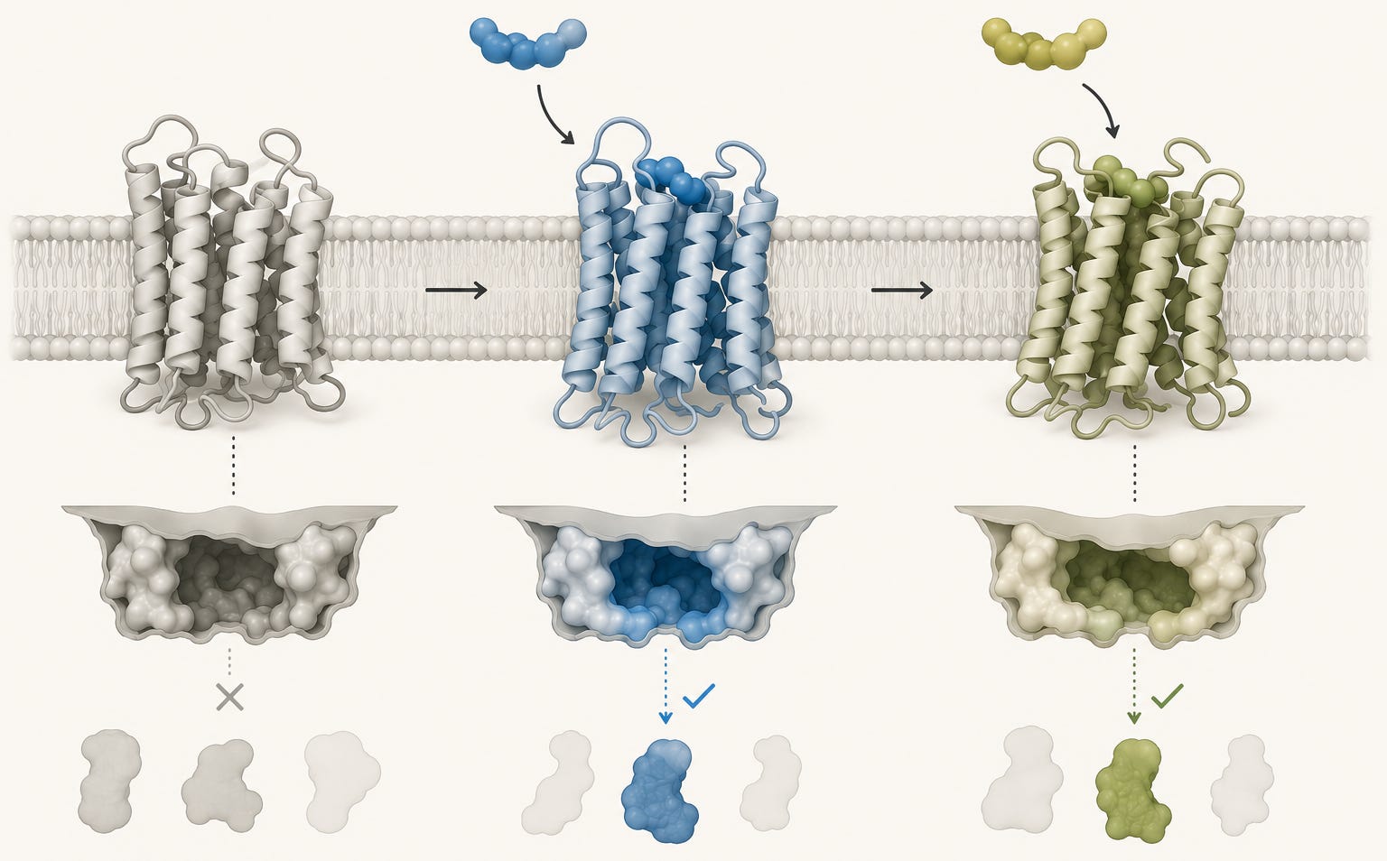
A GPCR is not a rigid switch with only one inactive and one active position. It is a flexible protein that can adopt several related active shapes.
Which shape is favoured depends partly on the ligand that binds.
This matters because the extracellular and intracellular parts of the receptor are physically connected. When ligand binding changes the shape of the receptor on the outside of the cell, it also changes the surface presented to signaling proteins on the inside. Some intracellular proteins may therefore bind more efficiently, while others bind less efficiently.
The receptor responds not only to whether a ligand is present, but also to how that ligand holds the receptor in an active state. This principle is known as ligand bias or biased agonism.
For GLP-1R, this is no longer just a theoretical concept.
Cryo-EM structures (a technique that captures molecular structures at near-atomic resolution) are available for GLP-1R bound to semaglutide, tirzepatide, and retatrutide. These snapshots reveal how each peptide fits into the receptor and highlight both shared and ligand-specific molecular interactions.
However, a structural difference does not by itself prove that a ligand produces a distinct functional signaling program.
A structure is a molecular snapshot.
It can show where a peptide sits, which parts of the receptor it contacts and which receptor shape is stabilized under the experimental conditions.
But it cannot, by itself, tell us how strongly a living cell generates cAMP, recruits β-arrestin, internalizes the receptor, or adapts during prolonged exposure, all of which are downstream events following GLP-1R activation.
Those questions require functional experiments.
The clearest functional evidence currently comes from tirzepatide. At GLP-1R, tirzepatide shows relative bias toward cAMP generation over β-arrestin recruitment, an intracellular regulatory process involved in receptor desensitization and trafficking. In the experimental systems studied, tirzepatide also produces less receptor internalization than endogenous GLP-1, meaning that fewer receptors are removed from the cell surface and transported into the cell.
This does not mean that tirzepatide activates only one pathway.
It means that the balance between the pathways is shifted. Relative to endogenous GLP-1, cAMP signaling is better preserved than β-arrestin recruitment and receptor internalization.
For retatrutide, receptor structures and cAMP potency have been characterized, but its GLP-1R-specific trafficking and biased signaling remain less comprehensively mapped.
Retatrutide therefore illustrates a second important point.
Knowing how a ligand sits within a receptor is not the same as knowing the full signaling program it produces. The structural map is emerging, but the functional map is still incomplete.
This distinction is important.
Structural biology tells us how the ligand binds to the receptor.
Functional biology tells us what happens next.
In other words, one explains how the receptor changes shape, while the other explains how that structural change is translated into intracellular signaling.
Together, these studies show that GLP-1 agonists should not simply be viewed as molecules that turn the receptor on.
They shape how the receptor communicates with the cell. And that influences the biological response.
The shape of the signal is only one part of the biology.
The other is time.
A receptor conformation may be induced by a brief physiological pulse, or repeatedly promoted for days while a long-acting agonist remains available in the circulation. These are not equivalent signaling conditions.
Endogenous GLP-1 is released after meals and is rapidly degraded, with a circulating half-life of approximately one to two minutes. By contrast, the approximate half-lives of semaglutide, tirzepatide, and retatrutide are measured in days—about seven, five, and six days, respectively.
GLP-1R therefore encounters a transient, fluctuating physiological signal rather than sustained systemic exposure.
Repeated meals generate repeated episodes of endogenous GLP-1 secretion. Even when these episodes occur relatively close together, each GLP-1 molecule remains short-lived and circulating concentrations continue to rise and fall.
This is fundamentally different from maintaining a long-acting pharmacological agonist in the circulation for days.
Because these drugs remain in circulation for several days, they create sustained systemic exposure and repeated opportunities for GLP-1R engagement rather than the rapidly rising and falling exposure produced by endogenous GLP-1.
This does not mean that one individual receptor remains continuously occupied for several days.
Instead, receptor populations continuously cycle through ligand binding, signaling, internalization, recycling and, in some cases, degradation while drug molecules remain available in the circulation.
The persistent feature is therefore not one permanently activated receptor, but a receptor system that remains exposed to ligand over time.
This fundamentally changes how the receptor system behaves over time.
Immediately after activation, GLP-1R primarily signals through Gs proteins, which activate adenylyl cyclase and increase cAMP. cAMP acts as an intracellular second messenger and activates downstream effectors, including PKA and EPAC.
This is the rapid signaling phase: information is transmitted from the receptor at the cell membrane to molecular effectors inside the cell.
As stimulation continues, the receptor itself begins to adapt.
One of the first steps is phosphorylation. Small phosphate groups are added to the receptor, changing which intracellular proteins can bind to it.
These events form part of a complex intracellular signaling cascade, but the central point is simple: they gradually change how the receptor communicates with the cell.
Depending on which ligand activates the receptor and in which cell type this occurs, arrestins may then be recruited to GLP-1R. Arrestins are regulatory proteins that bind to the activated and phosphorylated receptor and can reduce its coupling to G proteins.
The receptor may also be internalized, meaning that it is removed from the plasma membrane and transported into the cell.
These processes are closely connected, but they are not identical. Receptor phosphorylation, functional desensitization, arrestin recruitment and internalization each describe a different molecular event.
Phosphorylation changes the receptor itself by adding small phosphate groups. Desensitization means that the receptor becomes less effective at generating the same intracellular response despite continued stimulation. Arrestin recruitment changes which proteins interact with the receptor. Internalization changes where the receptor is located.
Importantly, internalization does not necessarily mean that signaling has stopped.
Once inside the cell, GLP-1R may enter small membrane-bound compartments called endosomes. From there, several things can happen.
The receptor may continue signaling from inside the endosome. It may be recycled, meaning that it is transported back to the plasma membrane and becomes available for stimulation again. Or it may be sent to lysosomes, cellular compartments that break down proteins, where the receptor is degraded.
Together, these processes determine how many functional receptors remain available at the cell surface, how quickly previously internalized receptors return, and how strongly the cell can respond to future stimulation.
The cell is therefore regulating more than the strength of the current signal. It is also adjusting its sensitivity to the next one.
Importantly, the ligand helps shape this process.
Different ligands may stabilize different receptor conformations and recruit intracellular signaling and regulatory proteins to different degrees. The signaling bias described above may therefore influence not only the immediate production of second messengers such as cAMP, but also whether the receptor is internalized, where it is sorted inside the cell, how quickly it returns to the membrane and how many responsive receptors remain available.
The identity of the ligand and the duration of exposure therefore work together.
A long half-life does not simply extend the original signal over a longer period. It exposes the receptor system to stimulation for long enough for these regulatory processes to develop.
Over time, the cell may change which intracellular proteins are coupled to the receptor, where the receptor is located, how quickly it is recycled and how many receptors remain able to respond.
This adaptation is a normal part of GPCR biology and should not automatically be interpreted as a loss of drug efficacy.
It means that cells continuously adjust their sensitivity when stimulation persists.
The final response is therefore shaped by three related questions:
What binds to the receptor?
How does it bind?
And how long does the receptor system remain exposed?
For all three, endogenous GLP-1, semaglutide, tirzepatide, and retatrutide differ.
Why should anyone care about receptor conformation, signaling proteins, and receptor trafficking?
Because these molecular events determine how cells receive, regulate, and adapt to a drug signal over time.
They may influence the balance between immediate signaling and longer-term receptor adaptation, how many receptors remain available at the cell surface and how cellular responses develop during repeated exposure.
A cell that retains more responsive receptors at its surface is not in the same molecular state as a cell that has internalized a larger proportion of them.
Likewise, a receptor that continues signaling from an endosome may create a different pattern of intracellular activity from one that signals mainly at the plasma membrane.
These differences can alter the timing, location and persistence of the signal, even before we consider the additional receptors activated by tirzepatide and retatrutide.
This provides a biological reason not to regard all GLP-1 agonists as interchangeable signals.
However, receptor pharmacology alone cannot currently tell us which drug will produce the greatest benefit or the fewest side effects for an individual.
Tirzepatide and retatrutide activate additional receptors and most of the mechanistic findings described here come from cellular or preclinical experiments rather than direct measurements in human tissues during treatment.
The point is therefore not that one signaling pattern can already be declared superior.
The point is that the label “GLP-1 agonist” identifies a shared receptor while concealing important differences in how that receptor is engaged, regulated, and exposed over time.
The shared label is therefore accurate, but incomplete.
Semaglutide, tirzepatide, and retatrutide all activate GLP-1R. Yet ligand structure, receptor conformation, signaling balance, trafficking and duration of exposure can all shape what that activation means inside the cell.
The receptor is the same.
The signal is not.
If prolonged GLP-1 receptor stimulation changes receptor behavior, another question naturally follows.
How does the receptor recover?
In many GPCR systems, receptor responsiveness can gradually return when agonist stimulation is reduced or removed.
The β₂-adrenergic receptor is one of the best-studied examples. During prolonged stimulation, the receptor can become phosphorylated, functionally uncoupled from its G protein, and internalized into the cell. Once agonist stimulation decreases, receptors can be dephosphorylated, recycled back to the plasma membrane and regain their ability to respond. In experimental systems, some of this recovery begins within minutes to hours, whereas recovery after more extensive receptor downregulation may require new receptor synthesis and take considerably longer.
Whether GLP-1 receptors behave in the same way during long-term treatment with semaglutide, tirzepatide, or retatrutide remains unknown.
This leads to an interesting biological question.
When GLP-1R is stimulated for a long time, the receptor system may adapt. Could a period of reduced stimulation allow some of that sensitivity to return?
At present, we do not have an answer.
The question follows naturally from what we know about GPCR biology, but it has not been tested as a therapeutic strategy for long-acting GLP-1 receptor agonists.
The question is also more complicated than it may first appear.
Because these drugs have half-lives of several days, stopping treatment does not immediately remove the agonist from the circulation or end receptor exposure. Drug concentrations decline gradually over multiple half-lives, meaning that receptor exposure decreases progressively rather than ending abruptly.
We therefore do not know whether a clinically practical treatment interruption would allow measurable receptor resensitization, whether different tissues would recover at different rates, or whether any molecular recovery would translate into meaningful clinical effects.
These are not recommendations for clinical practice.
They are unanswered biological questions.
Understanding not only how GLP-1 receptors are activated, but also how they recover from prolonged stimulation, may become one of the next important questions in GLP-1 biology.
Zhang Y, Sun B, Feng D, et al. Cryo-EM structure of the activated GLP-1 receptor in complex with a G protein. Nature. 2017.
Liang YL, Khoshouei M, Glukhova A, et al. Phase-plate cryo-EM structure of a biased agonist-bound human GLP-1 receptor–Gs complex. Nature. 2018.
Wootten D, Simms J, Miller LJ, et al. Polar transmembrane interactions drive formation of ligand-specific and signal pathway-biased family B G protein-coupled receptor conformations. Proceedings of the National Academy of Sciences. 2013.
Zhang X, Belousoff MJ, Zhao P, et al. Structure and dynamics of semaglutide- and taspoglutide-bound GLP-1R–Gs complexes. Cell Reports. 2021.
Zhao F, Zhou Q, Cong Z, et al. Structural insights into multiplexed pharmacological actions of tirzepatide and peptide 20 at the GIP, GLP-1 or glucagon receptors. Nature Communications. 2022.
Li W, Zhou Q, Cong Z, et al. Structural insights into the triple agonism at GLP-1R, GIPR and GCGR manifested by retatrutide. Cell Discovery. 2024.
Willard FS, Douros JD, Gabe MBN, et al. Tirzepatide is an imbalanced and biased dual GIP and GLP-1 receptor agonist. JCI Insight. 2020.
Jones B, Buenaventura T, Kanda N, et al. Targeting GLP-1 receptor trafficking to improve agonist efficacy. Nature Communications. 2018.
Novikoff A, O’Brien SL, Bernecker M, et al. Spatiotemporal GLP-1 and GIP receptor signaling and trafficking/recycling dynamics induced by selected receptor mono- and dual-agonists. Molecular Metabolism. 2021.
Baggio LL, Kim JG, Drucker DJ. Chronic exposure to GLP-1R agonists promotes homologous GLP-1 receptor desensitization in vitro but does not attenuate GLP-1R-dependent glucose homeostasis in vivo. Diabetes. 2004.
Deacon CF, Nauck MA, Toft-Nielsen M, et al. Both subcutaneously and intravenously administered glucagon-like peptide-1 are rapidly degraded from the amino terminus in type 2 diabetic patients and in healthy subjects. Diabetes. 1995.
Lau J, Bloch P, Schäffer L, et al. Discovery of the once-weekly glucagon-like peptide-1 analogue semaglutide. Journal of Medicinal Chemistry. 2015.
Coskun T, Sloop KW, Loghin C, et al. LY3298176, a novel dual GIP and GLP-1 receptor agonist for the treatment of type 2 diabetes mellitus: From discovery to clinical proof of concept. Molecular Metabolism. 2018.
Coskun T, Urva S, Roell WC, et al. LY3437943, a novel triple GIP, GLP-1, and glucagon receptor agonist for glycemic control and weight loss: From discovery to clinical proof of concept. Cell Metabolism. 2022.
Urva S, Coskun T, Loghin C, et al. LY3437943, a novel triple GIP, GLP-1, and glucagon receptor agonist in people with type 2 diabetes: A phase 1b, multicentre, double-blind, placebo-controlled, randomised, multiple-ascending dose trial. The Lancet. 2022.
Pippig S, Andexinger S, Lohse MJ. Sequestration and recycling of β₂-adrenergic receptors permit receptor resensitization. Molecular Pharmacology. 1995.
No posts
Comments
Nothing yet. Say the first thing.
Sign in to join the conversation.