
Another day, another shift. You’ve barely had time to put your bag down and take a half sip of coffee before you see Shannon, your charge nurse, rush over to you.
“Oh good, you’re here!” she tells you. “I need you to run down to room 28 and help Kara. There’s an arrest coming in too, but the teaching team can handle that case. I’ll give them a hand with the code; Kara just radio’ed back asking for an attending stat to room 28.”
“Are we breaking up?” you ask Shannon as she rolls her eyes at you and gives you an indignant point towards room 28. “Such a drama queen” she mutters as she runs off to the code.
“How’s my favorite nurse?” You ask Kara as you walk into room 28. She’s hustling around the room, clearly worried about the patient.
“Busy. I could use some help here.” She tells you. “Let me catch you up. This is Ms. Flemming. She’s a 32-year-old female who came in for UTI symptoms. No medical problems, no surgeries, no heart problems. Takes no medications. She was in a hallway chair when she got up to give a urine sample. When she came back from the restroom, she called me over and said she felt extremely unwell and as though her heart was trying to beat out of her chest. I have no monitors or anything in the chair hallway, but, luckily, one of the respiratory therapists was walking by and had a portable pulse ox in her pocket. We put it on her finger and it was blinking a heart rate of 285. I called an emergent resus and Shannon told me to bring her to bed 28 and you’d meet us here. Now you’re up to date, go do doctor things!”
“Hi Ms. Flemming! How are you doing?” you ask.
“Terrible. My heart is trying to explode. I think it’s trying to beat out of my chest. Don’t just stand there; do something!” She tells you, clearly in distress.
“Here’s her EKG, BP cuff is cycling now. I have a crash cart bedside and she’s hooked up to the Zoll.” Kara tells you.
“What if you...” You open your mouth to say something but someone cuts you off.
“Wow Kara’s really crushing it today! Maybe that’s so you can focus on reading that ekg and doing doctor stuff…” you hear Nicole, one of the other nurses quip.
“Ok. Ok… Taking a look now. Please continue to gang up on me while I try to do critical care; Kara, yell when the BP finishes cycling?”
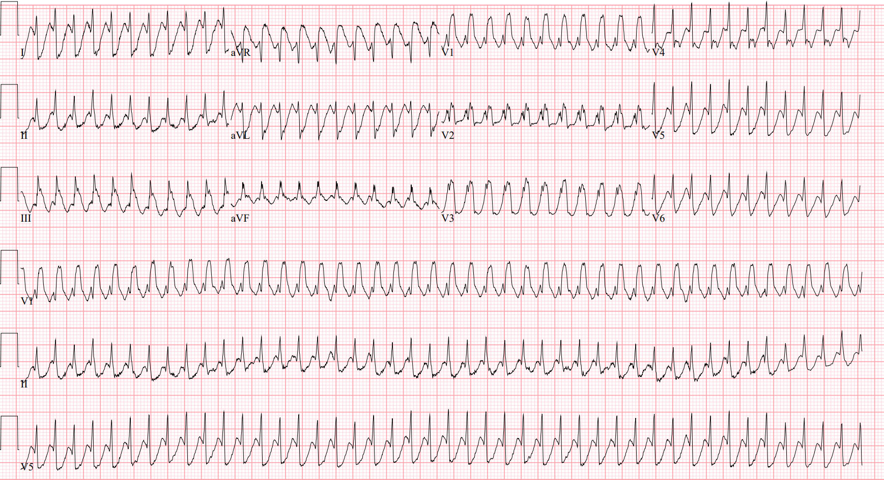
Rate: 290s, trying really hard to be 300 beats per minute. This heart is beating quickly!
Rhythm: Definitely not sinus rhythm
Axis: Normal
Intervals: The big question here is, ‘are the QRS complexes wide or narrow’ and I’m going with narrow, which we’ll get into below. There is also right bundle branch morphology, likely secondary to the rate.
Morphology: Look at aVR, those ST segments are quite elevated. You can see diffuse depressions in the lateral leads too, this heart is under a LOT of strain.
Final Read: This heart is beating VERY quickly. Not a lot of things make the heart try and beat 300 times per minute. Let’s zoom in on a few things here:
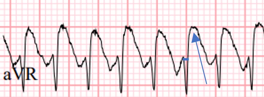
Take a look at a very, very magnified aVR. The thick blue line (on the 6th QRS complex) is the duration of the QRS complex. Notice that it’s definitely less than 2 small boxes (0.80 sec), probably between 1 to 2 small boxes (0.40 - 0.80 sec) so it’s consistent with a narrow QRS. The blue arrow points out the ST segment. Notice how there is ST segment elevation, likely due to the rate.
Let’s check ourselves with another lead. Here’s a lateral lead this time, take a look at V5:
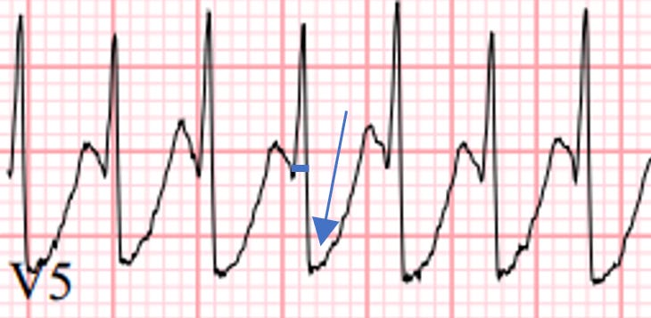
Again, blue line (on the 4th QRS complex) marks out the QRS duration, the arrow shows the (significantly depressed) ST segment.
Ok, we’re comfortable with a narrow complex, regular tachydysrhythmia, but what is it?
When I think of narrow and regular tachydysrhythmias, there are 3 that I think of right away. Sinus Tachycardia, Atrial Flutter, and some kind of Supraventricular Tachycardia (like AVRT, AVNRT, etc). I think it’s pretty obvious that this is not Sinus Tachycardia. Which leaves SVT and Aflutter.
How do you tell the difference here between the two? It’s really hard to tell.
Sometimes you can see a rhythmic pattern to the baseline, suggestive of flutter waves. Take a look at lead III:
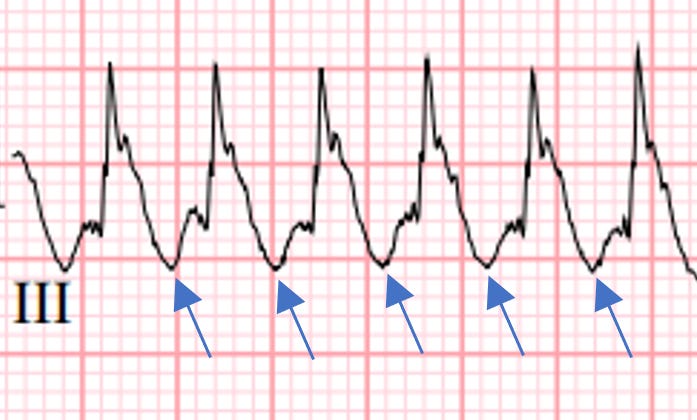
I’ve marked out, what appear to be regular dips in the baseline, with blue arrows. Are these definitely flutter waves? Absolutely not. But they certainly could be. They also could be part of the T wave and just have caught my eye. This person needs an EP study to figure out exactly what is going on. But the highlighted waves are certainly interesting and worth noticing.
“DOC! Blood pressure is 60 over palp. It’s getting a little hard to wake Ms. Flemming up! I need less EKG talk and more real life help here!” Kara tells you, pulling you out of the EKG.
“That’s not good. Charge the zoll, get ready to deliver a synchronized cardioversion. Ms. Flemming, you still with us?”
“I uhhh…. Ummm…. Uhhhhh…” she mutters, eyes closed and no longer making purposeful movements.
“Now Kara!”
ZAP! “Ouch!” Ms. Flemming yells. “What the hell did you just do to me? Damn that hurt!”
“Good to see you back Ms. Flemming! Your heart was beating too fast. We had to emergently slow it down. How are you feeling?”
“Sore and angry! That hurt!”
“Vital signs now show heart rate of 85, blood pressure 126/82. Ms. Flemming you’re looking much better. Here’s the repeat ECG, doc,” Kara says.
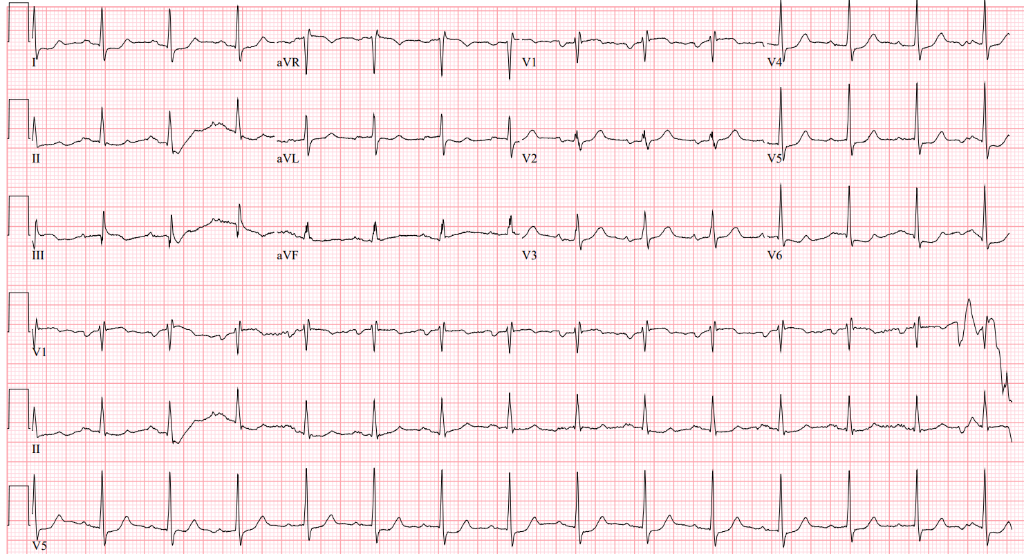
Rate: 80-ish beats per minute
Rhythm: Sinus rhythm
Axis: Normal
Intervals: Narrow QRS, PR maybe slightly long. On the boarder of a first degree AV block, QTc looks ok
Morphology: The significant ST segment changes have mostly resolved, but there is still some lingering ST-T wave changes, such as some slight ST segment depression in V4-V6 and I and some slight ST segment elevation in V1. I’d put this in the non-specific ST-T wave changes bucket.
Final Read: Normal sinus rhythm with a rate in the 80s and some non-specific ST-T wave changes.
Case Wrap Up:
“Hello! EP? Hi! It’s me again. I have a cool ECG for you and a patient you’ll probably want to take to the EP lab. Ms. Flemming in room 28. 32-year-old female coming in for a uti. Suddenly went into a narrow complex tachycardia after using the restroom. It sure looks like 1-1 flutter, but could also be 1-1 atrial tachycardia, AVNRT or AVRT. Really not 100% sure. Her BP dropped and she became minimally responsive, so I emergently cardioverted her. She’s not thrilled with me, but she’s back in sinus rhythm. I’ll have the ECGs bedside for you when you come see her. Thanks!
Recap:
Remember, when confronted with a tachydysrhythmia, start ask yourself if the R-R interval is regular or irregular and if the QRS complexes are wide or narrow
In this case, we saw a regular, narrow complex tachydysrhythmia
That points us to SVT, Aflutter, and sinus tachycardia
At rates approaching 300, it is very challenging to tell the difference between 1:1 Flutter, 1:1 Atrial tachycardia, AVRT, or AVNRT
This patient likely needs an EP study to ablate whatever pathway is letting her heart beat like this
Remember, when clinical instability is due to a dysrhythmia, the patient should be emergently cardioverted
We’ve had a string of interesting tachydysrhythmias recently, more coming your way soon!
Hit me up with any questions,
Lloyd
**Looking for a dynamic guest lecture on Emergency Cardiology for your Grand Rounds? Reach out here for a lecture request by Dr. Tannenbaum. If you have reached out already, I have emailed you back. If you haven’t gotten it, please message me directly on Substack or just reply to this email.**
_________________________
Lloyd Tannenbaum, MD
Comments
Nothing yet. Say the first thing.
Sign in to join the conversation.