
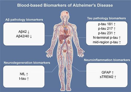
The approach to Alzheimer’s disease (AD) has changed dramatically over the past decade. Rather than waiting until dementia develops, clinicians are now able to identify the biological changes associated with Alzheimer’s disease years—even decades—before significant cognitive impairment occurs. This has shifted the emphasis toward prevention, early diagnosis, aggressive risk-factor modification, and targeted therapy.
Current evidence suggests that Alzheimer’s disease progresses through three major pathological stages:
Amyloid accumulation
Tau pathology
Neurodegeneration
The ideal biomarker panel evaluates each of these processes. CHM is now offering select specialized single test and panels with our new FullScript platform on very affordable lab screening. Please inquire for more information and how you can get these tests done.
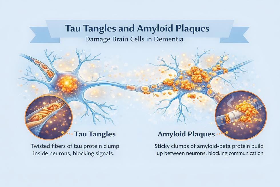
These are rapidly becoming the preferred first-line laboratory evaluation because they are relatively inexpensive, minimally invasive, and increasingly accurate.
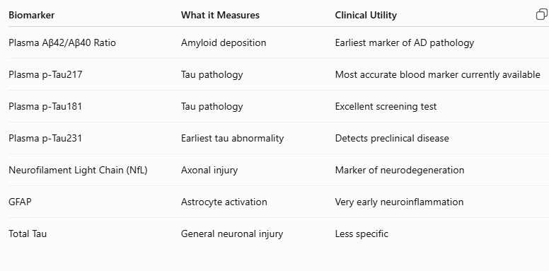
The amyloid beta proteins are produced normally by neurons.
Healthy brains clear these proteins efficiently.
Patients developing Alzheimer’s disease gradually accumulate:
Amyloid Beta 42
Amyloid plaques
As plaque formation increases:
Blood Aβ42 decreases
Aβ40 remains relatively stable
The resulting decline in the Aβ42/Aβ40 ratio is one of the earliest detectable biological changes.
Sensitivity in recent studies approaches 85–90% compared with amyloid PET imaging.
This has become the single best blood biomarker for Alzheimer’s disease.
Advantages include:
Excellent diagnostic accuracy
Detects disease before symptoms
Correlates closely with tau PET imaging
Predicts progression from mild cognitive impairment (MCI) to dementia
Recent studies report:
Sensitivity: approximately 90–95%
Specificity: approximately 90–95%
Many memory clinics now consider p-Tau217 their preferred screening biomarker.
Although slightly less accurate than p-Tau217, p-Tau181 is widely available and well validated.
It is particularly useful for:
Early diagnosis
Monitoring progression
Distinguishing Alzheimer’s disease from frontotemporal dementia
This appears to rise even earlier than p-Tau181.
It may detect disease before substantial amyloid deposition and may become increasingly important in prevention clinics.
NfL is released when neurons are damaged.
It reflects:
Axonal degeneration
White matter injury
Neurodegeneration
Elevated NfL is not specific for Alzheimer’s disease.
It is also elevated in:
ALS
Multiple sclerosis (MS)
Parkinson disease (PD)
Traumatic brain injury (TBI)
Vascular dementia
Nevertheless, increasing NfL suggests ongoing neuronal injury.
GFAP reflects activation of astrocytes.
Research suggests it may increase:
before p-Tau
before amyloid PET becomes positive
It may therefore become an excellent biomarker for identifying individuals in the earliest preclinical stages.
Lumbar puncture (LP aka spinal tap) remains one of the most accurate methods for diagnosing Alzheimer’s disease. More invasive than serum/blood tests.
The classic CSF profile includes:
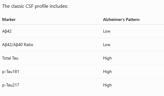
Diagnostic accuracy exceeds 90–95% in experienced centers.
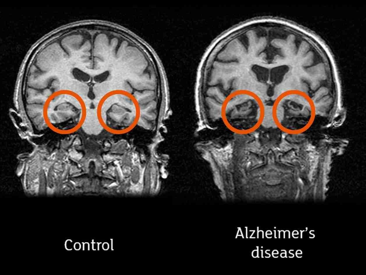
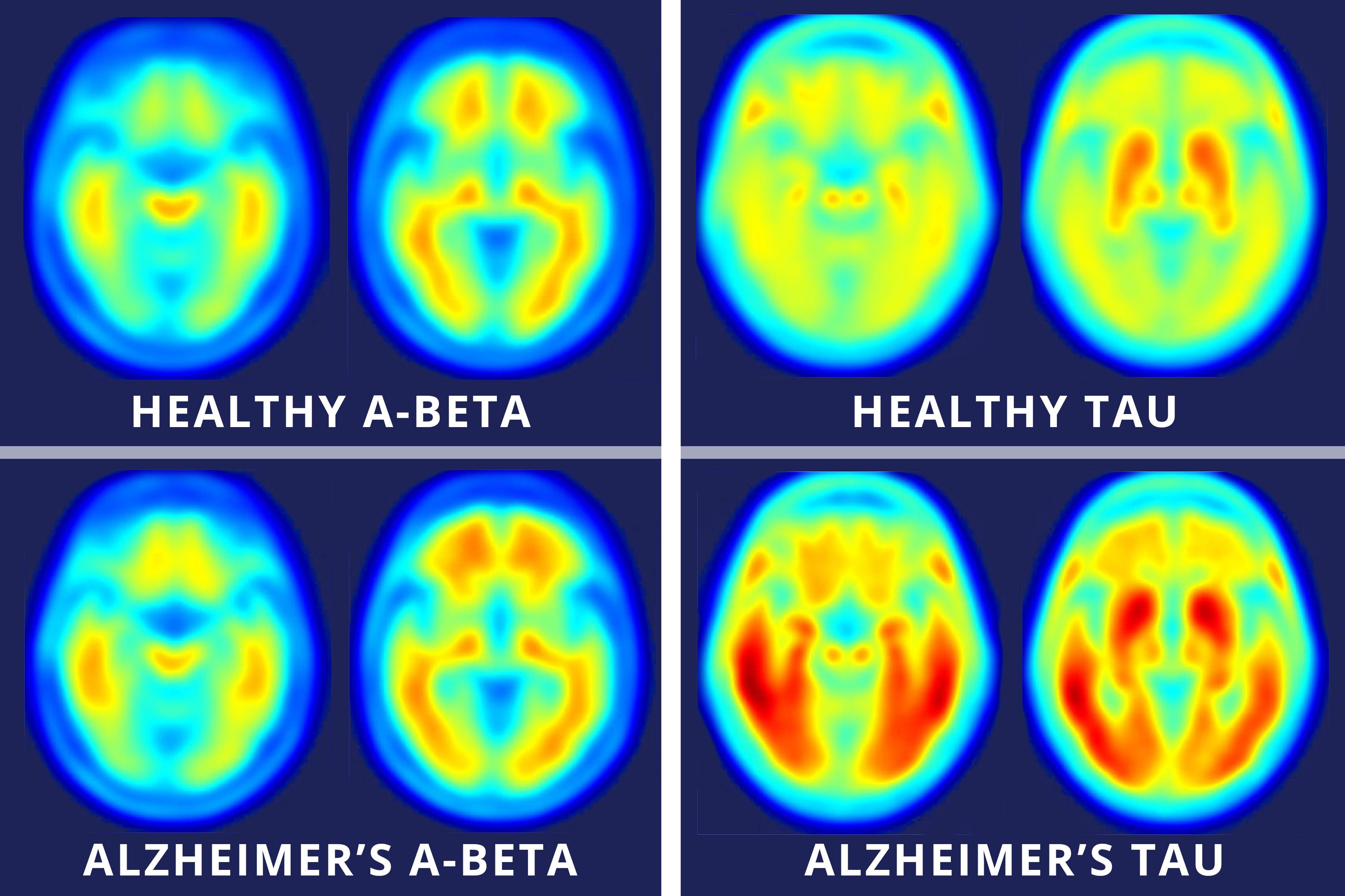
MRI evaluates structural changes.
Important findings include:
Hippocampal atrophy
Medial temporal lobe atrophy
Cortical thinning
Enlarged ventricles
White matter disease (vascular contribution)
MRI also excludes other causes of cognitive decline, such as tumors, normal pressure hydrocephalus, or significant cerebrovascular disease.
Amyloid PET identifies cerebral amyloid plaques.
Advantages:
Detects pathology years before dementia
Confirms diagnosis when blood tests are equivocal
Often required to determine eligibility for anti-amyloid therapies
A positive scan indicates amyloid deposition but does not alone confirm symptomatic Alzheimer’s disease. Does require exposure to ionizing radiation (x-rays)
Tau PET visualizes neurofibrillary tangles.
It correlates closely with:
disease severity
cognitive impairment
future progression
Tau burden generally correlates more strongly with clinical symptoms than amyloid burden.
Measures cerebral glucose metabolism.
Typical Alzheimer’s pattern:
bilateral temporoparietal hypometabolism
posterior cingulate involvement
precuneus involvement
The APOE ε4 allele is the strongest common genetic risk factor for late-onset Alzheimer’s disease. Usually see Apo E3/E3 as normal or native but higher risk with Apo E3/E4 or E2/E4 and even higher risk with Apo E4/E4.
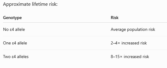
APOE genotyping is not diagnostic, but it is useful for:
Risk stratification
Counseling
Eligibility assessment for some disease-modifying therapies (because ε4 carriers have a higher risk of amyloid-related imaging abnormalities, or ARIA); Early lifestyle modifications like diet and select dietary supplements.
Many reversible or modifiable contributors to cognitive decline should be assessed:
CBC
Comprehensive metabolic panel
Vitamin B12 (with methylmalonic acid if borderline)
Folate
Vitamin D
TSH and advanced thyroid tests including BBT measurements
Cortisol
Sex Hormones (E, P, T, DHEA-s)
HbA1c
Fasting insulin (if assessing insulin resistance)
Lipid panel
High-sensitivity CRP (hsCRP)
Homocysteine (HCY)
Ferritin and iron studies (when indicated)
Magnesium (preferably RBC magnesium if available)
Liver and kidney function tests
Depending on the clinical context, additional tests may include:
Sleep evaluation for obstructive sleep apnea (OSA)
Depression screening
Hearing assessment
Selected infectious, autoimmune, or toxicology testing
Rule out stealth infections and TBDz (Lyme, others)
Several promising biomarkers are under investigation:
Plasma α-synuclein
TDP-43 biomarkers
Neurogranin
YKL-40
Soluble TREM2
Exosomal neuronal proteins
Plasma inflammatory cytokine panels
Advanced proteomic and metabolomic signatures
These remain primarily research tools at present.
For patients with a family history of Alzheimer’s disease, subjective cognitive complaints, mild cognitive impairment, or other elevated risk:
Baseline cognitive screening (e.g., MoCA).
Blood biomarkers: plasma Aβ42/Aβ40 ratio, p-Tau217 (or p-Tau181 if p-Tau217 is unavailable), NfL, and GFAP.
MRI of the brain with volumetric analysis if available.
Comprehensive laboratory evaluation for reversible contributors.
APOE genotyping after appropriate counseling, if the results would influence management or treatment decisions.
If blood biomarkers are abnormal or diagnostic uncertainty remains, consider CSF biomarkers or amyloid PET (and, in selected cases, tau PET), particularly in consultation with a memory disorders specialist.
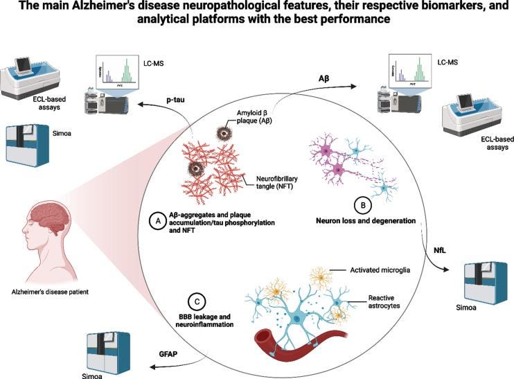
The current evidence supports a blood-first strategy for Alzheimer’s disease prevention and early detection. Among blood tests, plasma phosphorylated (p)-Tau217 has emerged as the most accurate single biomarker, while combining Aβ42/Aβ40 ratio, p-Tau181, NfL, and GFAP provides a comprehensive assessment of amyloid deposition, tau pathology, neurodegeneration, and neuroinflammation. Genetic predisposition checking ApoE for select alleles (SNPs) is helpful too.
When combined with cognitive testing, MRI, and evaluation of modifiable risk factors, these biomarkers offer the best available approach for identifying individuals at risk before significant cognitive decline develops.
Comments
Nothing yet. Say the first thing.
Sign in to join the conversation.