
See also my previous article on Intracellular Hypercalcinosis.
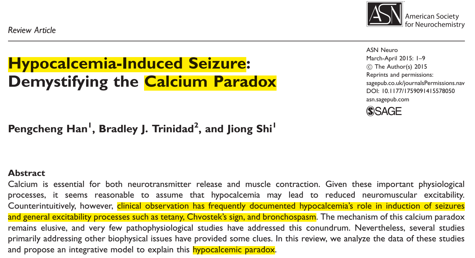
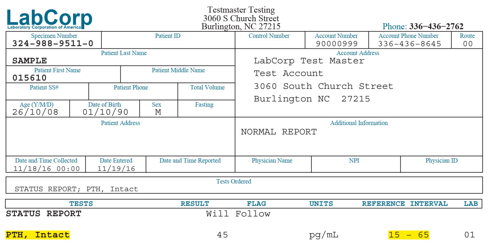
As I described in my previous article on Intracellular Hypercalcinosis, vitamin D deficiency and other factors contribute to Intracellular Hypercalcinosis which has several neurotoxic and anti-metabolic consequences. The mechanism by which mild-moderate vitamin D deficiency promotes neurotoxicity is via increased intracellular calcium via increased compensatory/secondary hyperparathyroisim (hPTH). Increased PTH causes increased intracellular calcium with its neurotoxic consequences. This can be measured clinically by simply measuring serum PTH levels as described in the PDF article provided below at the end of this page.
See sample lab report in the image below.
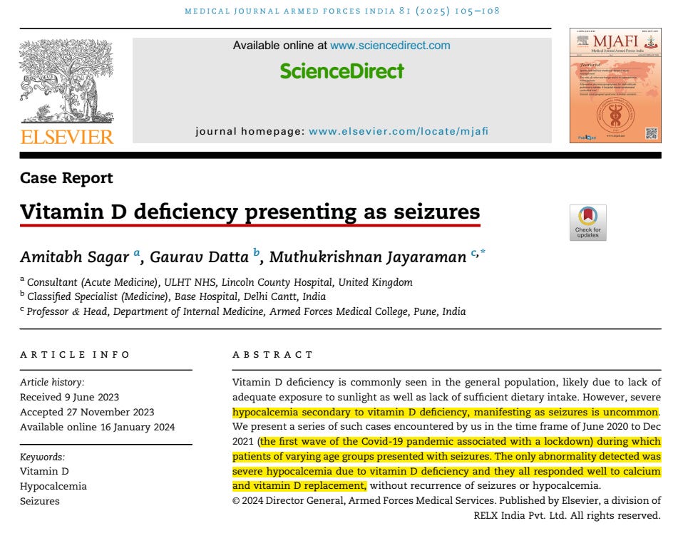
Severe vitamin D deficiency causes hypocalemia—low calcium in the blood and therefore low calcium in the extracellular fluid. As you can see in more detail in the PDF article provided below, the authors detail the mechanisms by which low extracellular calcium causes neuroexcitation including seizures (which are obviously neurotoxic). The mechanism is by increased firing/depolarization rather than the increased intracellular inflammation caused by intracellular hypercalcinosis described previously.
Mild vitamin D deficiency = secondary hPTH = increased intracellular calcium = neurotoxic consequences
Severe vitamin D deficiency = hypocalcemia = increased firing/depolarization = neurotoxic consequences
Trousseau’s sign is a clinical indicator of low blood calcium (hypocalcemia) or high nerve and muscle sensitivity (neuromuscular excitability). It causes the hand and wrist to spasm into a flexed position when a blood pressure cuff is inflated above the systolic pressure for two to three minutes.
Chvostek Sign: How to test: Tap gently on the facial nerve, located right in front of the ear just below the zygomatic arch (over the masseter muscle). Positive result: A brief twitch or spasm of the facial muscles (such as the corner of the mouth or the side of the nose) on the side being tapped. Clinical note: It is less reliable because roughly 10–25% of healthy people can test positive, and some patients with true hypocalcemia may not show it.
Note the very impressive laboratory results reported in this article: the two tests that I highlighted are tests that clinicians should be using in the evaluation of these more severe cases, in addition to the usual serum calcium and 25ohD.
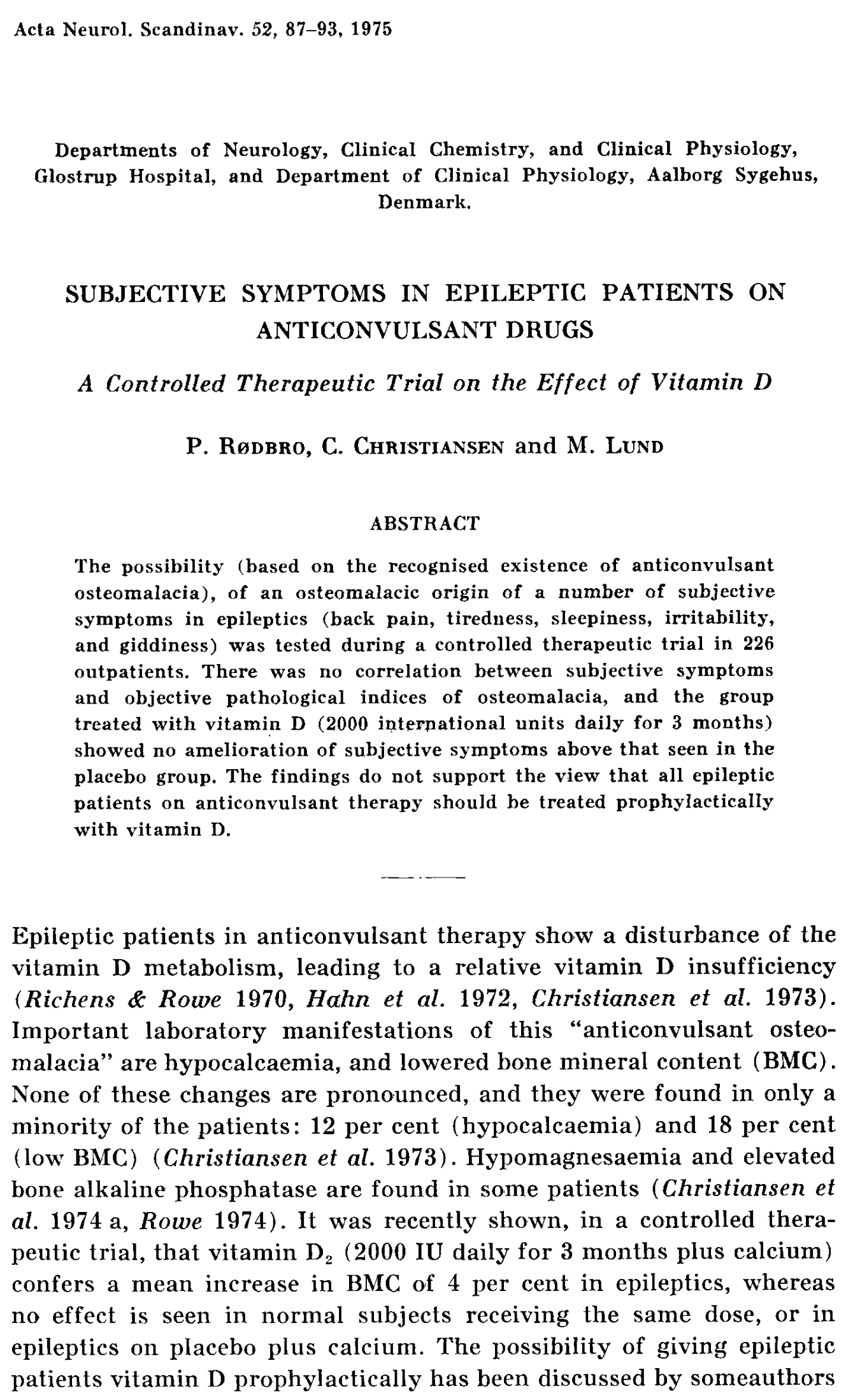

Dr Alex Vasquez 2004 2008 2024 Vitamin D 2008 Jcem Paradigm 2004 Jama 2004 Bmj 2005 Pharmacology 2024
4.89MB ∙ PDF file
Comments
Nothing yet. Say the first thing.
Sign in to join the conversation.