
Throughout the years in my journey attempting to dissect SARS-CoV-2 and its complexity to the best I can, many aspects surprised me, but among the few at the top resides receptor usage. As a refresher, receptors are like intricate Lego pieces on top of cells, and viruses, bacteria, our own immune cells, and many proteins must “connect” to either hijack the cell in the case of pathogens or trigger a reaction in the case of everything else. Sometimes these Lego pieces need intermediary pieces called “ligands”.
To attempt to understand all its nuances in using so many different receptors, in so many different cells I thought of the concept of “hierarchical receptor usage”, the virus has its primary, preferred receptor. On the lack or low expression of said receptor (ACE2), it will use different ones, creating distinct cascading effects.
This conceptual framework is useful for me to understand and produce deeper research, but it doesn’t explain what is actually occurring at a molecular level, how the virus infects cells that, in theory, it shouldn’t, because they lack the necessary receptors.
How does SARS-CoV-2 infect not only respiratory epithelial cells, but also alveolar macrophages, endothelial cells, and pericytes, macrophages being critical immune cells, and the last 2 critical for vascular integrity and barrier function. Under normal (healthy) conditions, all these cells in the lung express little to no ACE2 or its critical priming protease TMPRSS2. They hypothesized this occurs via EVs.
What are EVs ? The acronym stands for Extracellular Vesicles, super-small particles secreted by cells for intercellular communication. This is how cells talk to each other and signal what to do, they play a central role in modulating immune responses, tissue regeneration, and also in cancer, diabetes, and many other diseases. As pointed out by the authors themselves, much of the research regarding EVs and SARS-CoV-2 is focused on infection, transmission, and EVs carrying viral components. And this is their reasoning for this research, what other roles EVs may possess.
As per most research, their first step is demonstrating within their hypothesis parameters if or how efficiently SARS-CoV-2 infects the cells they are using, or samples. Here, they used a lung organoid model, which is created from human stem cells, and represents actual lung architecture, rather than just being a bunch of cells. They used two infection approaches, one of which was using a pseudoviral model (pretty close to the last article), which depends entirely on TMPRSS2 rather than Cathepsin L.
But they also used live SARS-CoV-2, interestingly, most cells remained viable 4 days after infection, even though they sustained viral damage. This confirms that the virus both infects and replicates in their lung organoid model, and replicates the actual viral infection. Their next step is groundbreaking, so I need to present you with context.
Until recently, TMPRSS2 has been classified as a transmembrane serine protease, in simpler terms, an enzyme that stays at the cell surface, and its role is breaking peptide bonds. It is abundant in the prostate, lung epithelium, and the gut. Staining the lung organoids to detect TMPRSS2 and the epithelial marker EPCAM revealed a dotting, vesicular pattern, suggesting the enzyme is localized inside the cell, close to the membrane (a. in the image below)
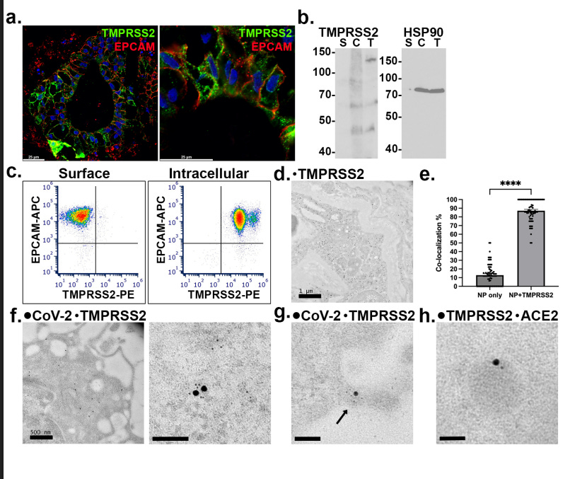
To further understand the localization of TMPRSS2, they used biotin to label cell surface proteins, and subsequently separated the cell surface and cytoplasmic (inside) portion of the cells using strepavidin beads, because these beads have an extremely strong and very specific affinity for biotin. TMPRSS2 was easily detectable in the lysate (when you release all the content from inside the cells to analyze it), and also in fractions of the cytoplasm, but absent from the surface fraction.
Next, they used an antibody test, using antibodies that work against the extracellular domain of TMPRSS2, and its expression was only detectable inside the cell, not outside, on the cell surface. Antibody binding can trigger proteolytic shedding, the cell cuts off the protein, and despite their tests, no evidence of antibody-induced shedding was found.
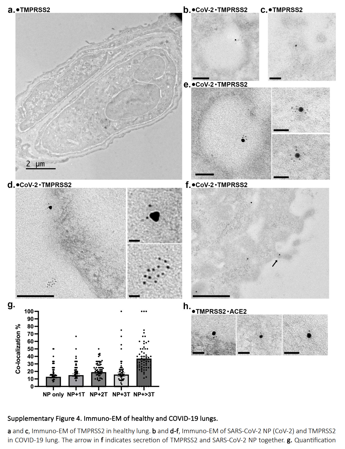
Using immuno-electron microscopy (immuno-EM) with normal and Covid-19 lung samples, they detected TMPRSS2 in the cytoplasm, or right beneath the plasma membrane, and also observed it within vesicle-like structures (b and e in the image above). They also observed that the majority of the SARS-CoV-2 nucleocapsid Protein was located in close proximity to TMPRSS2 inside the cell. 87% of the nucleocapsid protein was associated with TMPRSS2, and TMPRSS2 was also found co-localized with ACE2. This section of the paper itself is remarkably important, and I will expand on it later.
Given the presence of TMPRSS2 inside the cell, localized with visible vesicles, the next step is to measure common EV’s markers. 52% of the enzyme was co-localised with CD9 (52%), CD63 (36%), and CD81 (38%). Further investigation with immuno-EM in lung organoids found TMPRSS2 located in close proximity to CD63 and TSG101 (another EV marker).
Isolating EVs from lung organoids, they detected TMPRSS2 in both EVs and cell lysates, but absent in the samples that were depleted of EVs. However, ACE2 was detected in EVs, in EV-depleted ones, and in lysates, so ACE2 can exist both inside EVs and as a soluble (free) protein. By testing the topology of the EVs, they found that TMPRSS2 has its extracellular domain oriented towards the inside of the EV, which makes perfect sense, as when the EV fuses or is taken up by a cell, the enzyme is delivered in its correct topology, with its domain outward. ACE2 was present at the EV surface, the outside.
Since both the enzyme and the main receptor the virus uses are present in EVs, they aimed to test if EVs did, in fact, participate in SARS-CoV-2 infection and transmission in the lung. After incubating purified EVs with SARS-CoV-2 pseudovirus, they added the mixture to lung organoids. SARS-CoV-2 transduction (understand this term as “artificial infection”) rate increases in a dose-dependent manner. Incubation with just EVs without the Spike (read my last article to understand the pseudovirus model used here) didn’t have an effect on transduction. These are Spike-centric effects.
To extend these findings, they analyzed human lung samples, from deceased patients. In healthy lungs, TMPRSS2 was essentially absent from the surface of epithelial cells, but in disease lungs, up to 10% of the same cells displayed surface TMPRSS2. This aberrant surface translocation is significant because patients with underlying pulmonary conditions such as Idiopathic Pulmonary Fibrosis, Covid-induced fibrosis, Interstitial Lung disease, and others are all more susceptible to severe infection and sequelae.
As within the trend of this paper, more surprises were to be found, as when the authors analyzed non-epithelial cells in these lungs, they carried TMPRSS2 on their surface despite being unable to actually produce either the enzyme, and these cells were alveolar macrophages, endothelial cells, and pericytes. These cells showed no detectable TMPRSS2 or ACE2 mRNA. Further testing from multiple lungs and their fluids showed that up to 90% of the endothelial cells and alveolar macrophages had TMPRSS2 on their surfaces.
To prove this, they used macrophages from human pluripotent stem cells and vessel organoids, containing endothelial cells and pericytes, which are new and therefore never exposed to epithelial ERVs. They lacked endogenous TMPRSS2 and ACE2 at both the mRNA and protein levels. After 24 hours of exposure to EVs purified from lung organoid-containing media, both TMPRSS2 and ACE2 were easily detectable. Flow cytometry showed that nearly all macrophages and endothelial cells now had surface TMPRSS2.
EVs can carry damage-associated molecular patterns (DAMPs) and TLRs, which are capable of increasing TMPRSS2 and ACE2 by themselves. No significant upregulation of either was found at 24 or 48 hours post-exposure. Even exposure to endotoxin, which is a potent TLR4 agonist, failed to induce transcription of both. Protein transfer is the only viable explanation.
So, how do the recipient cells take up these EVs ? Using multiple inhibitors, they found that blocking actin polymerization with cytochalasin D, or depleting membrane cholesterol with methyl-Beta-cyclodextrin strongly impaired EV internalization. Other inhibitors targeting clathrin-mediated endocytosis, caveolae, and macropinocytosis only partially reduced the uptake. In simpler terms, EVs rely mostly on direct membrane fusion, or lipid raft-mediated pathways to deliver its cargos.
Their last step was testing whether both macrophages and vessel organoids derived from stem cells primed with EVs would exhibit increased susceptibility to live SARS-CoV-2 infection. Both exhibited higher viral titers compared to their unprimed counterparts (without EVs). This increase suggests a transfer of both TMPRSS2 and ACE2 protein via EVs, facilitating viral entry, and demonstrating EVs act as viral infection enhancement factors in previously “resistant” cell types.
To isolate the specific contribution of TMPRSS2, they generated a TMPRSS2-knockout (KO) cell and lung organoid model, KO means without, so these organoids still contained ACE2 and largely normal levels of other proteins, but lacked TMPRSS2. When used to prime macropahges, the KO’d EVs reduced the viral titers by approximately 70% compared to wild-type EVs. This confirms TMPRSS2 is the critical rate-limiting factor for efficient EV-mediated infection enhancement.
This paper presents distinct but parallel mechanisms. The first is the direct hitchhiking model observed during the pseudovirus assay. The Spike Protein binds to ACE2 displayed on the outer surface of EVs, effectively using the vesicle as a ferry towards a recipient cell. Because TMPRSS2 is oriented with its catalytic domain towards the inside of the EV, priming likely does not occur during this transport phase, it likely occurs when the EV encounters a cell that never expresses its main receptor.
The second mechanism is the long-term one. EVs are actively taken up by many cells, including macrophages, endothelial cells, and pericytes, depositing both ACE2 and TMPRSS2 onto said cells. The cell is then rendered susceptible to viral infection through its canonical receptor-mediated entry, as if it always harbored the receptor and the critical enzyme similar to the epithelial cells the virus targets. But it goes much further.
The implications are significant because, per this paper, even in healthy lungs, there is a constitutive secretion of TMPRSS2/ACE2 EVs that creates a baseline susceptibility in macrophages and vascular cells that explains how SARS-CoV-2 didn’t and doesn’t cause just pneumonia, but systemic vascular inflammation, thrombosis, endothelial dysfunction, and immune dysregulation. Learning how to target specifically EV biogenesis, or block their uptake by specific cells, may enable us to limit the systemic damage.
In addition.
EVs can be considered biologically active but immunologically inert payloads. EVs will travel systemically, an EV from the lung can end up in the heart. EVs travel both through the blood and the lymphatic system. EVs can carry not only receptors, enzymes, and other proteins, but also viruses and DAMPs, they can carry an assortment of different molecules. This means EVs can carry viral fragments and act as “reservoirs”. The fact that the nucleocapsid was found so close to TMPRSS2 is important.
EVs can carry SARS-CoV-2 proteins and are associated with both severity and lasting damage. Since EVs carrying SARS-CoV-2 fragments can last months inside the body, it makes the clearance and inflammation aspect of the disease a lot more complex (as seen in this paper, I will cover during the fragment article). EVs are very contextual, and they can both aid repair or induce injury, especially when mediating “cleaner cells” activity, such as macrophages.
The paper we just went through gives significant background and understanding on why SARS-CoV-2 and its fragments have an abnormally long tendency to last in the body, and how/why fragments have particularly novel effects. Which is the theme of my next article, either that or a short update on my (infamous?) Brain Stack, which incidentally targets many of the Covid brain-related changes.
Thank you for your continued support, and consider becoming one if you can. If you can’t, I hope my posts help you in some way.

No posts
Comments
Nothing yet. Say the first thing.
Sign in to join the conversation.