
I had another article in mind for today, but sometimes life gives you “gifts” in the way Nurgle gives gifts.
I have shared the following in a few other articles before, but since I wrote about it sparsely, I will remind you. Back in the early days of the pandemic (between November 2019 and March 2020), a lot of research work was done by anonymous people on imageboards, specifically, scientific research, as all the anons were trying to foretell which pathogen was spreading in China at an alarming pace, something that would cause severe pneumonia, so quickly. The following is for contextual reference.
It is in the nature of imageboards ever since their inception to give rise to conspiracy theories, and it was not different with SARS-CoV-2. Many of the “theories” were confirmed over time. Ralph Baric's involvement, EcoHealth, mRNA vaccines, and some of their side-effects, a lot of that information came from imageboard, and most imageboard users knew it was a coronavirus by early December, likely engineered. The Dengue side of it (Dengue antibodies can make a Covid infection worse and induce more vascular damage) “Airborne AIDS”, which, for reference, it isn’t, is a lot more complex and somewhat (arguably to me) worse.
But one of the more outlandish conspiracy theories remained unconfirmed, but for some reason or another, I always remembered it. One anon stated, “SARS-CoV-2 spreads like fungal spores (such as Candida Auris) and infects you via skin”. The mechanism is not accurate, but the overall idea…
The authors mention that while the skin is an efficient physical and immunological barrier, it can be directly infected by Herpes Simplex, Varicella Zoster, Molluscum Contagiosum, and, not mentioned by them, but mentioned by me, Candida Auris. If you are curious, C. Auris is ascomycetous, a fancy word for spore shooting.
Like all respiratory viruses, SARS-CoV-2 enters the body through the respiratory tract, and while the virus will invariably reach the gut and other organs because its preferred receptor is expressed in so many cells, over time, we have found it can also enter via the eyes. But both viral RNA and vaccine mRNA/Spike protein can be found in skin lesions, frequently preceded by dermatological reactions such as chilblains, urticaria, and vasculitis. I am mentioning this because it will be important later on.
Patients with pre-existing skin pathologies such as atopic dermatitis and psoriasis have an increased risk of developing SARS-CoV-2 infection, and so far, it has been hypothesized that this is due to inflammatory conditions and immunological changes associated with these diseases, rather than viral interaction with the skin.
They used two models to test whether skin is susceptible to infection and which changes exactly can lead to a higher chance of infection. A 3D human skin organoid (HSO), which is a lab-made high-fidelity representation of the skin (generated from human epidermal keratinocytes) and neonatal skin explants.
In the HSO model, they used African American (AA) and White Non-Hispanic (WNH) skin models, because there is a significant disparity in both Covid infection and risk of inflammatory skin conditions between populations. Both ACE2 and TMPRSS2 were found to be highly expressed in both human skin explants and HSO, with a higher expression of TMPRSS2 in African American HSO. Their experiments also revealed a big inter-individual variability in the expression of both ACE2 and TMPRSS2.
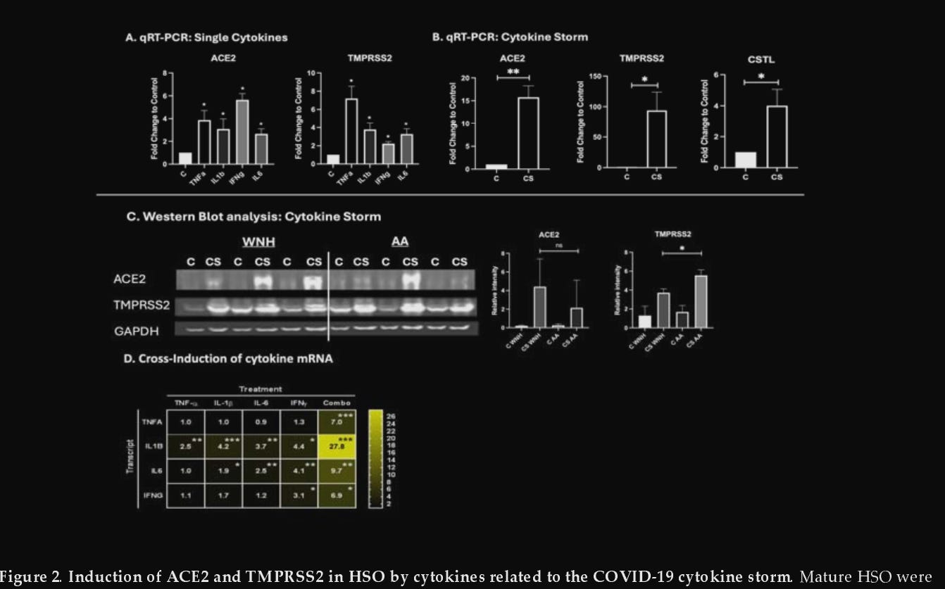
Next, they chose 4 pro-inflammatory cytokines that serve a dual purpose here. They play central roles in acute and chronic skin conditions and are also a core aspect of severe SARS-CoV-2 infection, response, and its infamous cytokine storm, the cytokines being TNF-α, IL-6, IL-1β, and IFN-γ.
Individually, each of these significantly increased ACE2 and TMPRSS at both mRNA and protein levels. But combining them created an even bigger increase. Each of these cytokines can increase the expression of another, such as TNF-α, which can increase IL-6 expression, which in turn can increase IFN-γ expression. Once again, significant individual variability was found.
They also chose to test the effects of cytokine exposure on the expression of other receptors that SARS-CoV-2 uses for viral entry, those being KREMEN1, ASGR1, AXL, NPL1, and lastly Cathepsin L (CTSL). Minimal to no change in expression was observed in all these except… CTSL, which was increased by 5 times upon combining multiple cytokines.
This matters a lot. Cathepsin L is a lysosomal protease (it breaks down proteins) that can cleave the SARS-CoV-2 Spike Protein at a very specific site in its Furin Cleavage Site, allowing for viral entry via an overlooked route (bypassing the TMPRSS-2 dependent membrane pathway), which is important when TMPRSS2 is absent or inhibited. While the study doesn’t test for CTSL-mediated entry, the 5x upregulation serves as an indication that inflamed skin possesses the molecular makeup for an alternative entry route.
The next step is testing how different skin conditions and the cytokines associated with them affect the expression of ACE2 and TMPRSS2. On the Th1 cytokines, they chose TNF-α + IL-17, which is a pro-inflammatory pro-psoriasis combination, on the Th2, IL-4 and IL-13, which is a pro-atopic dermatitis (Th2 is often referred to as the “allergic” pathway).The Th1 cytokines predominantly induce ACE2 expression, and Th2 primarily induce TMPRSS2 in HSO. The Th1 cytokine combination effects on ACE2 expression were more pronounced in African-American HSO. This demonstrates that inflammatory skin conditions such as psoriasis can be particularly more vulnerable to SARS-CoV-2 entry because ACE2 expression is higher, whereas conditions such as atopic dermatitis, which bear higher TMPRSS2, have blunted ACE2 expression as a limiting factor.
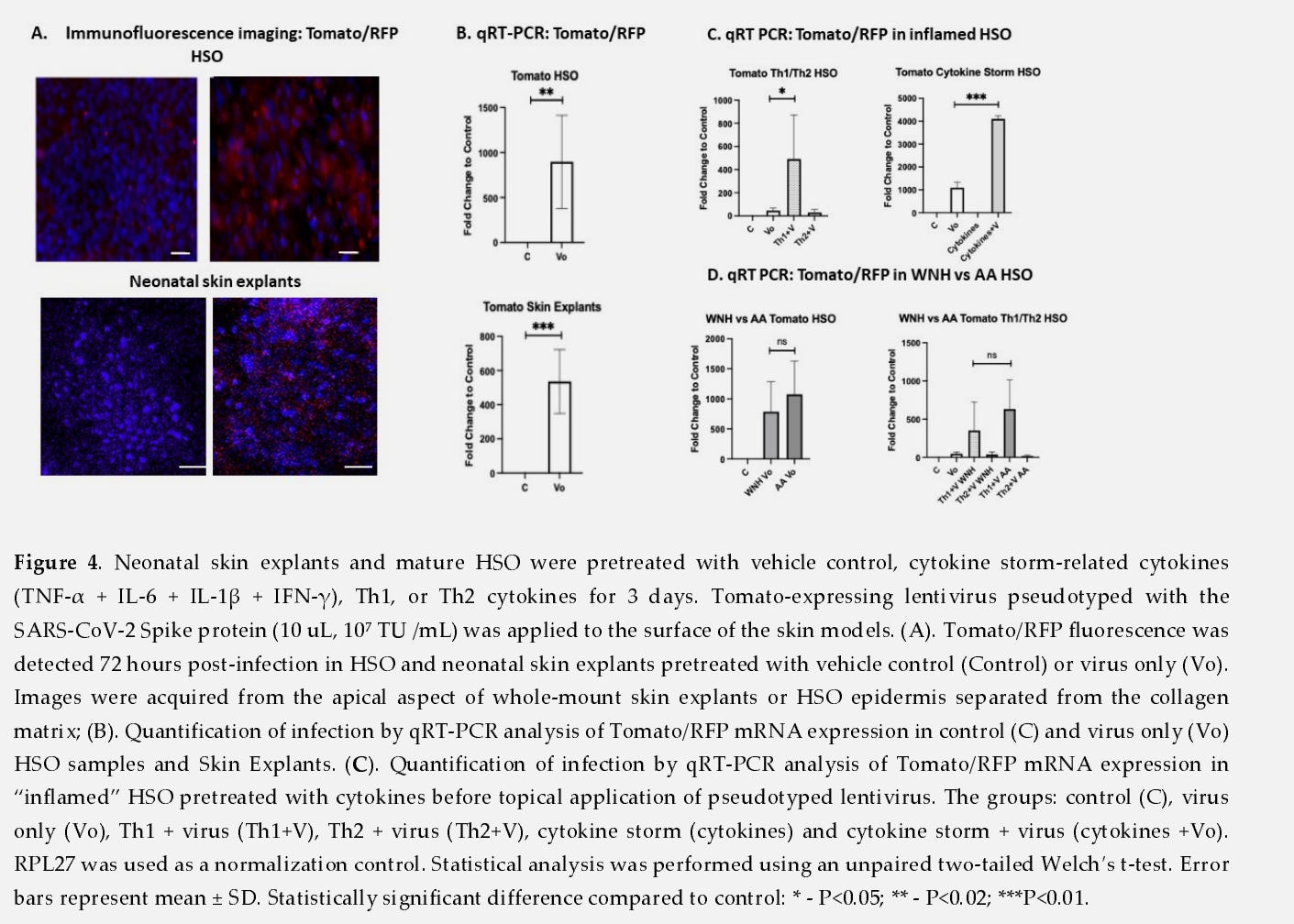
To test actual viral entry without using live virus, which requires BSL-3, the authors developed a BSL-2 compatible, non-replicating approach, using a lentiviral reporter pseudotype with the Wuhan-strain Spike Protein. This lentiviral pseudotype approach is fairly common. They inserted a Tomato/RFP fluorescent marker, so successful entry lights up in the cells.
Even without cytokine pretreatment, both HSO and neonatal skin explants showed detectable fluorescent gene (they added the gene to be able to observe successful interaction) 2 to 3 days post-infection. This shows even healthy skin is able to support SARS-CoV-2 entry. Pretreatment with the cytokine storm combination dramatically increased infection rates compared to controls, with Th1 significantly increasing infection efficiency. The Th2 cytokines did not, demonstrating how critical ACE2 is (sadly, they didn’t test for Cathepsin L entry route).
The lack of TMPRSS2 in certain cells that still get infected by SARS-CoV-2 will be covered in the coming article, because yes, they have found how the virus is able to infect cells that lack its main receptors. Outside of the alternative Cathepsin L pathway.
As mentioned a few paragraphs above, one of the limiting factors, brought up by the authors themselves, is how they tested the viral interaction with skin cells. It mostly replicates Spike Protein-ACE2 interactions, rather than a full viral cycle that activates all metabolic and immune pathways. Limiting, but illuminating.
They analyzed the gene expression signature of lungs from deceased Covid patients with their skin models treated with the cytokine storm combination, and subsequently infected with the pseudotype virus. There were 148 common genes that were differentially regulated, significantly enriched, and these genes were related to Inflammation, innate and adaptive immunity, and SARS-CoV-2-induced activation of immune pathways.
This study provides a good framework to understand multiple aspects of SARS-CoV-2 that have been completely overlooked in relation to our skin. It provides a mechanistic explanation for how a percentage of patients can experience skin reactions, beyond the viral infection, as many topical reactions can now be explained not as a successful infection, but as an immunological and inflammatory reaction to the virus and its Spike Protein.
While the adult skin is more mature, therefore resilient to a successful viral entry and replication (meaning getting infected by the skin), it wouldn’t surprise any of my readers to know that in the past 4 years alone, skin conditions have been increasing. This means the barrier is compromised, and someone with a skin condition is susceptible to further reactions, and to a minor degree, infection.
But inflammation and drastic immunological changes don’t end when the virus clears. One of the most lasting patterns, covered in my Substack for a long time, is the lasting immunological shift occurring in both mild to severe infections, with Long Covid acting as a pillar to a deeper understanding. The body lives, transiently, in a Th2 state, the body forgets to shut off its allergic response for most people, and many people (up to 20%) become suddenly allergic for months.
Both Th2 cytokines tested here, IL-4 and IL-13, are known to upregulate TMPRSS2, and these same cytokines are known to be released by mast cells. This means that effectively the virus can maintain the skin in a higher TMPRSS2-state for a while, making it permissive for viral entry.
The specific role of histamine in this dynamic remains unexplored. Histamine is not just a mediator of itchness and vasodilation, through its receptors (H1 and H4) in keratinocytes, it can directly disrupt the skin integrity and impair healing. Patients with visible skin issues or biomarkers for histamine/mast cell dysregulation have a higher risk of having further inflammatory signaling from their skin.
This closing paragraph mentioning histamine and mast cells was written for a reason, while behind my publishing, it is coming. But first, the next article, which some may think it is as impactful or more than this one, on how SARS-CoV-2 can infect cells that do not express its main receptors.
Consider becoming a paid subscriber, and if you are not, thank you. It is extremely helpful.

No posts
Comments
Nothing yet. Say the first thing.
Sign in to join the conversation.