
It’s George.
The topic of intracellular calcium might initially sound too technical, boring and mundane, yet it is far more fundamental to brain health than is commonly appreciated.
For example, failed or “sluggish” calcium clearance is often one of the core physiological hallmarks of bipolar disorder.
The same underlying disturbances such as elevated basal free Ca2+ for example, also appear in ADHD, where it introduces background noise into prefrontal circuits that govern attention and impulse control.
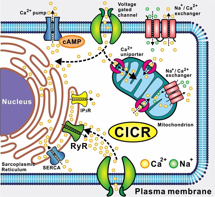
Then in epilepsy, it can tip neurons into uncontrolled calcium-induced calcium release and synchronized hyperexcitability.
Or in anxiety disorders, it impairs the precise synaptic plasticity required to extinguish fear responses in amygdala–hippocampal networks.
In each case the molecular players are largely the same: L-type voltage-gated calcium channels, the smooth endoplasmic reticulum store, buffering proteins, mitochondria, and calcium sensors such as calmodulin that we will break down later in this article.
When their coordinated function is compromised, the result is a loss of signaling fidelity that can manifest as very different clinical pictures depending on which brain regions and cell types are most affected.
Overall, calcium signaling is one of the primary “languages” the neuron uses inside the cell to convert an incoming signal (whether from a neurotransmitter, an action potential or other stimuli) into a concrete downstream action.
So, neglecting calcium signaling for the people who are interested in improving their brain health is foolish.
Now unlike cyclic nucleotides (cAMP, cGMP) or lipid-derived second messengers (IP3, diacylglycerol, arachidonic-acid metabolites), which have to be enzymatically made and then broken down, Ca2+ is simply an elemental ion that cells move in and out of storage instead of building and destroying it chemically.
That’s it.
So its concentration can be changed within microseconds just by opening or closing membrane channels.
This lets neurons produce extremely fast, high-amplitude, and tightly localized signals and why in almost every eukaryotic cell, free cytoplasmic Ca2+ acts as a universal second messenger.
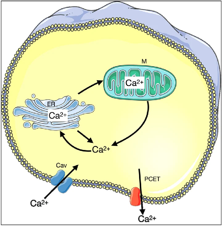
In order to grasp the importance of this better, changes in free cytoplasmic Ca2+ for example, control:
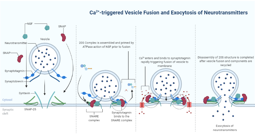
Synchronous vesicle fusion and neurotransmitter release at presynaptic terminals.
When an action potential reaches the end of a neuron, voltage-gated calcium channels open and a brief pulse of Ca2+ enters.
This calcium binds to proteins (that we will break down later in this article) on synaptic vesicles, causing them to fuse with the membrane and release neurotransmitter into the synapse within less than a millisecond.
A calssic example is in a motor neuron, where this precise Ca2+ signal triggers the release of acetylcholine that makes a muscle contract.
If the calcium signal is too weak or poorly timed, the muscle doesn’t move properly, if it’s excessive, too much neurotransmitter is dumped.
Postsynaptic calcium-dependent kinases and phosphatases that drive long-term potentiation or depression.
In the receiving neuron, calcium that enters through NMDA receptors or other channels activates enzymes that either strengthen or weaken the synapse.
In case you have read the previous articles (go here if you haven’t: https://healthlibrary.substack.com/archive), a practical example is in the hippocampus, where a strong, high-frequency burst of activity causes a large Ca2+ rise that activates CaMKII.
This leads to long-term potentiation (LTP) aka the synapse becomes stronger, which is one of the cellular bases of learning and memory.
So a weaker, prolonged Ca2+ signal activates phosphatases such as calcineurin and produces long-term depression, weakening the synapse instead.
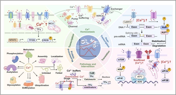
Excitation–transcription coupling that turns brief electrical activity into lasting changes in gene expression (through CREB, NFAT, and other transcription factors).
Calcium that enters through L-type channels (more about these in a moment) can travel to the nucleus or activate signaling cascades that reach transcription factors.
For example, repeated firing of a neuron causes Ca2+ to activate CaMKIV and other pathways that phosphorylate CREB which then turns on genes such as BDNF so the neuron starts producing more BDNF protein.
Intrinsic excitability by modulating calcium-activated potassium channels and other conductances.
Calcium inside the cell can open or close certain ion channels that control how easily the neuron fires.
For example, after a burst of action potentials, Ca2+ activates BK or SK potassium channels.
Potassium flows out, temporarily hyperpolarizing the cell and making it harder to fire again right away (spike-frequency adaptation).
This prevents the neuron from over-firing.
In some prefrontal neurons, this calcium-dependent braking mechanism is crucial for stable working-memory activity.
Long-range circuit connectivity and dendritic integration.
Calcium signals in dendrites help decide whether inputs from different parts of the brain are added together or ignored, and they shape which connections are maintained.
For example, in the prefrontal cortex, precise Ca2+ signals in the dendrites of layer III pyramidal cells allow the neuron to integrate information from many sources while holding information during a working-memory task (a basic example of this would be remembering a phone number long enough to dial it).
In the amygdala, dendritic calcium signals help strengthen or weaken fear-related connections, influencing whether a neutral sound becomes linked to a feeling of anxiety or remains neutral.
Now if these sound too complicated, keep in minf that free cytoplasmic Ca2+ acts like a fast, local “decision-making” signal that tells the neuron when to release transmitter, whether to strengthen or weaken a synapse, which genes to turn on, how excitably it should behave, and how to combine information from different brain regions.
All of this is extremely fascinating and far more important than we might think.
We can demonstrate this from many different angles that are quite easy to understand.
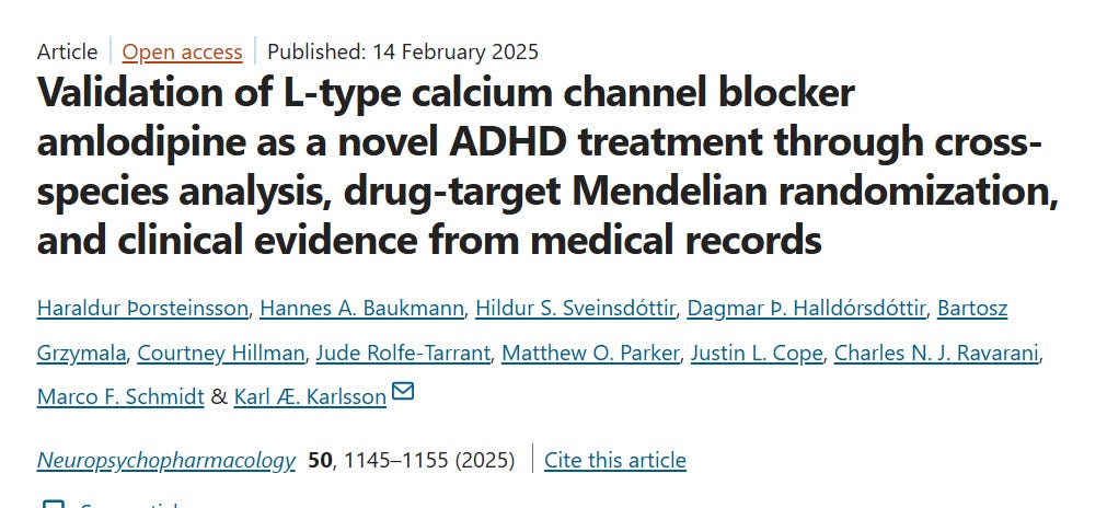
Let’s say that you have issues with ADHD for example.
Then since intracellular calcium (Ca²⁺) influx triggers the release of dopamine and norepinephrine, this obviously means that impaired calcium handling directly interferes with this signal transmission.
So instead of artificially forcing neurotransmitter levels to increase in order to monitor your symptoms, by restricting excess calcium influx, you might be able to lower baseline neuronal excitability and restore natural autoreceptor feedback loops in order to calm overactive brain regions like the telencephalon.
Now, at rest, the concentration of free Ca2+ inside a neuron is only 50–100 nM (nanomolar) and outside the cell it is 1–2 mM (millimolar).
That is a 10,000 to 20,000-fold difference(*).
Because of this huge concentration gradient plus the electrical force (more than 100 mV), Ca2+ ions are desperate to enter the cell.
So when a neuron fires and opens just a few dozen L-type or other calcium channels, local Ca2+ inside the cell can jump 10- to 100-fold in less than one millisecond.
This rapid spike is what triggers neurotransmitter release or starts the molecular cascades for learning.
Immediately afterward, three main pumping systems go to work:
PMCAs (plasma-membrane calcium ATPases) push Ca2+ out of the cell
NCX (sodium-calcium exchangers) also move Ca2+ out
SERCAs pump Ca2+ back into the smooth endoplasmic reticulum store
(*) Maintaining that gradient is energetically expensive and relies on two continuous extrusion systems: the high-affinity plasma-membrane Ca2+-ATPases (PMCAs), which use ATP to pump calcium out, and the high-capacity sodium–calcium exchanger (NCX), which trades three sodium ions for one calcium ion.
Within tens to hundreds of milliseconds, the free Ca2+ is back down to the tiny resting level.
This is exactly what makes this both powerful and dangerous.
The same system that lets the neuron send a fast, precise signal becomes harmful if the Ca2+ stays high for too long (in the micromolar range).
An excess of Ca2+ activates enzymes that “chew up” proteins and lipids, produces toxic free radicals, and can open pores in mitochondria that cause the cell to die.
That is why neurons use multiple overlapping safety layers such as carefully controlled channels, internal stores, buffering proteins that quickly grab free Ca2+, and sensor proteins such as calmodulin that turn the calcium signal into specific actions.
If any of these layers is damaged (for example by a genetic change in a calcium channel, weak SERCA pumps, or low levels of buffering proteins), the neuron can no longer keep the streets dry.
You get:
Chronically higher baseline Ca2+
Noisy, unreliable signals
Slow clean-up after each signal
Or uncontrolled chain-reaction calcium release
These cellular problems appear as neurons that fire too easily, synapses that cannot strengthen or weaken properly, stressed internal stores, and in extreme cases cell death.
This kind of calcium dysregulation is linked to the mood swings of bipolar disorder, the attention and impulse-control problems of ADHD, the cognitive symptoms of schizophrenia, persistent anxiety circuits, and the uncontrolled firing of epilepsy.
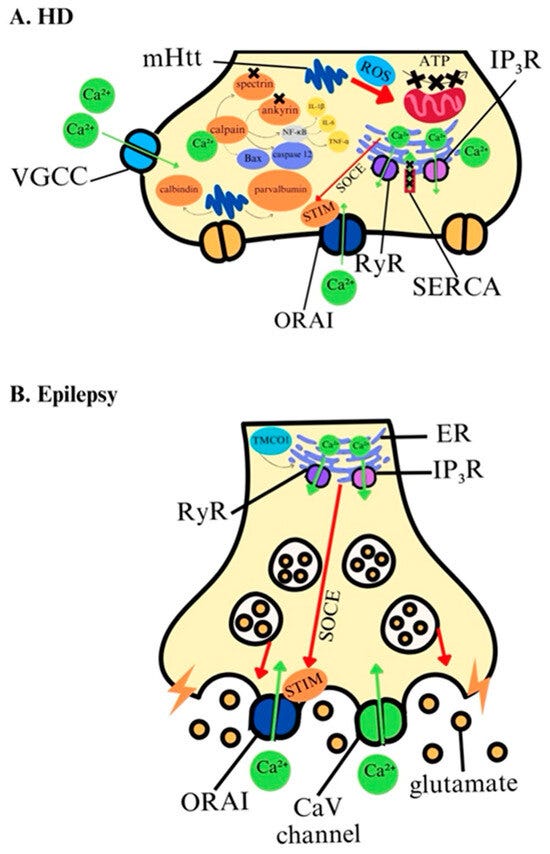
So the extreme concentration gradient gives neurons speed and precision, but it also means that even small failures in the control systems can produce large and harmful effects on brain function.
But hold on, where does intracellular calcium even comes from?
Well, cells do not synthesize calcium.
Every calcium ion present in the human body originates from the external environment, primarily the food and water we consume.
Once ingested, calcium is absorbed across the intestinal epithelium by two complementary routes.
The active transcellular pathway is tightly controlled by 1,25-dihydroxyvitamin D₃ (calcitriol).
Calcitriol increases the expression of the apical calcium channel TRPV6 (the main entry door on the gut side), the small intracellular buffer calbindin-D9k (which shuttles calcium across the cell), and the basolateral extrusion pumps PMCA1b and NCX1 that push calcium into the blood.
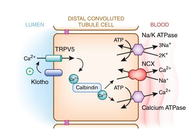
At the same time, parathyroid hormone (PTH) helps maintain whole-body balance by increasing calcium reabsorption in the kidneys and stimulating the final activation step of vitamin D.
Comments
Nothing yet. Say the first thing.
Sign in to join the conversation.