
By Dr. David Traster, DC, MS, DACNB
Co-owner, The Neurologic Wellness Institute
Boca Raton • Chicago • Waukesha • Wood Dale
www.neurologicwellnessinstitute.com
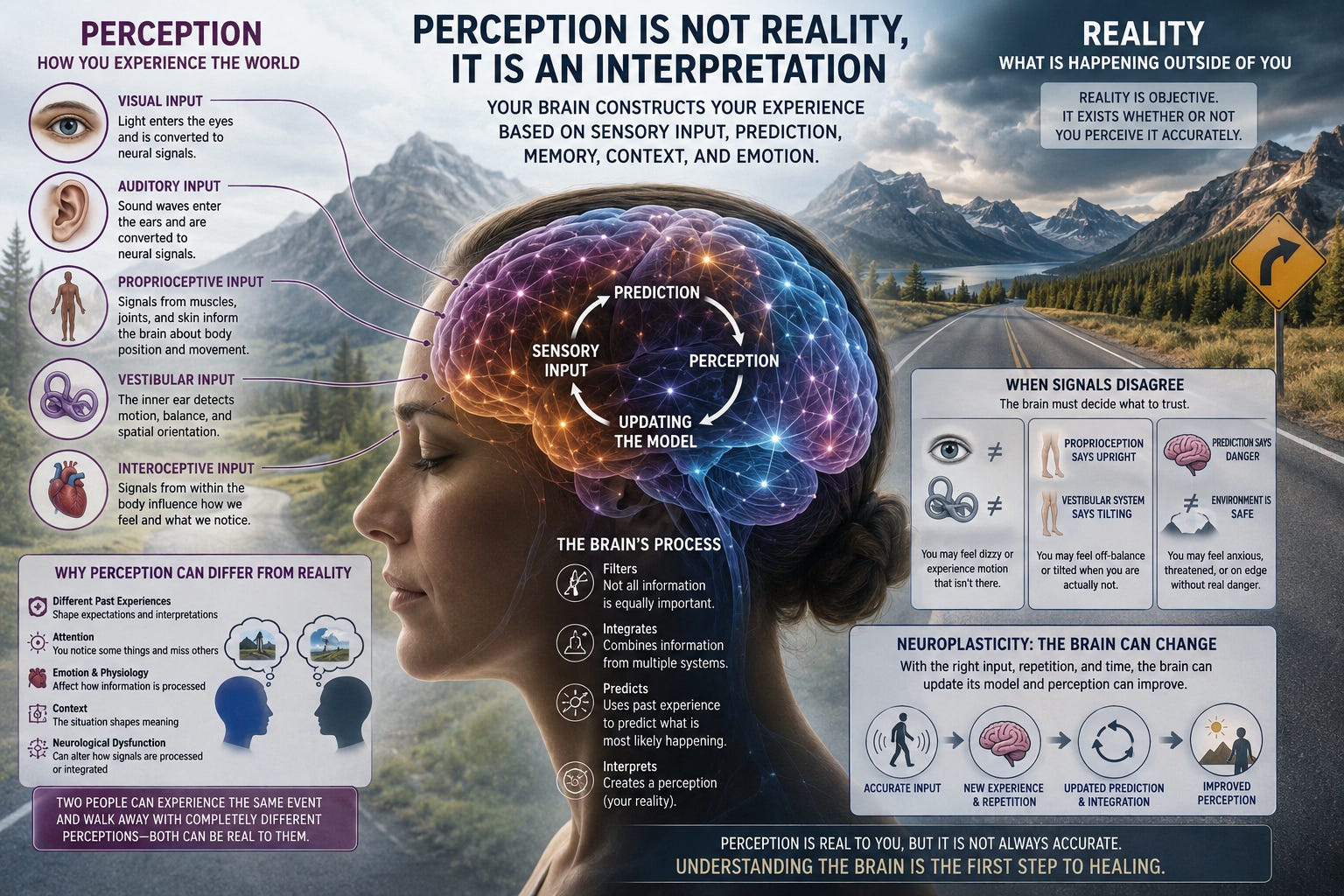
One of the most fascinating concepts in neuroscience is that the human brain does not experience reality directly. What we experience is the brain’s interpretation of reality. At every moment, enormous amounts of information are arriving from the eyes, ears, skin, muscles, joints, vestibular system, internal organs, and environment. Yet what ultimately enters our conscious awareness is not a perfect recording of all of this information. The nervous system filters it, prioritizes it, compares it with previous experiences, predicts what it believes is happening, and then constructs a perception.
This distinction is extremely important. Reality is what occurred. Perception is the nervous system’s interpretation of what occurred. Most of the time, perception corresponds reasonably well with external reality. It has to. Otherwise, we could not successfully navigate through the world. But perception is never completely neutral. Two people can participate in exactly the same conversation, witness the same event, hear the same sound, or experience the same movement and walk away with remarkably different interpretations.
Neither person’s brain necessarily received a perfect representation of what actually occurred. Understanding why requires understanding that perception is not a single neurological event. It is a continuously evolving construction involving sensation, attention, prediction, memory, emotion, autonomic physiology, and previous experience.
The distinction between sensation and perception is fundamental. Sensation begins when specialized receptors detect some form of physical or chemical information. Photoreceptors in the retina respond to light. Hair cells in the cochlea respond to sound-induced mechanical forces. Vestibular hair cells respond to angular and linear acceleration. Mechanoreceptors in the skin respond to touch and pressure. Muscle spindles, Golgi tendon organs, and joint receptors provide information related to body position, movement, and force.
These signals travel through the peripheral nervous system toward the spinal cord, brainstem, thalamus, cerebellum, and cerebral cortex. But receiving a signal does not automatically tell the brain what that signal means. Meaning is constructed. The brain must determine which information deserves attention, whether different sensory signals agree with one another, whether the information represents a threat, whether it resembles something encountered previously, and what response should follow. Consequently, two nervous systems receiving similar sensory information may construct different perceptual experiences from it.
Modern neuroscience increasingly describes perception as an interaction between incoming sensory information and internally generated predictions. The brain does not simply wait passively for information to arrive. It continuously predicts what it expects to see, hear, feel, and experience.
Incoming sensory information can be thought of as bottom-up information traveling from sensory receptors toward higher neurological networks. Expectations, memories, beliefs, context, and previous experiences generate top-down influences that shape how those signals are interpreted. The resulting perception emerges from the interaction between the two.
If sensory information strongly contradicts the brain’s prediction, the brain may update its internal model. But when sensory information is incomplete, ambiguous, noisy, or uncertain, previous expectations may have considerably more influence over perception. This helps explain why context can dramatically alter what we believe we saw or heard. The brain is not merely asking: “What information am I receiving?” It is also asking: “Based upon everything I already know, what is most likely happening?” That distinction changes our understanding of reality.
Vision provides perhaps the easiest example. We commonly assume that our eyes operate like cameras and that the brain simply watches the resulting picture. In reality, visual perception requires enormous neurological processing. Light reaches the retina, where photoreceptors begin transforming electromagnetic information into neural signals. Those signals undergo extensive processing within the retina before traveling through the optic nerves and pathways toward structures including the lateral geniculate nucleus and visual cortex.
From there, distributed cortical networks process features such as orientation, contrast, color, depth, motion, object identity, spatial location, and relationships between objects. The brain then integrates these features into what appears to us as one continuous visual world. But that world is constructed. Visual illusions beautifully demonstrate this. The physical stimulus can remain unchanged while our perception of its size, color, movement, or orientation changes because the surrounding context changes.
In neurological and vestibular patients, this difference can become even more obvious. A person may report that the environment appears to move, bounce, tilt, rotate, or become unstable even though the external environment is objectively stationary. The patient’s perception is real, even though the movement being perceived may not exist in external reality. That distinction is essential in clinical neurology.
The same principle applies to hearing. Sound waves enter the ear and ultimately create mechanical displacement of hair cells within the cochlea. Neural signals then travel through complex brainstem auditory pathways toward the thalamus and auditory cortex. But recognizing speech requires much more than detecting frequencies.
The brain must identify phonemes, separate speech from background noise, determine where sounds originate, recognize voices, process timing, and integrate what is heard with language, attention, memory, facial expression, and context. This is why two people can hear exactly the same sentence and interpret it differently.
Imagine someone saying:
“That’s interesting.”
Depending upon tone, facial expression, context, relationship history, and expectation, the listener might perceive that statement as genuine curiosity, sarcasm, criticism, dismissal, or excitement. The acoustic information may be identical or nearly identical. The perceived meaning may be completely different.
This concept becomes particularly important when considering human relationships. Two people can have the same conversation and later provide dramatically different descriptions of what happened. One person may remember the conversation as calm and constructive. The other may remember the person as aggressive or dismissive.
Why?
Because the brain processes much more than words. It analyzes facial expressions, eye movements, vocal tone, volume, body posture, timing, interpersonal distance, previous interactions, emotional significance, and expectations.
The amygdala contributes to evaluating emotional salience and potential threat. The hippocampus helps relate the current interaction to previous experiences and memory. Prefrontal networks contribute to contextual interpretation, behavioral regulation, and decision-making. The insula contributes to awareness of internal bodily states. Temporal and parietal networks participate in language, social cognition, and interpretation of other people’s intentions.
At the same time, the autonomic nervous system is changing. Heart rate may increase. Breathing may accelerate. Muscles may tense. Pupils may dilate. Sympathetic activity may increase. Those internal physiological signals themselves become additional sensory information for the brain. Therefore, we are not merely perceiving the other person. We are simultaneously perceiving our own body’s reaction to the other person. That reaction can influence how the interaction is interpreted.
Perception does not stop when an event ends. Memory is also reconstructive. When we remember an event, the brain does not necessarily retrieve an untouched recording stored somewhere in the nervous system. Memories can be reconstructed from components of the original experience combined with emotional state, later information, expectations, and existing beliefs.
Every time a memory is recalled, it can potentially be modified before being stored again. This means two people can sincerely remember the same event differently. They do not necessarily have to be lying. Their nervous systems may have attended to different details, assigned different emotional significance to those details, encoded different aspects of the event, and reconstructed those memories differently afterward. This does not mean objective reality does not exist. It means our neurological access to reality is imperfect.
This same phenomenon occurs with our perception of our own body. Proprioception provides the nervous system with information about body position, muscle length, tension, joint position, and movement. Information from muscle spindles, Golgi tendon organs, joint receptors, cutaneous receptors, and other sensory systems continuously informs the brain about where the body is positioned in space.
Yet proprioception is also an interpretation. A person can feel that their body is leaning when they are objectively upright. They may feel that one leg is longer than another, that their head is rotated, or that their body is moving when objective measurements show something different.
Neurological injury can magnify these discrepancies. Following concussion, vestibular disorders, peripheral nerve injuries, stroke, or other neurological conditions, the brain’s internal representation of the body may become less accurate. The subjective body and the objective body can temporarily become different things.
The vestibular system offers one of the clearest demonstrations that perception and physical reality can diverge. The semicircular canals detect angular acceleration of the head, while the otolith organs—the utricle and saccule—provide information related to linear acceleration and gravitational orientation. This information is integrated with visual and proprioceptive signals throughout the brainstem, cerebellum, thalamus, cortex, and other neurological networks.
Normally, these sensory systems agree. Your eyes indicate that the room is stationary. Your vestibular system indicates that your head is stationary. Proprioceptive information indicates that your body is stationary.
The brain therefore concludes:
“I am not moving.”
But what happens when those signals disagree? The brain must decide which information to trust. A person with vestibular dysfunction may objectively be sitting perfectly still while experiencing rocking, swaying, spinning, floating, falling, or translation. The movement is not occurring in the external environment. Yet the experience of movement is neurologically real.
This is why telling a dizzy patient, “You’re not moving,” does very little to eliminate the sensation. Their conscious brain may intellectually understand that they are stationary while sensory integration networks continue generating the perception of motion.
The nervous system continuously determines how much importance—or weight—to assign different sensory inputs. This is called sensory weighting. Imagine standing on a firm floor with your eyes open. Visual, vestibular, and somatosensory information all contribute to maintaining orientation.
Now close your eyes. Visual information disappears, and the nervous system must increase its reliance on vestibular and somatosensory information.
Now stand on an unstable foam surface. Reliable proprioceptive information becomes reduced, forcing the brain to place greater emphasis on visual and vestibular information.
A healthy nervous system continuously performs this reweighting automatically. However, neurological dysfunction can produce inappropriate sensory weighting. A patient may become excessively visually dependent. Another may rely disproportionately on proprioception. Someone else may have difficulty trusting vestibular information.
Their external environment has not changed. Their neurological interpretation of the environment has changed. This concept may help explain symptoms such as visual motion sensitivity, dizziness in grocery stores, difficulty in crowds, instability in darkness, motion intolerance, or symptoms when walking across visually complex surfaces.
The thalamus plays an important role in this process. Nearly every major sensory system interacts with thalamic nuclei before information reaches higher cortical networks. Rather than functioning as a simple relay station, the thalamus participates in filtering, gating, synchronizing, and prioritizing information.
The brain cannot consciously process every signal reaching the nervous system. Some information must be amplified. Some must be suppressed. Some must be ignored. Attention therefore becomes another filter separating reality from perceived reality.
You can sit in a room for an hour without consciously noticing the sensation of your clothing against your skin. The sensory information exists, but the brain considers it irrelevant. The moment someone mentions your shirt touching your shoulder, you may suddenly become aware of it. Nothing changed in the external stimulus. Your attention changed. And therefore your perception changed.
Emotional state can further influence sensory processing. Fear provides a powerful example. When the nervous system believes danger may be present, sensory thresholds and attentional priorities can change. Ambiguous information may be interpreted more readily as threatening.
A neutral facial expression may appear angry. An unexpected sound may seem louder. A minor bodily sensation may suddenly become extremely noticeable. Someone’s tone of voice may seem hostile. The nervous system is attempting to protect the organism.
From an evolutionary perspective, this makes sense. When survival may be threatened, failing to detect danger can be much more consequential than occasionally interpreting a harmless stimulus as dangerous. But this also means emotional and autonomic states can influence the reality that we consciously experience.
Perception is not limited to the external world. The brain also continuously receives information from inside the body. This process is known as interoception. Signals related to heartbeats, breathing, gastrointestinal activity, temperature, blood pressure, chemical environment, and other physiological processes reach central networks involving structures such as the brainstem, hypothalamus, thalamus, insula, anterior cingulate cortex, and other regions.
These signals contribute to our perception of how we feel. Two individuals can have similar changes in heart rate but perceive those changes completely differently. One person may barely notice them. Another may experience intense awareness of pounding in the chest. Again, objective physiology and subjective perception are related but are not identical.
One useful way to think about neurological dysfunction is that perception can become excessively influenced by prediction rather than current sensory evidence. If the brain repeatedly predicts instability, danger, dizziness, pain, or motion, it may become increasingly sensitive to sensory information consistent with that prediction.
This does not mean the symptoms are imaginary. Quite the opposite. The symptoms represent real activity within neural networks responsible for generating conscious experience. The distinction is between the source of the perception and the reality of the experience. Someone can genuinely feel motion without physically moving. Someone can genuinely experience visual instability while the external scene remains stationary. Someone can genuinely perceive a conversation as threatening even when another observer considers it neutral.
The perception is real. The interpretation may not perfectly correspond with external reality. Those are two very different statements.
Perhaps the most encouraging aspect of this discussion is that perception is plastic. If perception were simply a passive recording of sensory input, there would be relatively little opportunity to change it. But because perception emerges from networks that continuously integrate sensory information, prediction, attention, memory, and experience, those networks can adapt. This is one of the foundations of neurological rehabilitation.
Vestibular rehabilitation may repeatedly expose the nervous system to carefully controlled combinations of head movement, visual information, balance challenges, proprioceptive feedback, and motion. Gaze-stability exercises can help recalibrate relationships between head movement and visual stability. Balance exercises can challenge sensory weighting. Optokinetic stimulation can alter responses to visual motion. Proprioceptive exercises can provide repeated information about body position. Dual-task exercises can modify how sensory processing interacts with cognition and attention.
Repeated appropriately dosed experiences can provide the nervous system with new evidence. Over time, the brain may update its predictions.
This creates a different way of thinking about neurological rehabilitation. Sometimes rehabilitation is not simply about making a muscle stronger or improving a reflex. It is about helping the brain recalibrate its internal model of reality. The nervous system may need to relearn what stable feels like. It may need to relearn what upright feels like. It may need to relearn that head movement does not necessarily predict dizziness. It may need to relearn that visual motion does not mean the body itself is moving. Through repetition and neuroplasticity, the difference between objective sensory information and subjective perception may gradually become smaller.
Human beings live within an extraordinary neurological paradox. There is an external physical world, but we never experience that world without the nervous system standing between it and our conscious awareness. Every color we see, sound we hear, movement we feel, conversation we interpret, and bodily sensation we experience has been processed by the nervous system before becoming part of our conscious reality.
That processing is influenced by sensory receptor function, brainstem integration, thalamic filtering, cortical processing, attention, memory, emotion, autonomic state, expectation, and previous experience. This does not mean reality is arbitrary. It means perception is biological.
Two people can stand in the same room, hear the same words, witness the same event, and experience different versions of it because their brains are interpreting the information through different neurological histories and physiological states. Perhaps one of the most important lessons neuroscience can teach us is therefore remarkably simple:
What we experience feels like reality because, to the brain, perception is reality.
But perception and objective reality are not always the same thing. Understanding the difference has profound implications not only for neurology and rehabilitation, but also for dizziness, chronic symptoms, pain, sensory processing, memory, emotional regulation, communication, relationships, and ultimately our understanding of human consciousness itself.
Friston K. The free-energy principle: a unified brain theory? Nat Rev Neurosci. 2010;11(2):127-138.
Rao RPN, Ballard DH. Predictive coding in the visual cortex: a functional interpretation of some extra-classical receptive-field effects. Nat Neurosci. 1999;2(1):79-87.
Angelaki DE, Cullen KE. Vestibular system: the many facets of a multimodal sense. Annu Rev Neurosci. 2008;31:125-150.
Cullen KE. Vestibular processing during natural self-motion: implications for perception and action. Nat Rev Neurosci. 2019;20(6):346-363.
Klingner CM, Axer H, Brodoehl S, Witte OW. Vertigo and the processing of vestibular information: a review in the context of predictive coding. Neurosci Biobehav Rev. 2016;71:379-387.
Fetsch CR, DeAngelis GC, Angelaki DE. Visual-vestibular cue integration for heading perception: applications of optimal cue integration theory. Eur J Neurosci. 2010;31(10):1721-1729.
DeAngelis GC, Angelaki DE. Visual-vestibular integration for self-motion perception. In: Murray MM, Wallace MT, eds. The Neural Bases of Multisensory Processes. CRC Press/Taylor & Francis; 2012.
Keshavarzi S, Velez-Fort M, Margrie TW. Cortical integration of vestibular and visual cues for navigation, visual processing, and perception. Annu Rev Neurosci. 2023;46:301-320.
Cullen KE, Chacron MJ. Neural substrates of perception in the vestibular thalamus during natural self-motion: a review. Curr Res Neurobiol. 2023;4:100073.
Ernst MO, Bülthoff HH. Merging the senses into a robust percept. Trends Cogn Sci. 2004;8(4):162-169.
No posts
Comments
Nothing yet. Say the first thing.
Sign in to join the conversation.