
By Dr. David Traster, DC, MS, DACNB
Co-owner, The Neurologic Wellness Institute
Boca Raton • Chicago • Waukesha • Wood Dale
www.neurologicwellnessinstitute.com
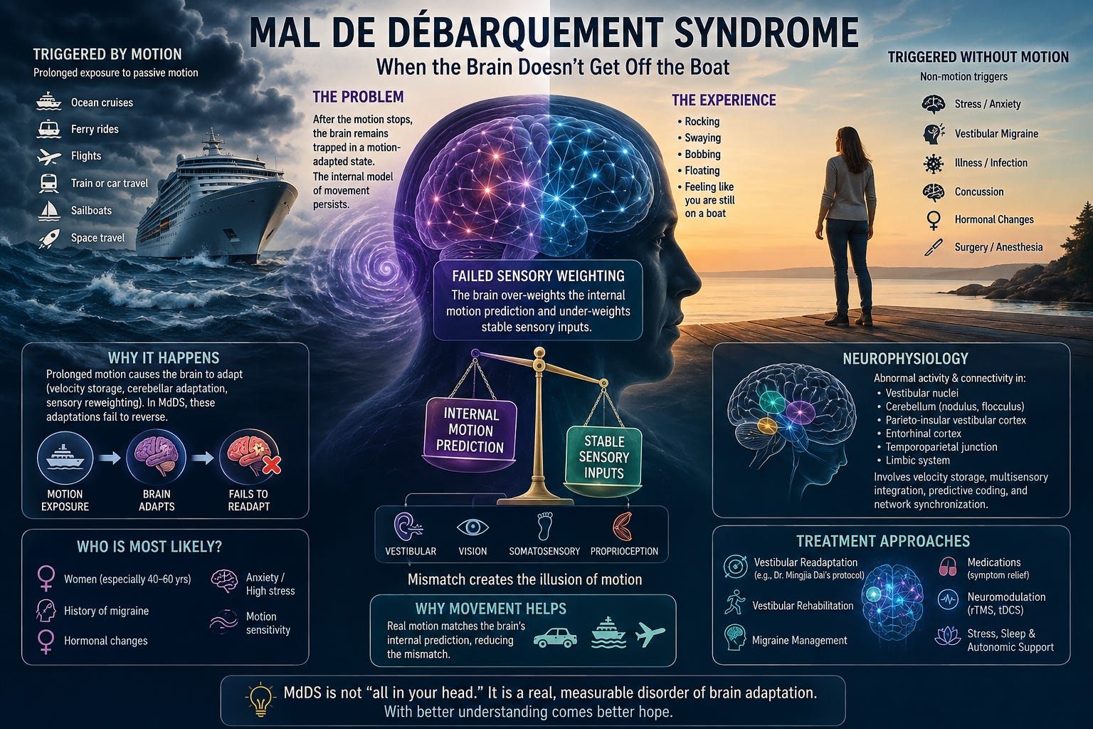
Nearly everyone has experienced the strange sensation of stepping off a boat and briefly feeling as though the ground is still rocking beneath them. For most people, this illusion disappears within minutes or hours as the brain recalibrates to stable ground. In a small percentage of individuals, however, this adaptation never fully resets. Days become weeks, weeks become months, and sometimes years. They continue to feel as if they are constantly rocking, swaying, bobbing, or floating despite standing perfectly still. This condition is known as Mal de Débarquement Syndrome (MdDS), which translates from French as “sickness of disembarkation.”
Unlike many vestibular disorders, MdDS is not caused by damaged inner ears, spinning vertigo, or obvious structural abnormalities on MRI scans. Instead, it appears to represent a disorder of persistent brain adaptation. The nervous system becomes trapped in a motion-adapted state long after the motion stimulus has disappeared. Understanding MdDS requires understanding how the brain normally adapts to movement—and how that remarkable ability for neuroplasticity can occasionally become maladaptive.
Every second of every day, the brain integrates information from multiple sensory systems to determine whether the body is moving or stationary. The vestibular organs detect angular and linear acceleration. Vision detects optic flow and environmental motion. The neck provides proprioceptive information regarding head position relative to the trunk. Somatosensory receptors in the feet and joints continuously monitor body position relative to gravity. Normally, these systems constantly recalibrate one another.
When someone spends several days on a cruise ship, the vestibular system is exposed to continuous low-frequency oscillatory motion. Initially this feels uncomfortable because the nervous system is not expecting these repetitive movements. After several hours to days, however, the cerebellum and vestibular nuclei gradually adapt. The rocking motion begins to feel normal.
This adaptation is actually beneficial. Without it, people would remain seasick throughout the voyage. The problem occurs when the brain fails to reverse this adaptation once stable ground is restored.
Patients with MdDS commonly describe feeling as though they are:
Walking on a floating dock
Standing on a trampoline
Bobbing in ocean waves
Rocking back and forth
Floating on water
Swaying while standing
Importantly, these sensations are internal perceptions rather than visible body movements. Objective balance testing may appear relatively normal, while the patient experiences overwhelming sensations of instability.
Unlike benign paroxysmal positional vertigo (BPPV), the room is not spinning. Unlike vestibular neuritis, there is usually no acute unilateral vestibular injury. Unlike cerebellar degeneration, neurological examination is frequently surprisingly normal. Instead, the brain continuously predicts motion that no longer exists.
The classic form of MdDS develops after prolonged passive motion exposure.
Common triggers include:
Ocean cruises
Sailboats
Long-distance ferry rides
Commercial flights
Helicopter travel
Long train rides
Extended automobile travel
Sleeping on watercraft
Space flight
One fascinating clinical feature is that symptoms often improve while riding in a moving vehicle again.
Many patients report:
“I feel almost normal while driving.”
This paradox provides one of the strongest clues regarding the underlying mechanism. The brain remains calibrated for continuous oscillatory motion. When real motion returns, the mismatch disappears temporarily.
Not every patient develops MdDS after travel.
Increasing evidence suggests that identical symptoms may develop following events such as:
Major psychological stress
Vestibular migraine
Viral illnesses
Concussion
Surgery
Hormonal transitions
Pregnancy
Menopause
Significant sleep deprivation
General anesthesia
Certain vestibular disorders
These patients experience identical rocking sensations despite no preceding boat ride or prolonged travel. Some researchers believe these patients possess similar central network dysfunction but require different initiating triggers.
Modern neuroimaging has dramatically changed our understanding of this disorder. Rather than representing a disease of the inner ear, MdDS appears to involve abnormal communication between multiple brain regions responsible for spatial orientation, prediction, and multisensory integration.
Areas implicated include:
Vestibular nuclei
Cerebellar nodulus and uvula
Flocculus
Fastigial nucleus
Parieto-insular vestibular cortex
Posterior insula
Temporoparietal junction
Entorhinal cortex
Hippocampus
Medial superior temporal cortex
Visual motion processing regions
Default mode network
Salience network
Functional MRI studies have demonstrated abnormal hypermetabolism within the entorhinal cortex and amygdala along with altered functional connectivity between vestibular, visual, cerebellar, and limbic regions. These findings suggest that MdDS is not simply a vestibular disorder but rather a disorder of persistent network synchronization involving spatial memory, motion perception, and predictive coding.
One of the most compelling hypotheses involves the vestibular velocity storage mechanism. Velocity storage is a neural network primarily involving the vestibular nuclei and cerebellar nodulus that prolongs vestibular signals beyond the brief responses generated by the semicircular canals. It helps stabilize vision during sustained rotation, contributes to perception of self-motion, and assists with orientation relative to gravity.
During prolonged oscillatory motion, this system adapts continuously. Normally, once motion stops, velocity storage gradually recalibrates. In MdDS, researchers believe this recalibration may fail. Instead of returning to baseline, the oscillatory neural activity persists indefinitely. The brain continues behaving as though it remains aboard a moving vessel.
Another leading theory suggests that MdDS represents abnormal synchronization of oscillating neural networks. The brain naturally contains rhythmic activity across numerous frequencies. Continuous boat motion provides rhythmic sensory input over hours or days.
For most individuals, these oscillations disappear after returning to land. In MdDS, however, the oscillatory networks may become abnormally entrained, remaining synchronized long after external motion has ceased. This persistent oscillation may explain why patients perceive rhythmic rocking even while completely stationary.
One of the most useful ways to understand Mal de Débarquement Syndrome (MdDS) is through the concept of sensory weighting. The brain is constantly deciding which sensory system should be trusted the most at any given moment. Rather than simply averaging information from the vestibular system, vision, proprioception, and somatosensation, the central nervous system dynamically adjusts the “weight” assigned to each source of information based on which one is most reliable. This process occurs largely within the vestibular nuclei, cerebellum, thalamus, parietal cortex, insula, temporoparietal junction, and other multisensory integration centers.
Imagine standing on the deck of a cruise ship. The vestibular organs continuously detect rolling, pitching, and heaving of the boat. The pressure receptors in your feet also move constantly because the deck is shifting beneath you. Even your visual world may move as the horizon rises and falls.
Initially these sensory signals conflict with your internal expectation of standing on stable ground, producing motion sickness in susceptible individuals. Over several hours or days, however, the brain performs an elegant adaptive process.
It begins to downweight the significance of these repetitive vestibular oscillations because they are now expected and predictable. Simultaneously, the cerebellum recalibrates the vestibulo-ocular reflex (VOR), the velocity storage mechanism adapts to continuous oscillation, and predictive models within the cortex begin anticipating rhythmic movement rather than treating it as an unexpected event.
This is healthy neuroplasticity. The nervous system has learned that the world itself is moving.
Once the individual steps back onto solid ground, the brain must perform the reverse adaptation. Now the vestibular system is relatively quiet. The feet provide stable pressure. Vision becomes stationary.
The cerebellum should immediately begin increasing the weighting of these stable sensory inputs while suppressing the previously learned oscillatory predictions. For most people this occurs within minutes to hours. The internal model of motion is discarded and replaced with one representing stable ground.
In MdDS, this second adaptation never fully occurs. Instead, the nervous system continues assigning excessive weighting to the internal prediction that the body is moving. The brain has learned an oscillatory state but fails to abandon it.
This means the brain’s predictive model continues telling higher cortical centers:
“You are still on a moving boat.”
The sensory information arriving from the feet, muscles, joints, and visual system all indicate that the body is standing still, yet the internally generated motion prediction continues to dominate perception. The result is a chronic sensory mismatch. Patients genuinely perceive rocking despite every external sensory cue indicating stability.
Modern neuroscience increasingly recognizes that perception is not simply driven by incoming sensory information.
Instead, perception represents a combination of:
Incoming sensory information (bottom-up processing)
Internal predictions (top-down processing)
Normally these continuously update one another. In MdDS, the top-down prediction becomes excessively strong. Rather than allowing new sensory evidence to update the model, the nervous system continues interpreting stable sensory input through the expectation of motion. The patient therefore experiences persistent rocking because the brain predicts rocking.
One way to conceptualize MdDS is that the nervous system has become “stuck” weighting an old sensory memory. The oscillatory vestibular adaptation that was appropriate aboard the ship remains highly weighted even after returning home.
Meanwhile, the reliable information coming from:
the plantar mechanoreceptors,
proprioceptors,
otolith organs,
visual motion system,
and cervical proprioceptors,
fails to regain its normal dominance.
The brain behaves as though the previous internal model remains more trustworthy than the current sensory environment.
One of the most remarkable clinical observations in MdDS strongly supports this theory.
Many patients report dramatic symptom improvement while:
driving a car,
riding in a train,
flying,
or returning to a boat.
This initially seems paradoxical. However, it makes perfect physiological sense. Their brain is still heavily weighting continuous motion. Once actual movement resumes, the incoming vestibular signals finally match the brain’s internal prediction. The sensory mismatch disappears. The nervous system no longer experiences conflict. Ironically, movement temporarily normalizes perception because the external world once again matches the maladapted internal model.
The cerebellum is central to sensory weighting. It continuously evaluates sensory prediction errors and determines how much trust should be assigned to each sensory modality. Normally, repeated prediction errors drive recalibration.
In MdDS, one hypothesis suggests that cerebellar adaptive mechanisms become locked into a persistent oscillatory state. Instead of recognizing that stable ground has returned, cerebellar learning continues reinforcing the previous adaptation. Rather than facilitating normal sensory reweighting, the cerebellum may inadvertently stabilize the maladaptive state.
The vestibular velocity storage mechanism further illustrates this concept. Velocity storage extends vestibular signals beyond the duration of semicircular canal activation, helping stabilize gaze and improve perception of sustained motion. During prolonged travel, velocity storage appropriately adapts to repetitive oscillatory motion.
Normally, once travel ends, velocity storage should gradually return to baseline. In MdDS, this recalibration appears incomplete. The velocity storage network continues generating an oscillatory expectation that becomes heavily weighted within the overall sensory integration process.
This framework explains why standard vestibular rehabilitation often produces inconsistent results. Traditional vestibular therapy frequently assumes the problem lies within weakened vestibular reflexes or peripheral vestibular loss. In MdDS, the issue is often not reduced vestibular function but incorrect sensory weighting. The nervous system is weighting the wrong information.
The therapeutic challenge therefore becomes convincing the brain to trust stable sensory information again while reducing reliance on the maladaptive internal prediction. This is precisely why treatments such as Dr. Mingjia Dai’s optokinetic readaptation protocol are thought to work. Rather than strengthening vestibular function, they attempt to recalibrate how the brain weights visual and vestibular inputs, reset the velocity storage mechanism, and restore an appropriate internal model of self-motion.
Ultimately, MdDS can be viewed as a disorder of persistent maladaptive sensory weighting. The brain successfully learned how to function in a moving environment but failed to learn that the motion had ended. As a result, an outdated internal model continues to dominate perception, overriding accurate sensory information from the vestibular system, vision, proprioception, and somatosensory receptors. The nervous system is not hallucinating motion—it is trusting the wrong sensory prediction. This perspective not only explains the characteristic rocking sensation but also provides a physiological framework for developing therapies that restore appropriate multisensory integration and adaptive neuroplasticity.
MdDS affects women far more frequently than men. Approximately 80–90% of diagnosed patients are female. The most common age of onset falls between 40 and 60 years.
Potential risk factors include:
Female sex
Perimenopause
Menopause
History of migraine
Motion sensitivity
Anxiety disorders
Hormonal fluctuations
Previous vestibular disorders
High sensory sensitivity
Repeated prolonged travel exposure
Not everyone exposed to long cruises develops MdDS, indicating that individual susceptibility likely depends upon underlying neurobiology rather than exposure alone.
Although no single cause has been identified, several biological predispositions have been proposed.
The overwhelming female predominance strongly suggests hormonal involvement. Many women report onset during menopause, pregnancy, postpartum periods, or while taking hormonal medications. Estrogen significantly influences vestibular nuclei, cerebellar plasticity, serotonin systems, and synaptic excitability, making hormonal changes a plausible contributor.
Vestibular migraine appears considerably more common among patients with MdDS. Shared abnormalities involving sensory gain, cortical excitability, thalamocortical processing, and multisensory integration may predispose certain individuals to persistent motion adaptation.
Anxiety does not appear to cause MdDS. Rather, persistent vestibular mismatch likely activates limbic networks, increasing anxiety, which then further amplifies sensory vigilance and symptom severity. This creates a self-perpetuating cycle.
Ultimately, MdDS appears to represent maladaptive neuroplasticity. The nervous system successfully learned motion adaptation—but failed to unlearn it.
Among the pioneers in MdDS research, Dr. Mingjia Dai has made one of the most significant contributions to treatment.
Working at the Icahn School of Medicine at Mount Sinai, Dr. Dai proposed that MdDS results from maladaptation of the vestibulo-ocular reflex (VOR) and the velocity storage mechanism rather than irreversible brain damage. Building on this hypothesis, his group developed a treatment protocol designed to “de-adapt” the vestibular system.
During treatment, patients are exposed to a full-field moving visual stimulus while the clinician performs carefully timed, low-frequency passive head oscillations. The direction and frequency of the head movements are individualized based on the patient’s pattern of rocking or swaying and findings on vestibular examination. The goal is to recalibrate velocity storage and restore normal integration between vestibular and visual inputs.
Clinical studies from Dr. Dai’s group have shown meaningful improvement in many patients, particularly those with motion-triggered MdDS, although not everyone responds and some require repeat treatments. Even so, this protocol remains one of the few interventions developed specifically for the underlying physiology of MdDS rather than simply treating associated symptoms.
No universally effective treatment exists, but several therapies may benefit selected patients.
Dr. Dai’s optokinetic/VOR readaptation protocol currently has the strongest physiological rationale and the best published outcomes for motion-triggered MdDS.
Traditional vestibular rehabilitation often produces mixed results because MdDS is not primarily a peripheral vestibular deficit. However, individualized rehabilitation that emphasizes visual dependence, postural control, habituation, and multisensory integration may help some patients, particularly when migraine, deconditioning, or persistent postural perceptual dizziness (PPPD) coexist.
For patients with overlapping vestibular migraine, migraine-directed therapy—including lifestyle modification and preventive medications—may reduce symptom burden.
Some patients experience partial symptom relief with medications such as benzodiazepines or selective serotonin reuptake inhibitors (SSRIs), although these agents generally reduce symptoms rather than correct the underlying maladaptive neural state.
Repetitive transcranial magnetic stimulation (rTMS), transcranial direct current stimulation (tDCS), and other forms of noninvasive brain stimulation are under investigation. Early studies suggest they may modulate dysfunctional cortical networks in selected patients, but optimal stimulation targets and protocols remain uncertain.
Because stress, sleep deprivation, migraine, and autonomic dysfunction frequently exacerbate symptoms, optimizing sleep quality, managing stress, and addressing coexisting dysautonomia may improve overall function even if they do not directly reverse MdDS.
Mal de Débarquement Syndrome illustrates one of the most fascinating principles in neuroscience: the very neuroplasticity that normally allows the brain to adapt to a changing environment can occasionally become trapped in an adaptive state that is no longer appropriate. The nervous system learns the rhythm of continuous motion but fails to relinquish it once the external stimulus is gone.
Rather than viewing MdDS as a mysterious dizziness disorder, it is more accurately understood as a disorder of persistent central adaptation involving vestibular processing, cerebellar learning, multisensory integration, predictive coding, and large-scale brain network synchronization. As research continues to uncover the mechanisms behind velocity storage, oscillatory network dynamics, and maladaptive plasticity, new opportunities are emerging to restore the brain’s ability to recalibrate itself. For patients living with the relentless sensation of rocking on solid ground, these advances offer growing hope that the nervous system can once again learn that the journey has truly ended.
Cha, Y.-H. (2015). Mal de debarquement. Seminars in Neurology, 35(5), 525–530.
Cha, Y.-H., Brodsky, J., Ishiyama, G., Sabatti, C., & Baloh, R. W. (2008). Clinical features and associated syndromes of mal de debarquement. Journal of Neurology, 255(7), 1038–1044.
Cha, Y.-H., Cui, Y., & Baloh, R. W. (2018). Comprehensive clinical profile of mal de debarquement syndrome. Frontiers in Neurology, 9, 261.
Dai, M., Cohen, B., Smouha, E., & Cho, C. (2014). Readaptation of the vestibulo-ocular reflex relieves the mal de debarquement syndrome. Frontiers in Neurology, 5, 124.
Hain, T. C., Cherchi, M., & Yacovino, D. A. (2020). Mal de debarquement syndrome. In Handbook of Clinical Neurology (Vol. 174, pp. 391–407). Elsevier.
Mucci, V., Canceri, J. M., Brown, R., Dai, M., Yakushin, S. B., Watson, S. R. D., & Cha, Y.-H. (2018). Mal de debarquement syndrome: A survey on subtypes, misdiagnoses, onset and associated psychological features. Journal of Neurology, 265(2), 486–499.
Mucci, V., Canceri, J. M., Brown, R., Dai, M., Yakushin, S. B., Watson, S. R. D., & Cha, Y.-H. (2020). Overview of the pathophysiology and management of mal de debarquement syndrome. Frontiers in Neurology, 11, 102.
Nigmatullina, Y., Hellyer, P. J., Nachev, P., Sharp, D. J., Seemungal, B. M., & Cha, Y.-H. (2017). Functional neuroimaging of mal de debarquement syndrome: Neural activity and connectivity alterations. European Journal of Neurology, 24(11), 1362–1368.
Van Ombergen, A., Van Rompaey, V., Maes, L. K., Van de Heyning, P. H., & Wuyts, F. L. (2016). Mal de debarquement syndrome: A systematic review. Journal of Neurology, 263(5), 843–854.
Yakushin, S. B., Dai, M., & Cohen, B. (2020). The role of velocity storage in the pathophysiology and treatment of mal de debarquement syndrome. Frontiers in Neurology, 11, 601.
No posts
Comments
Nothing yet. Say the first thing.
Sign in to join the conversation.