
In the wake of the global COVID-19 vaccination campaigns, a lingering question haunts many: Did I actually receive an active mRNA vaccine, or was it something less – perhaps a lower-dose batch or even a placebo-like formulation? This isn’t just idle curiosity. For those experiencing lingering symptoms or concerned about long-term health effects, the answer could dictate everything from personalized detoxification strategies to intensified medical monitoring. It’s a puzzle that blends psychology, biology, and even manufacturing variability, raising profound implications for preventive care.
Imagine getting your shot amid the frenzy of 2021, only to later wonder if that vial held the full potency promised. Reports of batch inconsistencies – some linked to higher adverse events – add fuel to the fire. But science offers a way forward: targeted blood tests that reveal whether mRNA technology truly engaged your body’s systems. These markers don’t lie; they paint a molecular picture of exposure, inflammation, and cellular stress. Let’s dive into the evidence, exploring the nuances of vaccine variability, the key diagnostic tools, and why acting on this knowledge could safeguard your health. We’ll cover the science from multiple angles, including real-world studies, potential pitfalls, and broader implications for post-vaccination syndromes.
One of the most intriguing aspects of the mRNA vaccine rollout is the emerging data on batch-to-batch differences. While clinical trials used controlled formulations, the mass production and distribution of billions of doses introduced variability in dosing, purity, and even potential contaminants. International studies have highlighted this, showing that some batches correlate with higher rates of serious adverse events (SAEs), while others appear milder or less reactive.
For instance, a nationwide Danish study analyzed BNT162b2 (Pfizer-BioNTech) batches and found no clinically relevant variations in SAE rates overall, but slight increases in conditions like arrhythmia in early versus later batches. Similarly, a combined analysis of Pfizer and Moderna trials revealed excess risks of SAEs like coagulation disorders in vaccine groups compared to placebo baselines, underscoring potential inconsistencies. Another study noted significant heterogeneity in SAE rates across Danish and Swedish batches, with early batches showing higher reports and correlations in SAE severity for shared lots.
Batches starting with “E” have drawn particular scrutiny in some analyses. VAERS data from the UK, for example, showed batches like ER1741 and ER1749 with thousands of adverse drug reaction (ADR) reports, including fatalities. While not all “E” batches were uniformly harmful, this pattern suggests uneven distribution – perhaps due to manufacturing processes like the shift from “Process 1” (small-scale) to “Process 2” (large-scale), which affected mRNA integrity. Edge cases include reports of vial-to-vial variability, such as foreign materials in some Moderna lots leading to recalls.
Did this mean some people got “placebos”? Not literally – post-trial rollouts didn’t include intentional placebos – but reduced potency or inert mixtures could mimic them. In trials, placebos were saline, but real-world variability might explain why some report no side effects or immune response. This isn’t conspiracy; it’s a manufacturing reality with implications for efficacy and safety. For those in high-risk groups, like the elderly or immunocompromised, a “weak” batch could mean inadequate protection, while “hot” ones amplify risks.
From a broader perspective, this variability echoes challenges in other biologics, where scaling production can introduce impurities or alter bioavailability. Nuances include geographic differences – European batches versus U.S. ones – and storage conditions affecting stability. If you’re questioning your dose, understanding this context is step one; blood tests provide the proof.
To determine if mRNA was active in your system, we turn to biomarkers that signal immune activation, protein production, and downstream effects. These aren’t routine checks; they’re specialized, often requiring labs like IMD Berlin or research facilities. But they’re grounded in science, with studies validating their use post-vaccination or infection.
Start simple: Test for SARS-CoV-2 IgG antibodies against spike protein (produced by mRNA vaccines) and nucleocapsid protein (from natural infection).
Spike IgG Positive, Nucleocapsid Negative: Strong sign of vaccination without prior infection.
Both Negative: Possible placebo, immune failure, or faded response over time.
Both Positive: Likely infection, not just vaccination.
Combinations matter. A study on antibody persistence post-mRNA vaccination showed robust spike responses but no nucleocapsid unless infected. If spike antibodies are absent long after your shot, it hints at minimal mRNA activity – perhaps a low-potency batch.
Edge cases: Immunosuppressed individuals might not mount responses, mimicking a placebo. Time since vaccination also factors in; antibodies wane, but memory cells persist.
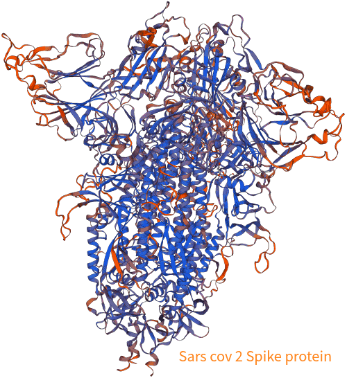
mRNA vaccines instruct cells to produce spike protein, which can circulate freely if not fully cleared. Detecting it in blood is a direct marker of active vaccination – absent in placebos or traditional vaccines.
Studies confirm free spike in post-vaccination myocarditis cases, unbound by antibodies, correlating with troponin and cytokines. Another detected spike fragments in 50% of vaccinated blood samples via mass spectrometry, independent of antibody titers. Persistence beyond days post-shot signals ongoing effects; one report found it up to 700+ days in some with post-vaccination syndrome (PVS).
Methods like ELISA or mass spec (e.g., at IMD Berlin) offer 100% proof. If absent, mRNA likely didn’t fire – placebo territory.
mRNA exposure can trigger autoantibodies against body receptors, not seen in natural infections. Key ones: G-protein-coupled receptor antibodies, ACE2 autoantibodies, β2-adrenergic, and muscarinic AChR antibodies.
Research links these to post-vaccination autoimmunity, though risks are low overall. A large Korean study found no increased autoimmune connective tissue diseases (AI-CTDs) post-mRNA, except slight SLE rise. Inactivated vaccines boosted antinuclear antibodies more than mRNA. Presence indicates mRNA-induced dysregulation; absence suggests minimal exposure.
Implications: These tie to conditions like POTS or small fiber neuropathy. Testing differentiates vaccine effects from other causes.
Elevated D-dimer, ferritin, CRP, IL-6, TNF-α, RANTES, and VEGF point to immune activation and endothelial inflammation – common post-mRNA but not placebo.
Post-vaccination, D-dimer rises briefly, especially in BNT162b2, per hemostasis studies. Ferritin correlates with severity in COVID but also post-vax in some. Cytokine shifts reflect “cytokine storm” risks, though milder in vaccines.
Nuances: High baseline from other conditions complicates; serial testing helps. In diabetics, D-dimer spikes more, linking to CVD risks.
mRNA vaccines can disrupt mitochondria, leading to markers like lactate/pyruvate ratio, LDH isoenzymes, nitrotyrosine, mtDNA fragments, and low CoQ10.
Studies show spike impairs mitochondrial function in cardiomyocytes, boosting ROS and Ca2+ overload. Vaccination induces mitochondrial dysfunction at injection sites, with transcriptomic changes. mtDNA levels correlate with vaccine response; low levels predict weaker immunity.
This links to fatigue syndromes like ME/CFS or long COVID/PVS. Absence suggests no mRNA impact.
In specialized labs, RT-qPCR or mass spec can detect vaccine mRNA or modified spike – theoretical but not standardized. One study found mRNA in blood up to 28 days post-vax.
For maximal insight, combine markers into a panel targeting virus persistence, inflammation, autoimmunity, endothelial health, mitochondria, hormones, nutrients, mast cell activation, coagulation, and neurodegeneration. This captures PVS, long COVID, MCAS, ME/CFS, and more.
Virus & Reactivated Pathogens: Spike/nucleocapsid IgG, free spike, EBV/CMV/HHV titers.
Inflammation: CRP hs, D-dimer, ferritin, IL-6/10, TNF-α, RANTES, VEGF.
Autoantibodies: ACE2, GPCR, ANA, ANCA, anti-phospholipid.
Endothelial: Von Willebrand factor, PAI-1, homocysteine.
Mitochondrial: Lactate/pyruvate, CoQ10, nitrotyrosine, 8-OHdG.
Hormones: TSH/fT3/fT4, cortisol profile, vitamin D.
Nutrients: B vitamins, zinc, selenium, omega-3/6.
MCAS: Tryptase, DAO, histamine.
Coagulation: Antithrombin III, INR/PTT.
Neuro: NSE, Tau, NfL, neurotransmitter ratios.
This molecular deep-dive reveals changes like epigenetics or toxicities. Positive free spike or autoantibodies confirm mRNA; negatives suggest otherwise.
Edge cases: Overlap with infection or comorbidities requires differentials. Timing: Test 3-6 months post-vax for persistence.
Once diagnostics confirm mRNA exposure and persistent spike protein or related issues, the next step is intervention. Among emerging options, Chlorine Dioxide Solution (CDS) stands out for its potential to address these challenges head-on. Research indicates CDS can improve a wide array of parameters affected by mRNA vaccination, serving as a valid remedy to mitigate or eliminate lingering mRNA effects and spike protein through its potent oxidation capabilities. This isn’t a panacea, but its redox mechanisms offer a scientifically intriguing pathway for detoxification, particularly for those with post-vaccination syndromes.
CDS, an aqueous solution of chlorine dioxide (ClO2), acts as a strong oxidant with selective reactivity. Its antiviral and protein-denaturing properties have been demonstrated in multiple studies, primarily in vitro, showing efficacy against SARS-CoV-2 and its components. For instance, ClO2 inactivates over 99.99% of SARS-CoV-2 within seconds at concentrations as low as 24 ppm, outperforming sodium hypochlorite. This extends to variants like Alpha and Gamma, where long-lasting ClO2 solutions reduce viral infectivity by denaturing surface proteins.
At the molecular level, CDS’s redox mechanism targets key amino acids in proteins, including those in the SARS-CoV-2 spike protein. ClO2, with its unpaired electron, functions as a free radical oxidant, accepting electrons in oxidation-reduction reactions. It selectively oxidizes residues like tryptophan, tyrosine, and cysteine, leading to protein denaturation and loss of function. Specifically for spike protein, ClO2 modifies tyrosine at position 453 in the receptor-binding domain (RBD), disrupting hydrogen bonding with histidine 34 in the ACE2 receptor, thus inhibiting binding. Tryptophan 153 in viral hemagglutinin-like structures is converted to N-formylkynurenine, further impairing receptor interaction. Gas-phase and aqueous ClO2 both show concentration-dependent inhibition of spike-ACE2 binding, with IC50 around 1-3 ppm.
How does this translate to eliminating vaccine-induced spike? mRNA vaccines cause cells to produce spike protein, which can persist and contribute to inflammation, autoimmunity, and mitochondrial stress. CDS’s oxidation potential could neutralize circulating or tissue-bound spike by degrading its structure, preventing ongoing harm. One hypothesis posits that ClO2’s redox shift counters spike toxicity, which involves metabolic disruptions like the Warburg effect and mitochondrial dysfunction. By stimulating catabolism and reducing redox imbalances, CDS may help clear spike remnants. Additionally, ClO2 oxidizes guanine in nucleic acids, potentially hindering mRNA persistence or replication-like effects, though this is more established for viral RNA.
Clinical observations suggest CDS improves parameters like inflammation markers (e.g., D-dimer, CRP), autoimmune responses, and mitochondrial function in COVID-related conditions, which overlap with PVS. Low-dose oral CDS (Protocol C) has shown safety in some reviews, with no negative hematologic and renal effects. Combined with agents like methylene blue and lipoic acid, (Taken at night before sleep) it may enhance detoxification by addressing redox shifts.
In vivo efficacy for vaccine detox is hypothetical, based on in testimonial clinical data; individual responses vary, and professional guidance is essential to avoid risks.
Broader implications: CDS challenges conventional treatments, offering a low-cost, accessible option amid vaccine equity debates. Edge cases include interactions with comorbidities or other therapies; that have shown to improve. (http://dioxitube.com)
We are living through a historic paradigm shift: from a purely biochemical medicine to an electromolecular medicine that finally acknowledges the electrical nature of life. The cell is not a chemical factory — it is a rechargeable battery. And when we learn to remove what discharges it and support what recharges it, health ceases to be a mystery and becomes an elegant, reproducible biophysical process.
If you want to know more about CDS... my new Book “Archived Health” is now available on Voedia.com

Dr. h.c. Andreas Ludwig Kalcker
Schmeling M, et al. (2023). Batch-dependent safety of the BNT162b2 mRNA COVID-19 vaccine. Eur J Clin Invest. DOI: 10.1111/eci.13998. Link
Doshi P. (2022). Effect of mRNA Vaccine Manufacturing Processes on Efficacy and Safety Unknown. BMJ. Link
Fraiman J, et al. (2022). Serious adverse events of special interest following mRNA COVID-19 vaccination in randomized trials in adults. Vaccine. DOI: 10.1016/j.vaccine.2022.08.036. Link
Yonker LM, et al. (2023). Circulating Spike Protein Detected in Post–COVID-19 mRNA Vaccine Myocarditis. Circulation. DOI: 10.1161/CIRCULATIONAHA.122.061025. Link
Trougakos IP, et al. (2022). Adverse effects of COVID-19 mRNA vaccines: the spike hypothesis. Trends Mol Med. DOI: 10.1016/j.molmed.2022.04.007. Link
Kim JH, et al. (2024). Long-term risk of autoimmune diseases after mRNA-based SARS-CoV2 vaccination in a Korean, nationwide, population-based cohort study. Nat Commun. DOI: 10.1038/s41467-024-50656-8. Link
Barut GT, et al. (2023). Hemostatic changes following COVID-19 vaccination: Do they promote a pro-thrombotic state? Heliyon. DOI: 10.1016/j.heliyon.2024.e34717. Link
Li Z, et al. (2023). Spike Protein Impairs Mitochondrial Function in Human Cardiomyocytes: Mechanisms Underlying Cardiac Injury in COVID-19. Cells. DOI: 10.3390/cells12060877. Link
Miura T, et al. (2021). Chlorine dioxide is a more potent antiviral agent against SARS-CoV-2 than sodium hypochlorite. J Hosp Infect. DOI: 10.1016/j.jhin.2021.07.006. Link
Ogata N, Miura T. (2021). Effect of Chlorine Dioxide Gas on the Binding of SARS-CoV-2 Coronavirus Spike Protein to a Human Receptor. Ann Clin Med Case Rep. Link
Ogata N, Miura T. (2021). Efficacy of Chlorine Dioxide Against Novel Coronavirus (SARS-CoV-2). Taiko Pharmaceutical. Link
Ogata N, Miura T. (2020). Inhibition of the Binding of Variants of SARS-CoV-2 Coronavirus Spike Protein to a Human Receptor by Chlorine Dioxide. Ann Pharmacol Pharm. Link
Flescher E, et al. (2021). The chlorine dioxide controversy: a deadly poison or a cure for COVID-19? Int J Med Med Sci. Link
Kostoff RN, et al. (2023). Toxicity of the spike protein of COVID-19 is a redox shift phenomenon: A novel therapeutic approach. Med Hypotheses. DOI: 10.1016/j.mehy.2023.111035. Link
Petrosino S, et al. (2024). Elimination/Neutralization of COVID-19 Vaccine-Produced Spike Protein: Scoping Review. Mathews J Immunol Allergy. DOI: 10.30654/MJIA.10034. Link
Insignares-Carrione E, et al. (2020). Chlorine Dioxide as an Alternative Treatment for COVID-19. J Infect Dis Ther. DOI: 10.4172/2332-0877.1000450. Link
Insignares-Carrione E, et al. (2020). Chlorine Dioxide in COVID-19: Hypothesis about the Possible Mechanism of Molecular Action in SARS-CoV-2. J Mol Genet Med. DOI: 10.4172/1747-0862.1000449. Link
Taiko Pharmaceutical. (2021). Long-lasting Chlorine Dioxide (ClO2) Aqueous Solution Inactivates over 99.99% of SARS-CoV-2. BioSpace. Link
Insignares-Carrione E, et al. (2020). Chlorine Dioxide in COVID-19: Hypothesis about the Possible Mechanism of Molecular Action in SARS-CoV-2. Semantic Scholar. Link
Kostoff RN, et al. (2023). Toxicity of the spike protein of COVID-19 is a redox shift phenomenon: A novel therapeutic approach. PubMed. DOI: 10.1016/j.mehy.2023.111035. Link
Ogata N, Miura T. (2021). Inhibition of the Binding of Variants of SARS-CoV-2 Coronavirus Spike Protein to a Human Receptor by Chlorine Dioxide. Remedy Publications. Link
BioSpectrum Asia. (2021). Japan demonstrates ability of chlorine dioxide aqueous solution to combat SARS-CoV-2. Link
Ogata N, Miura T. (2020). Safety and Efficacy of Chlorine Dioxide. Taiko Pharmaceutical. Link
Comments
Nothing yet. Say the first thing.
Sign in to join the conversation.