
Intratumoural chlorine dioxide ablation versus surgery — starting with the dimension where surgery wins
By now, most people who follow this work have stopped asking whether the tumour dies.
They have seen the fifteen-minute photographs: four dogs, four oral or lingual tumours, photographed under general anaesthesia from the same viewpoint before and after injection, recorded before the animal had woken up. At 20,000 ppm, a working solution injected into a large tumour produces gross structural change within minutes. The colour varies between cases — black-brown, tan-brown, pale yellow-white, depending on how much blood the tumour holds — but the structural reading is identical in every case: inside a sharply demarcated boundary, the tissue no longer looks like living tissue, and the vascular appearance is gone.
So the question has moved on. The question I now get from clinicians, and from patients, is the one that follows: nothing was removed. Where does all that dead tissue go?
It is a fair question, and it deserves a straight answer rather than a reassuring one.
Here is the frame I would ask you to hold through everything below.
Every effective local treatment of cancer has to solve two entirely different problems. The first is how to kill the tumour. The second is what to do with what is left of it afterwards.
They are not the same problem and they do not have the same difficulty. The first is oncology. The second is wound management.
Surgery solves both in a single operation. It kills the tumour and carries the dead mass out of the room in a specimen pot, in one act. That is an enormous convenience, and it is the reason surgery has been the default for a century.
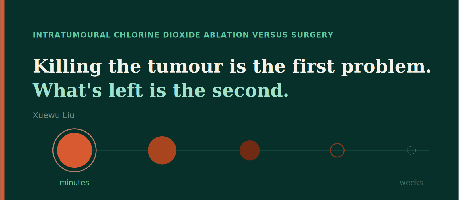
Intratumoural ablation solves them sequentially. The tumour dies in minutes. What is left of it leaves over days to weeks, and the body does most of that work.
Almost every difference between the two techniques — in both directions — follows from that single structural fact. Surgery pays for its clean exit by having to cut a path in, take a margin of healthy tissue with it, control the blood supply of everything it crosses, and then close under tension. Ablation pays for its minimal entry by leaving the second problem unfinished at the end of the procedure.
That is the trade. Everything below is an attempt to price it.
I will state this plainly, because it is the one place where surgery is straightforwardly better, and pretending otherwise would waste your time.
Ablating a large tumour creates a large volume of dead tissue inside a living body, and that volume is a burden. It has to be cleared. Clearance is not free. It can mean an open wound, a period of exudate, a route for bacteria, a risk of bleeding where the lesion sat against a large vessel, and — in an oral or pharyngeal tumour — a period where swallowing is compromised and nutritional support is needed. A surgeon who removes a 350 mL mass hands the owner a closed wound and a discharge sheet. I hand them a schedule.
How the clearance happens depends entirely on where the tumour sits, and in our experience it falls into three patterns.
Exposed tumours separate. An oral mass, a skin mass, an ulcerated breast tumour — these demarcate along a sharp line and the coagulated tissue detaches by itself, typically around day four or five. In several documented cases no debridement or excision was performed at all. In one cat with a subcutaneous mass, the tumour separated on day seven and the wound was fully epithelialised by day twenty, with hair regrowth and no scar, achieved without antibiotics and without bandaging. In a canine lingual tumour where the veterinary surgeon had recorded pre-operatively that the tumour had eroded the root of the tongue and that loss of the tongue was unavoidable, the necrotic mass separated on day four with no incision and no sutures.
Tumours that open into a cavity are expelled. A nasal mass has nowhere to slough to except out. In one Samoyed with a nasal mast cell tumour, the owner reported the dog sneezing out fragments of dark necrotic tissue around day ten.
Deep tumours are resorbed, and this is the slow one. The cell death is just as fast — the chemistry finishes in minutes — but there is no route of egress, so the body has to break the mass down and carry it away. Contour change on imaging typically takes on the order of ten days, and full resolution longer. This is the case where the burden is real and the patient simply has to carry it for a while.
There is one control worth knowing about, because it turns the burden from a fixed property of the technique into a variable the operator sets. The necrotic load per session is a dosing decision. In a 1,300 mL sarcoma in a corgi we injected 52 mL — about 4% of tumour volume — at six sites. In a 500–700 mL sarcoma in a golden retriever, 32 mL, about 5–6%. In a 350 mL oral melanoma, roughly 10%. Deliberately low fractions, delivered in stages, keep the amount of tissue dying at any one moment within what the animal and the wound can handle. The cost is that clearance takes several sessions instead of one, and that cost belongs on surgery’s side of the ledger.
But notice what has happened by this point in the sequence. Once the tumour inside the treated volume is dead, the problem that remains there is no longer cancer. It is a wound. Infection, slough, exudate, debridement, healing time — these are the oldest and best-understood problems in surgery, and they are problems every practice already knows how to solve. They are not a failure of cancer treatment. They are what success looks like before it finishes.
That distinction matters for how you read the rest of this.
This is the question I am asked most often, and it is usually asked the wrong way round — as though the volume of necrosis were the variable.
Necrosis does not create infection. Bacteria create infection. But devitalised tissue is a permissive substrate: it has no blood supply, which means neither neutrophils nor systemic antibiotics reach its interior, so where bacteria do have a route in, the inoculum required is far lower than in living tissue. The determining variable is therefore not how much necrosis there is. It is whether the necrosis is open to a colonised surface.
That splits the cases cleanly.
Closed deep necrosis rarely becomes infected. A visceral or intramuscular mass under intact skin has no route of entry. In the golden retriever with the 500–700 mL sarcoma, day one showed no acute inflammatory swelling, no oedema and no bleeding, and by day ten the mass had collapsed with the surrounding skin pale and dry rather than red and swollen. In the corgi with the 1,300 mL sarcoma, the skin at day twelve was intact — no ulceration, no discharge, no discolouration, no inflammatory swelling.
Open or contaminated necrosis carries real risk and needs a plan. An ulcerated skin tumour, anything in the mouth, anything in the nasal cavity — these sit in a colonised field from the start, and the slough phase is when the field is most exposed. This is where wound management earns its place. It is also, so far, where it has worked: the cat healed to full epithelialisation by day twenty without antibiotics and without bandaging, and the ulcerated breast tumour in a human patient, which had been actively bleeding before treatment, showed markedly reduced exudate and no secondary infection.
The practical rule that follows is the ordinary surgical one: antibiotics for documented infection, not prophylactically for the mere presence of dead tissue.
And it is worth saying the obvious about the comparator. Surgical site infection is among the commonest complications of oncological surgery, because a surgical wound is also a route in — one created deliberately, then closed over a field that has been retracted, cauterised and partially devascularised. Surgery does not avoid this problem. It makes it earlier and shorter.
While I am being honest about the ledger, there are two further entries on surgery’s side, and any oncologist will name them within thirty seconds if I do not.
A specimen. Excision produces tissue: grade, margins, lymph node status, the whole staging apparatus. Ablation produces a necrotic mass that separates onto a towel at home, and in real-world veterinary practice it does not get sent for histopathology. That is a genuine information loss. My working position is that in a treatment where the operator watches the treated volume form in real time and can simply treat again if anything remains, margin status carries less decision weight than it does when the operation cannot be repeated — but that is an argument, not a fact, and it is a gap I am not promising to close.
Maturity of evidence. Surgery is standardised across a century and thousands of centres. This technique rests on roughly 25 human cases and a companion-animal series in the low double digits. The companion-animal cohort is almost entirely end-stage and unselected: within the first ten Chinese cases, three animals died within days of treatment. My assessment is that these deaths reflect terminal disease rather than treatment toxicity — every animal observable beyond five days showed tumour necrosis and volume loss, including one that died on day ten — but that is my assessment, and a series of this size cannot settle it. Anyone comparing these two options should weigh a century of surgical outcome data against a few dozen cases and price the uncertainty accordingly.
Having conceded all of that, here is the other side. It is the longer side.
Comments
Nothing yet. Say the first thing.
Sign in to join the conversation.