
Beyond the Rigid Cage… #NeoArmory:
What do you think about a traditional interbody cage? It’s a bit like forcing an immovable object into an ever-changing environment. If you want the ideal implant, you’d probably want the elasticity of Mr. Fantastic and the strength of The Thing.
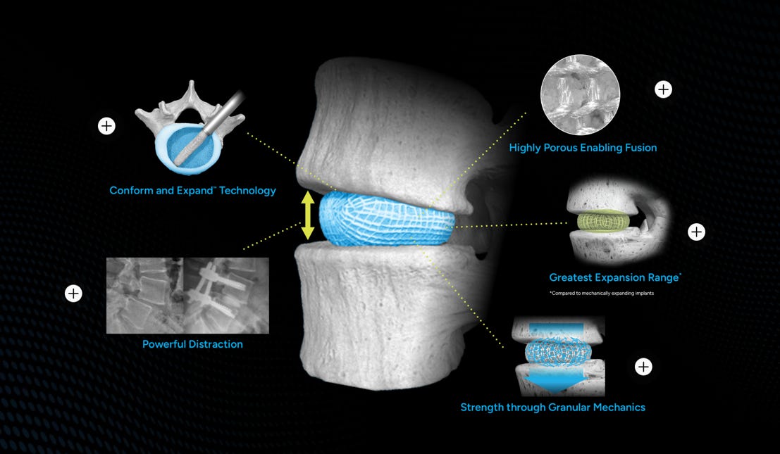
Enter Spineology’s OptiMesh® - Instead of a solid, rigid structure, this system uses an expandable, knitted PET mesh that goes in small and blooms in situ. It’s designed to “conform and expand,” filling the disc space completely and molding to the unique contours of the patient’s endplates rather than forcing them to match the cage.
The data is surprisingly solid, too. The SCOUT IDE trial reported a 99% fusion rate and 92% patient satisfaction at the two-year mark. By enabling an ultra-minimally invasive (OptiLIF) approach, it manages to provide significant distraction without the heavy-duty tissue trauma of traditional open techniques.
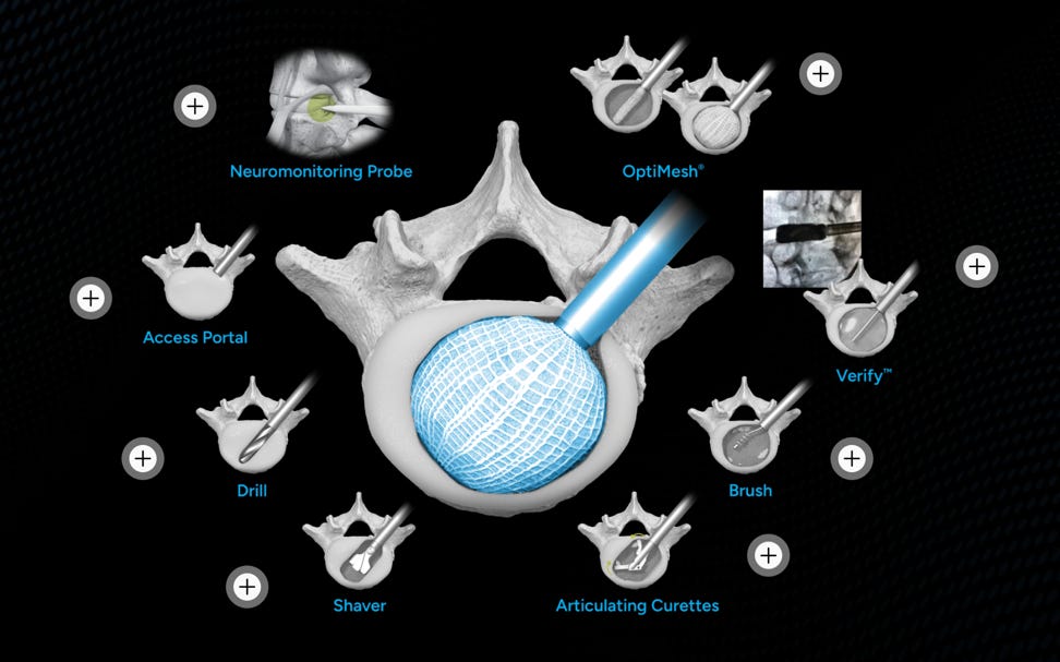
Their articulating cutters are a work of art
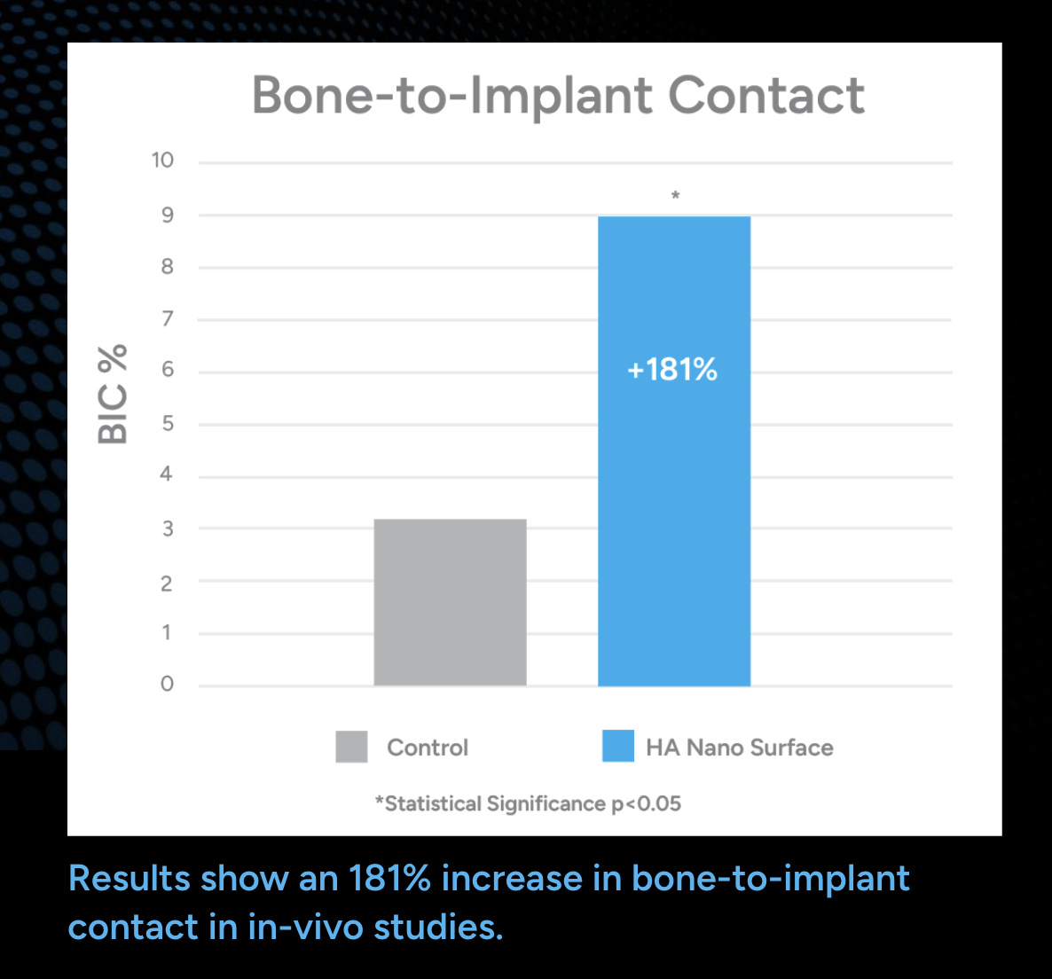
With the newer HA Nano version hitting the scene to boost osseointegration, it’s a great example of how our implants are finally getting a little more… flexible.
Victor Horseley – Father of Modern Neuro(spine)surgery #FromTheHistory:
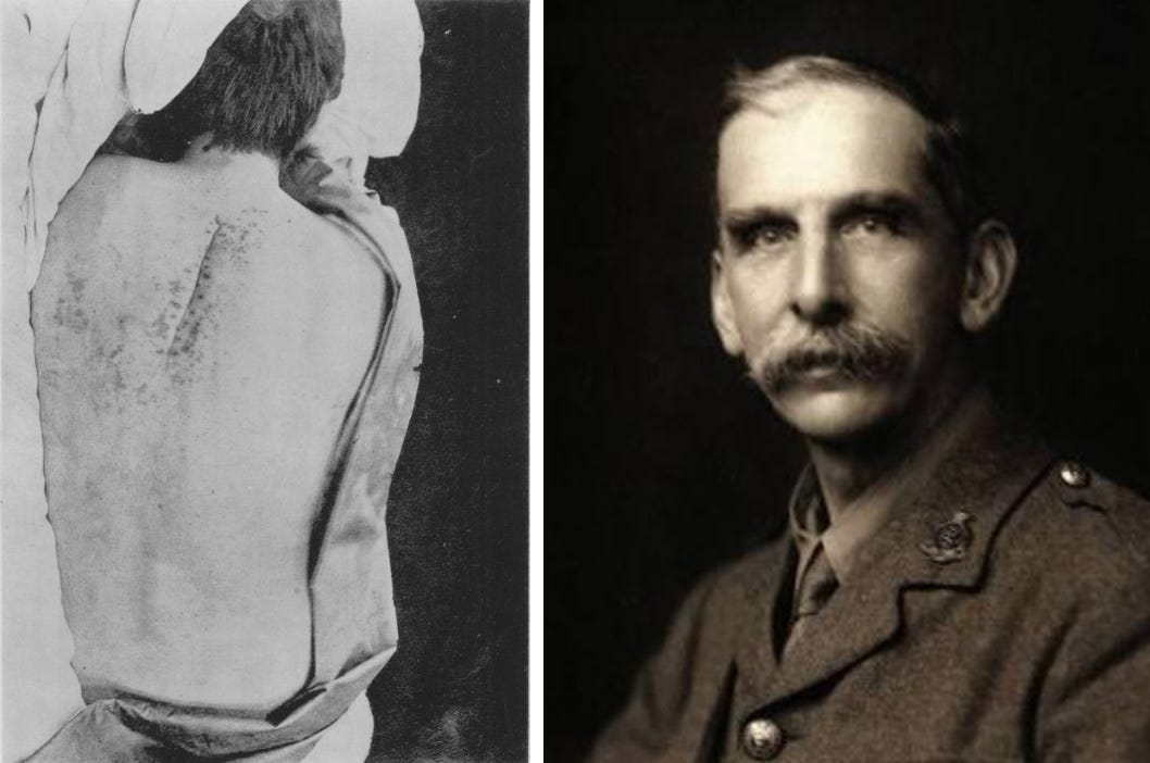
Sir Victor Horseley, born in Kensington, London in 1857 was named after Queen Victoria! He was gifted surgeon and social reformer as well. The curious case of Captain Gilby is beautifully described by Horseley in the article he published in 1888 is nothing short of a thriller story!
Captain Gilby started having back pain in 1883 and he went on to be treated by various people from different types of medicine in umpteen different treatments and none were successful for the next 3 years. By the time Horseley saw our patient G, he was almost paraplegic and bed bound. Dr. Gowers helped localize the lesion, a probable tumor compressing the spinal cord in the upper dorsal spine. After thinking through thoroughly, in discussion with the patient (who almost gave up and is ready to try any treatment, even experimental and dangerous ones to get rid of the pain and to stand on his two feet), ventured to do the unthinkable task, an elective spine surgery! He went ahead and opened up the patient’s back in semiprone position, under Ether anaesthesia and cut of 4th – 6th thoracic lamina but could not find any tumor. With his keen eye along with the precise knowledge of anatomy, he noted that the dura was pressed on the left side close to the neural canal. Then, he went on to extend the incision proximally and removed two more laminae and successfully removed that culprit causing the patient’s symptoms. All this he did in an era without imaging, good anaesthesia, positioning tools, sterile instrumentation or a sterile field and above something no one has ever even attempted. The success actually was that Captain G was able to walk within a year of the procedure and went on to live for 2 decades from surgery without the symptoms which crushed his spirit and body for 3 years.
Coming to the socialist in our surgeon, he was sent to assess the rabies vaccine developed by Louis Pasteur to Paris by the British government. He went there, validated Pasteur’s work and eventually spearheaded the mission which mandated muzzling and quarantine of dogs which eventually and effectively wiped out rabies from UK!
Turn the Ligament, Not the Facet #Surgical Pearl:
One of the biggest challenges during biportal endoscopic foraminal decompression is achieving adequate nerve root decompression without sacrificing the facet joint. A recent technical note in the Asian Spine Journal introduces an elegant solution—the Non-Facetectomy Ligamentum Flavum (LF) Turn-Down Technique.
Position the patient prone on a Wilson frame under general or spinal anesthesia, ensuring the abdomen hangs free to reduce venous congestion.
Create two small portals about one pedicle width lateral to the pedicle. The cranial portal is used for viewing, while the caudal portal serves as the working portal.
Dock on the transverse process and use a radiofrequency probe to expose the key landmarks—the transverse process, superior articular process (SAP), lateral isthmus, and most importantly, the accessory process.
Identify the accessory process. Think of it as your surgical GPS—it reliably guides you to the foramen while keeping you away from the exiting nerve root.
Detach the soft tissue from the medial side of the accessory process using a freer elevator. Since the nerve root lies deeper, this step can be performed safely.
Slide a curette beneath the SAP, keeping it in constant contact with bone to undermine the ligamentum flavum.
Rotate the curette to perform the “turn-down” maneuver, peeling the ligamentum flavum away instead of removing it piecemeal.
Expose the exiting nerve root and use it as a guide for any remaining decompression. Remove residual ligament or bone with a Kerrison punch or burr only if necessary.
Perform a discectomy if required for foraminal or extraforaminal disc herniations, followed by a ball-tip probe check to confirm the nerve root is completely free.
Preserve the facet whenever possible. The whole philosophy of this technique is to achieve adequate decompression with minimal bone removal, reducing the risk of postoperative instability while maintaining excellent visualization.
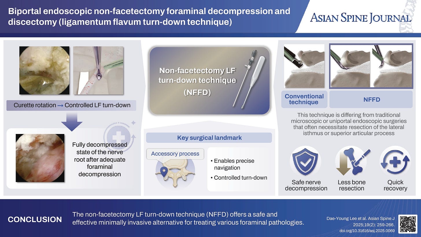
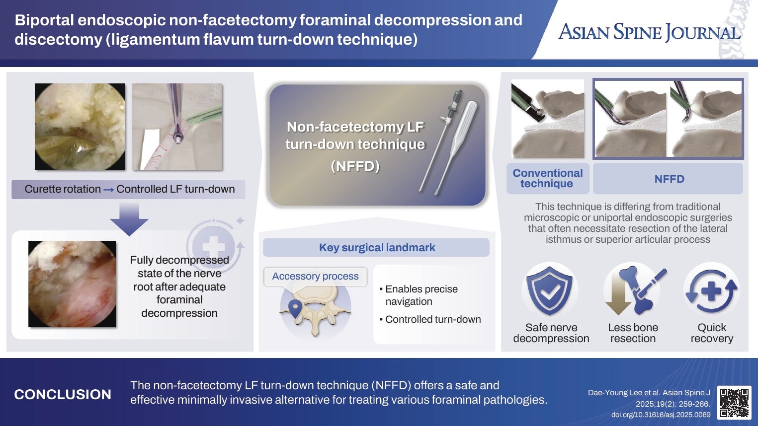
Dont hesitate to check the full paper on ASJ
Too much, or Too long — Intraoperative Hypotension isn’t as harmless as it seems!
Managing intraoperative hypotension (IOH) has been a long-standing challenge in spinal deformity surgery. For years, induced hypotension was the go-to strategy to keep the surgical field clean and blood loss in check.
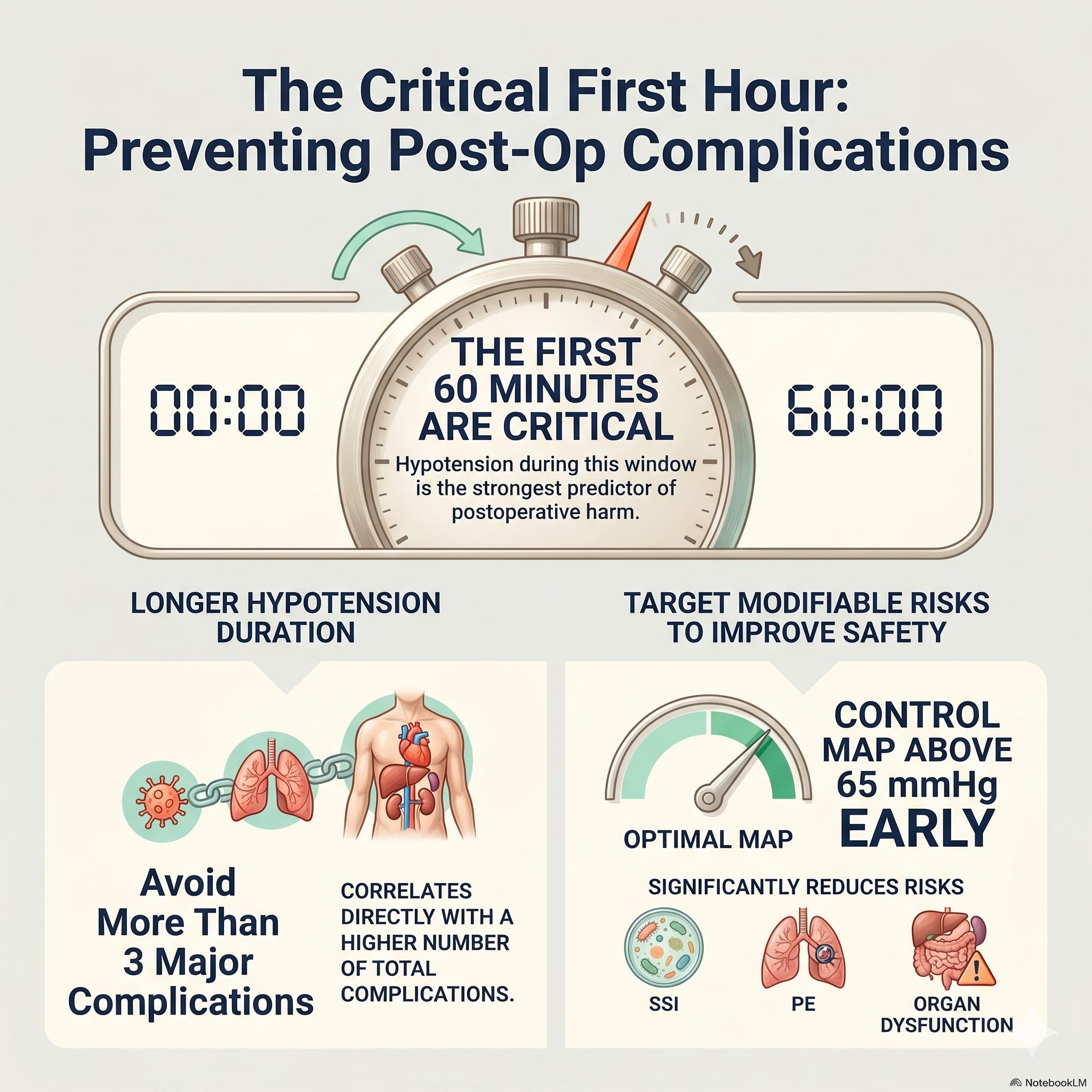
Glassman et al. looked at a retrospective cohort of 539 thoracolumbar fusion cases (>6 levels, >3 hours) and found that the longer the stretch of intraoperative hypotension (MAP <65 mmHg), the higher the risk of postoperative complications. Patients who clocked three or more complications had notably longer hypotensive episodes in the first hour of surgery and required more vasopressor support. The complications weren’t trivial either — SSI, pulmonary failure, PE, ileus, and cognitive dysfunction were all on the list.
The key takeaway? Early intraoperative hypotension is a modifiable risk factor in multilevel lumbar fusion, and one worth taking seriously. So, the next time you’re in theatre, don’t push your anaesthetist for a deeply hypotensive field — especially in that critical first hour. It may cost more than it saves.
Events to check out:
AOSSM Annual Meeting 2026 08 – 11th July 2026 Seattle, United States
35th Annual Conference of the European Society for Biomaterials – ESB 2026 07 – 11th Sept 2026 Antwerp, Belgium
46th SICOT Orthopaedic World Congress Kyoto 2025 29th – 01st Oct 2026 Kyoto, Japan
Comments
Nothing yet. Say the first thing.
Sign in to join the conversation.