
Imagine a fleet of microscopic traps in your bloodstream that silently sweep up and concentrate molecules, shielding them from elimination by the body.
Characterizing the molecules within these biological ‘black boxes’ might provide important clues about disease pathology previously unknown to science. This would be especially true if the circulating proteins and protein fragments were otherwise rapidly cleared, degraded, or too dilute to detect in standard blood tests.
A recent pre-print article from the same research group that discovered and characterized the microclots found in those with Long COVID revealed that these microscopic traps are also present in patients who developed Postural Orthostatic Tachycardia Syndrome (POTS) before the COVID-19 pandemic.
By cracking open these biological black boxes, scientists are now analyzing not just which proteins are trapped inside, but how those proteins have been physically altered, revealing disease-specific signatures that may explain symptoms despite normal blood work results.
Before we summarize the results of this manuscript, let’s first summarize what is known about microclot size, structure, and composition in those with Long COVID.
What Are Microclots and How Do They Differ from Typical Blood Clots?
Physiological clots form when a blood vessel is broken. The clot serves as a temporary scaffold that seals a breach in the vessel wall, preventing blood from leaking from the damaged artery, vein, or capillary.
However, pathophysiological (harmful) clotting occurs when clots form inside vessels, blocking blood flow and thus oxygen (and nutrient) delivery to tissues. This can cause life-threatening complications, such as a stroke, heart attack, or a pulmonary embolism if the clot travels to the lungs.
The size of a typical clot that forms inside a blood vessel varies from pea-sized to grape-sized (millimeters to a centimeter in diameter). However, since the SARS-CoV-2 pandemic, work from Dr. Etheresia Pretorius’s Lab at Stellenbosch University in South Africa has revealed that microscopic (1-200 µm on their longest axis) clots, termed microclots, are found in individuals with COVID-19 and those who developed Long COVID.
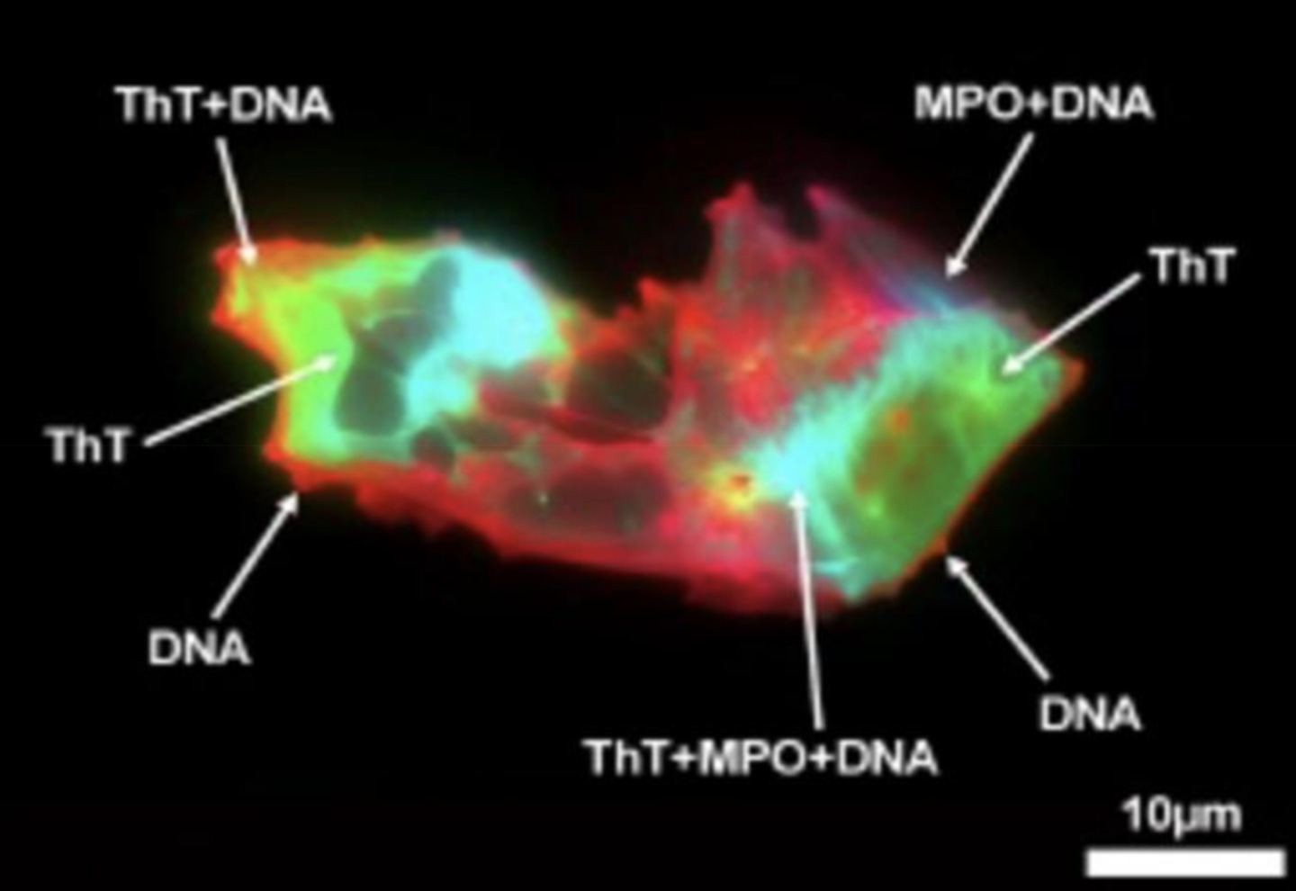
Microclots in Human Health and Disease
Microclots are not exclusive to Long COVID or COVID-19; they have also been observed in patients diagnosed with ME/CFS, Alzheimer’s, Parkinson’s, and rheumatoid arthritis. These microclots are predicted to contribute to symptoms such as fatigue, cognitive impairment, or organ damage by restricting blood flow in the body’s smallest blood vessels, the capillaries. They can promote inflammation, though their exact role in different disease contexts remains to be investigated.
Interestingly, these microclots are also found at low levels in healthy individuals, but elevated numbers (and size) of the microclots correlate with disease severity in Long COVID. Specifically, recent work from Dr. Pretorius’s Lab found that patients with long COVID have 19 times as many microclots as healthy controls.
The composition of the microclots is surprisingly different from that of typical blood clots. One key difference lies in the structure of the most abundant protein in clots, fibrin.
Microclots Are Reported To Contain Amyloid Structures
Blood clots form when a free-floating protein called fibrinogen is cut by molecular scissors into a smaller protein called fibrin. Fibrin sticks to itself, forming mesh-like webs known as clots. These clots can be easily dissolved by a process called fibrinolysis, which describes the destruction of a fibrin clot.
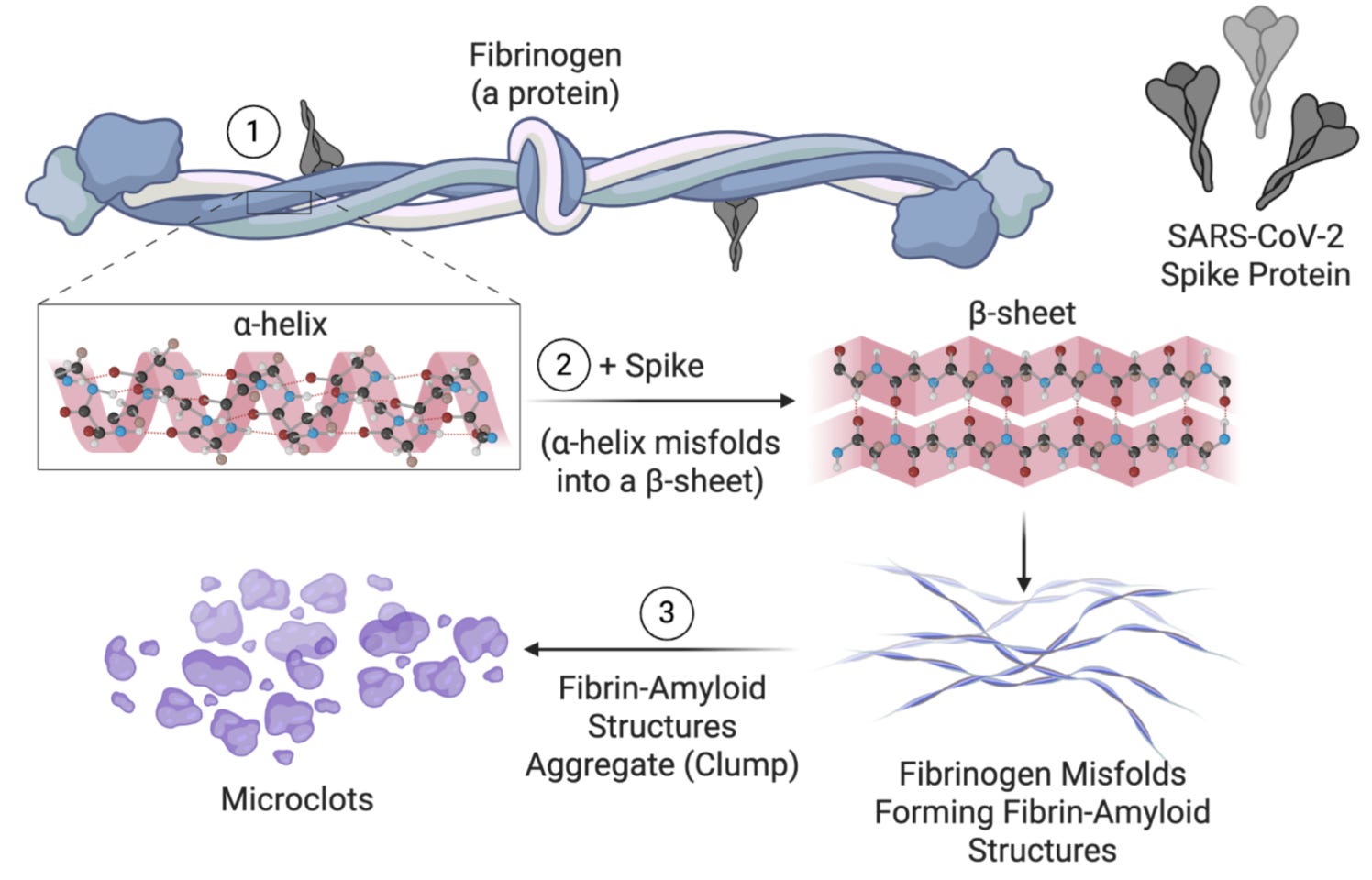
However, the fibrin in microclots is structurally different, making the clots more difficult for the body to break down naturally. An earlier 2022 paper from the Pretorius Lab describes microclots containing an amyloid form of fibrin, called amyloid-fibrin microclots, or fibrinaloids.
This type of fibrin is folded differently due to inflammatory factors, such as the SARS-CoV-2 spike protein, which has been demonstrated to physically interact with fibrinogen, creating amyloid-fibrin-like structures.
The molecular scissors that dissolve clots do not work on amyloid forms of fibrin. This makes the microclots very difficult for the body to clear.
Microclots Are Wrapped In DNA
The microclots from Long COVID patients not only contain amyloid fibrinogen, but they are also wrapped in DNA, making them even more challenging for the body to naturally break down.
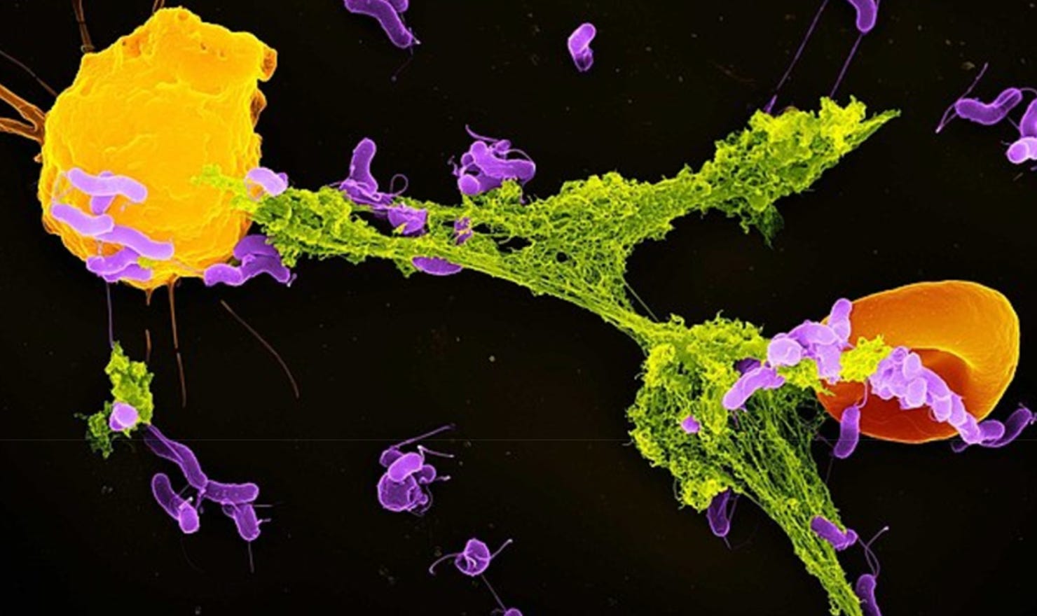
In 2023, Dr. Alain Thierry‘s team was among the first to identify elevated levels of neutrophil extracellular traps (NETs) in patients with COVID-19 and Long COVID. These NETs are webs of DNA (green) and proteins from white blood cells called neutrophils (yellow). The DNA is cast from the neutrophil like a net that traps pathogens, much like an angler casts a net to trap fish.
Microclots As Biological Traps
When a blood clot forms, it traps nearby proteins, protein fragments, and small molecules. The same logic applies to microclots.
Thus, in a recent preprint article, the same South African research group that investigated Long COVID microclots, in collaboration with POTS expert Dr. Sathish Raj, sought to assess the utility of analyzing microclot composition. Specifically, they determined what specific disease biomarkers are trapped within microclots and determined whether the entrapped components could serve as biomarkers to help clinicians identify those with:
POTS only
Long COVID only
POTS and Long COVID (LCPOTS)
They reasoned that conventional protein blood tests that measure free-floating (soluble) biomarkers frequently return normal results despite severe, debilitating symptoms in Long COVID patients and those with LCPOTS (and POTS).
Could the ‘normal’ blood test results be at least partly attributed to microclots acting as biological traps that embed biomarkers inside, making them inaccessible (insoluble) and undetectable in standard blood tests?
Comments
Nothing yet. Say the first thing.
Sign in to join the conversation.