
Practicing medicine is the systole to the diastole of research and evidence appraisal. Hospital medicine, in particular, is a fast-paced, intellectually demanding exercise in solving complex, deeply human problems—often with near-immediate feedback.
Every stretch on service teaches me something new (usually several somethings new) despite nearly two decades in practice caring for thousands of patients. Clinical presentations are endlessly variable. Knowledge evolves. General medicine is vast. There are thousands of ways to be sick enough to require hospitalization. Few careers offer that kind of breadth and pace.
This post is a potpourri of lessons from the wards: evidence-based pearls and diagnostic reflections with a few consequential misses. The goal is simple, to share what I learned with others. Tales from the Wards will be a recurring series.
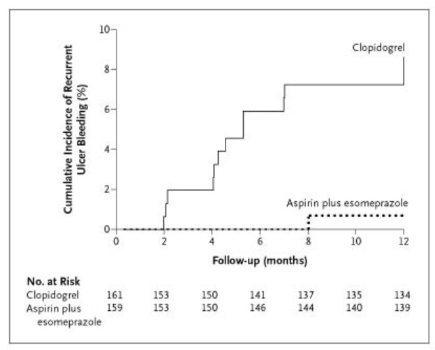
I long believed aspirin both causes peptic ulcers and increases bleeding risk, whereas clopidogrel only does the latter, and is thus safer with respect to lower the risk of an upper GI bleed. My intuition was wrong. A colleague recently pointed me back to a 2005 NEJM trial showing that continuation of aspirin (with a proton pump inhibitor) was safer than switching to clopidogrel. (see the Kaplan Meier curve below)
The key caveat is that the aspirin group received a PPI. Whether clopidogrel plus a PPI would perform similarly remains less clear. But the practical takeaway is straightforward: if a patient has a strong indication for aspirin, add a PPI rather than reflexively switching agents.
Radiation necrosis after treatment for brain metastases is something I’ve encountered infrequently. Most patients improve with time and corticosteroids. This patient did not. After a week of maximal steroid dosing, my patient remained functionally hemiplegic.
We gave a single dose of bevacizumab. The next day, he could grip my fingers, flex his elbow, and lift his leg slightly off the bed. While not a home run by any means, his progress was at least a single. And it’s far too early to judge. Time will tell.
Mechanistically, the rationale is strong. VEGF drives vascular permeability, leading to capillary leak and vasogenic edema. Bevacizumab, a VEGF inhibitor, targets that pathway. Observational data support benefit, though these study designs are too often biased. However, in this case the bias could cut both ways: either selecting patients least likely to respond since patients treated in the wild have the most severe disease, or capturing natural regression toward improvement since they are so severely affected.
Randomized data are limited but directionally supportive. The largest RCT is an open-label, multicenter trial of 112 patients that showed reduced edema on imaging and improved clinical symptoms with bevacizumab vs steroids (62% vs 43%), though recurrence rates at six months were similar (~20%). The open-label design introduces potential bias in symptom reporting, although radiographic assessments were blinded.
While the evidence base is not certain, it is strongly suggestive of benefit and one worth trying in patients who are refractory to steroids.
My practice has been largely within safety-net hospitals affiliated with major academic institutions. These are thought to be of the highest-quality health care systems by most conventional metrics of reputation, training, and scholarship. And yet diagnostic errors are routine.
Roughly 20–30% of my patients carry some degree of diagnostic inaccuracy. Most are low stakes without direct harm. Yet, even these still lead to waste, unnecessary treatment, and near misses. For example, the diagnostic error I most commonly encounter is treating asymptomatic bacteriuria as a urinary infection. This can lead to a non-trivial risk of adverse drug effects from unnecessary antibiotics (like a rash or kidney injury), and a driver of antibiotic resistance.
My recent experience was no exception. Two cases stood out.
Not everything red is infected
A man presented with a red, painful, swollen leg which was diagnosed as cellulitis. He did not respond to oral antibiotics at home. He then did not improve after nearly two weeks of broad-spectrum intravenous antibiotics. Meanwhile, the swelling worsened, extending proximally and involving the scrotum, ultimately leading to a broader diagnostic search for causes of venous insufficiency.
A CT scan eventually revealed iliac vein compression between the artery and spine (known as May Thurner Syndrome). The interventional radiologist confirmed the compression and found an extensive thrombus with near-complete obstruction. After thrombectomy and stenting, the swelling improved
Hindsight is dangerous in diagnostic error reviews. But the clues were there early. He never had systemic signs of infection such as fevers or chills. He did not have the expected inflammatory response for what was considered severe, worsening non-responsive infection (a normal WBC). His edema extended more proximally than the upper demarcation of erythema, even at the outset of treatment. So even if cellulitis explained the erythematous portion of his leg, it did not explain why the rest of his leg was edematous. He did not respond to appropriate oral antibiotics or expectedly to broad spectrum IV antibiotics. Antibiotic failure without abscess should force reconsideration. Bacteria do not particularly care whether antibiotics are oral or IV if bioavailability is adequate. When the clinical course does not fit, the diagnosis is usually the problem.
After we ultimately treated the actual disease, his leg improved quickly.
Not all neurologic symptoms can be explained anatomically
A patient with a known L5-S1 disc herniation presented after a fall with worsening back pain. Imaging showed progression of the disc bulge. That part fit. Everything else did not.
The patient reported inability to move their left leg, bilateral numbness, and toe contractures. But across repeated exams by different clinicians from different disciplines, the examination findings were inconsistent. Different clinicians noted effort-variable strength with the ability to stand and bear weight intermittently; myoclonus that diminished with distraction; and sensory deficits that did not map to the L5-S1 neuroanatomic distribution. The patient had a positive Hoover’s sign. (Coincidentally, I was able to compare this maneuver with a patient with hemiplegia and it clearly distinguished the two!)
This is the pattern of functional neurologic disorder (FND), not structural disease due to disc herniation. I made that diagnosis, while acknowledging much uncertainty around potential secondary gain. In practice, the distinction often matters less than clinicians think since management often converges to physical therapy and psychiatric care, often with treatment of underlying mental illness and cognitive behavioral therapy.
Shorty after disclosing this diagnosis, we deprescribed an extensive medication list including topiramate, baclofen, gabapentin, benzodiazepines, hydromorphone, and morphine. The patient’s symptoms improved, although they did not abate. The most important benefit was diagnostic clarity.
No posts
Comments
Nothing yet. Say the first thing.
Sign in to join the conversation.