
The goal of this post is to pick out some key points about breast anatomy. (1,2)
In the developing embryo, the milk lines appear at about 4-5 weeks. While most of it eventually regresses, milk tissue can persist anywhere along the milk lines. These parallel lines run from the axilla to the groin on both sides.
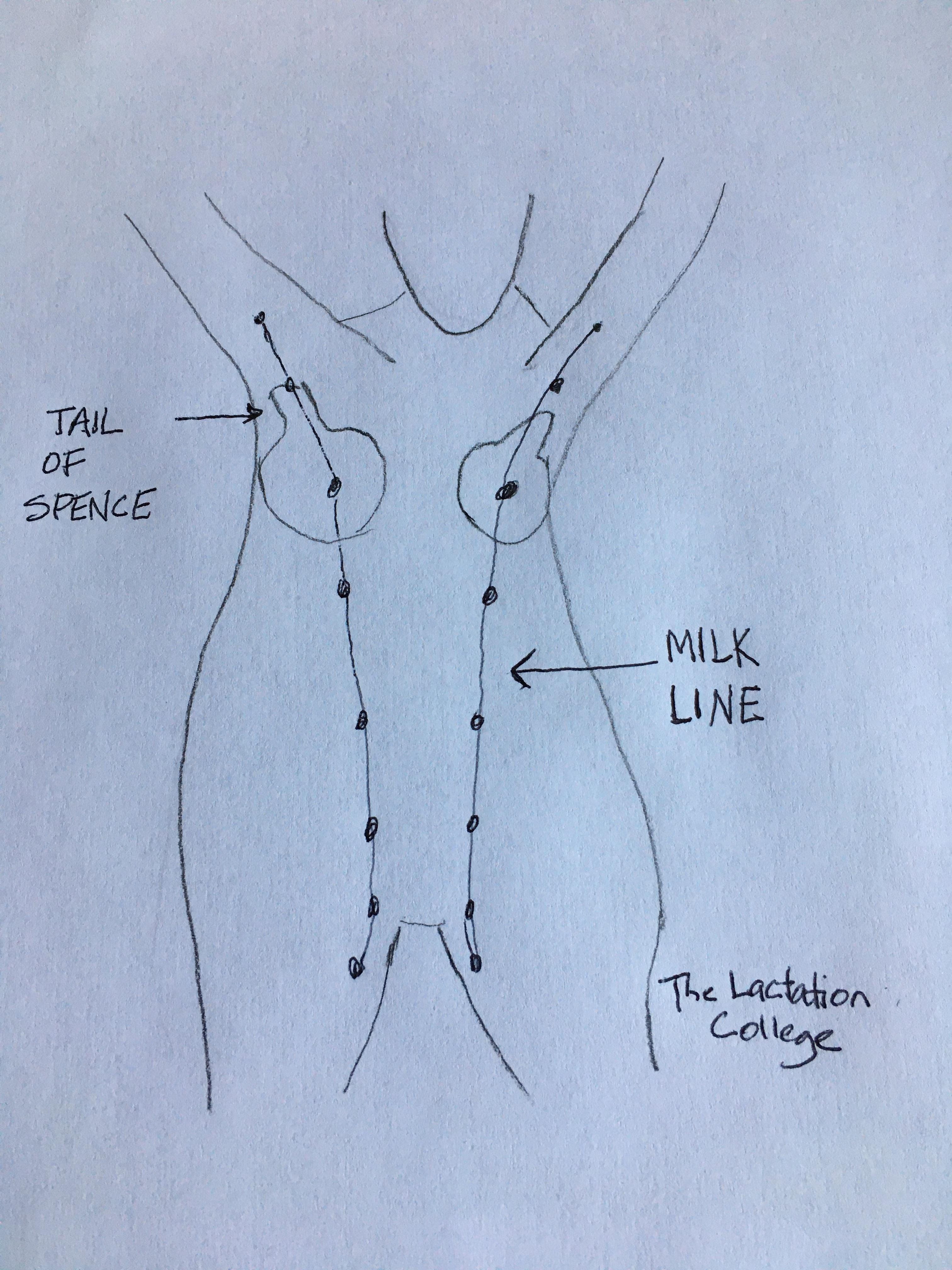
The extra milk tissue can include:
Just one nipple or multiple extra nipples
Breast tissue with no nipple and
Breast tissue with a nipple
Extra tissue is estimated to occur in 1-6% of women. It can also occur in men. Enlargement of the extra milk tissue is often seen as the mother’s milk is coming in; the extra tissue can also leak milk.
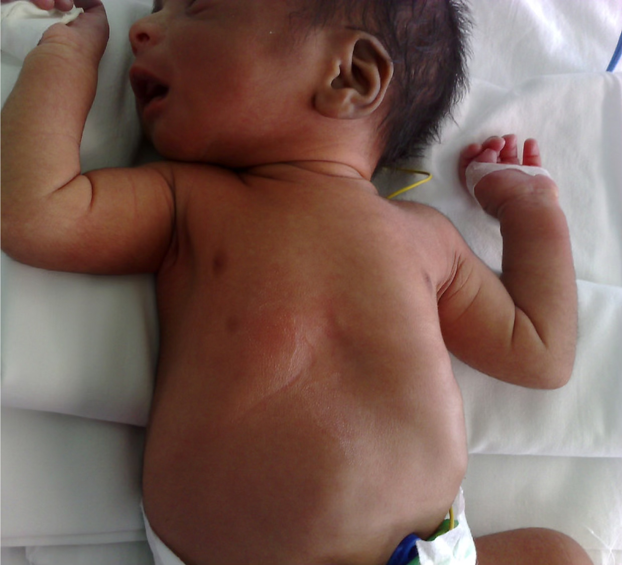
An extra nipple (also called a supernumerary nipple) can look like a mole. For test takers, if a test question asks about a picture that looks like a mole, think extra nipple. In practice, remember location, location, location – is the “mole” along the milk line? If so, think extra nipple. In the picture below, the little dark spot below the right nipple is an extra nipple.
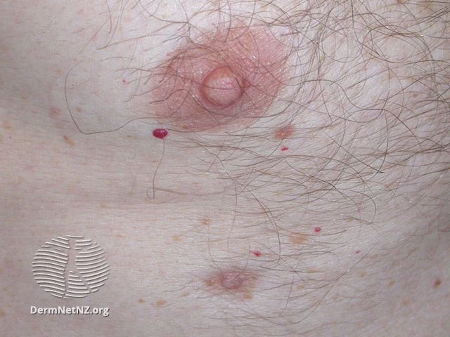
The picture below is from DermNet NZ. The extra nipple in the picture is located at 12 o’clock above the person’s left breast.

I received this picture from a new mother who was wondering what the bumps were in her armpit. Her baby was 3 days old and her milk was “coming in” nicely. Recall that the Tail of Spence is mammary glandular tissue that projects into the axillary region. The Tail of Spence is not extra milk tissue. It is differentiated from extra or supernumerary tissue in that it connects to the primary breast ductal system. I suspected that this was Tail of Spence tissue. The plan was for her to apply cool packs if it became troublesome. Yes, the Tail of Spence can be affected by mastitis.

The average diameter of a nipple is 1.6 cm. The average length is 0.7 cm.
The nipple is made up of smooth muscle fibers and is densely innervated with sensory nerve endings.
The average diameter is 6.4 cm (or 2.5 inches)
Contains smooth muscle and elastic connective tissue
Circular, darkly pigmented area surrounding the nipple
Darkens and enlarges with pregnancy
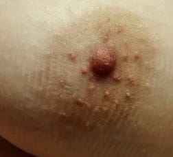
Contains little bumps which are the ductal openings of sebaceous (oily), lactiferous (milky), and sweat (sweat) glands - see picture below
So, YES, Montgomery tubercles can secrete milk
Function of Montgomery tubercles: lubrication and bacteriostatic (keep pathogen count down) but most important is that it might produces a scent to help the infant find the nipple
The exterior breast is located between the second rib and the sixth intercostal space.
60% from the internal mammary artery
30% from the lateral thoracic artery
Surges of estrogen – stimulate growth of ducts (remember Estrogen – Ducts ED)
Surges of progesterone – stimulate growth of glandular tissue (remember progesterone – glandular tissue PG)
Lymphatic system drains to the axillary lymph nodes.
Breast innervation is primarily from the 4th intercostal nerve.
Alveolus. The functional unit of the breast where the milk is made and secreted from.
Areola. Scent organ of the breast
Breast capacity is determined by the amount of milk that can be stored in the breast at any time.
Lactocytes. Cells that line the alveolar unit, make breast milk and secrete it into the alveolar lumen.
Hypermastia – extremely large mammary glands (breasts)
Hypoplasia – underdeveloped breast tissue, described by quadrants
During milk ejection: the ducts shorten and widen, which increases their diameter ad milk flows through them.
Myoepithelial cells. Muscular cells that surround the alveolus and contract under the influence of oxytocin.
Parenchyma – contains the functional parts of the breast (alveoli, myoepithelial muscle cells)
Polymastia – extra mammary tissue along the milk line
Polythelia – extra nipple along the milk line
Prolactin. Milk is produced under the influence of prolactin. Prolactin receptors are found in the basement membrane of the alveolus.
Stroma – contains the supporting tissues of the breast (connective tissue, fat, blood vessels, nerves, lymphatics)
References
Core Curriculum for Interdisciplinary Lactation Care. Edited by Suzanne Hetzel Campbell, Judith Lauwers, Rebecca Mannel, and Becky Spencer. LEAARC (Lactation Education Accreditation and Approval Review Committee). Jones & Bartlett Learning. 2019
Wambach K, Spence B. Breastfeeding and Human Lactation, 6th edition. Jones & Bartlett Learning. 2021
Estoy tratando de escoger algunos puntos clave sobre la anatomía del pecho. (1, 2)
En el embrión en desarrollo, las líneas mamarias aparecen a las 4-5 semanas aproximadamente. Aunque la mayor parte acaba atrofiándose, el tejido mamario puede persistir en cualquier parte de las líneas mamarias. Estas líneas paralelas van desde la axila hasta la ingle en ambos lados del cuerpo.
El tejido mamario extra puede incluir: un solo pezón o múltiples pezones adicionales, tejido mamario sin pezón y tejido mamario con pezón. Se estima que el tejido adicional se presenta en el 1-6% de las mujeres; también puede presentarse en los hombres. El agrandamiento del tejido mamario extra se observa a menudo cuando la leche de la madre “sube”; el tejido extra también puede gotear leche.
Un pezón extra (también llamado pezón supernumerario) puede parecer un lunar. Si en una pregunta del examen preguntan por una imagen que parece un lunar, piensa en un pezón adicional. En la práctica, recuerda la ubicación, la ubicación, la ubicación: ¿el “lunar” está a lo largo de la línea mamaria? Si es así, piensa en un pezón adicional.
Vea la imagen #1 anterior.
Un pezón extra observado en el examen de recién nacido de un bebé. Es la mancha oscura debajo del pezón derecho del bebé.
La imágene que sigue son de DermNet NZ. El pezón extra está situado a las 6 en punto justo debajo del pezón primario.
Recibí esta foto de una nueva madre que se preguntaba qué eran los bultos en su axila. Su bebé tenía 3 días y la leche le “bajaba” muy bien. Recordemos que la Cola de Spence es tejido glandular mamario que se proyecta hacia la región axilar. La Cola de Spence no es tejido extra de leche. Se diferencia del tejido extra o supernumerario en que se conecta al sistema ductal primario de la mama. Sospeché que se trataba de tejido de Cola de Spence. El plan era que ella aplicara compresas frías si se volvía problemático. (La cola de Spence puede verse afectada por la mastitis).
El diámetro medio de un pezón es de 1,6 cm. La longitud media es de 0,7 cm.
Está formado por fibras musculares lisas y está densamente inervado con terminaciones nerviosas sensoriales.
El diámetro medio es de 6,4 cm.
Contiene músculo liso y tejido conectivo elástico
Zona circular y de pigmentación oscura que rodea el pezón
Se oscurece y aumenta de tamaño con el embarazo
Contiene pequeñas protuberancias que son las aberturas ductales de las glándulas sebáceas (aceite), lactíferas (leche) y sudoríparas (sudor)
Así que, SÍ, los tubérculos de Montgomery pueden secretar leche
Función: lubricante y bacteriostática (mantiene baja la cantidad de patógenos), pero lo más importante es que puede producir un olor para ayudar al bebé a encontrar el pezón
La parte exterior del seno está situada entre la segunda costilla y el sexto espacio intercostal.
60% de la arteria mamaria interna
30% de la arteria torácica lateral
Picos de estrógeno – estimulan el crecimiento de los conductos (recuerda Estrógeno - Conductos EC)
Picos de progesterona – estimulan el crecimiento del tejido glandular (recuerda Progesterona - tejido Glandular PG)
El sistema linfático drena hacia los ganglios linfáticos axilares.
La inervación de la mama proviene principalmente del 4º nervio intercostal.
Alveolo. Unidad funcional de la mama donde se produce y secreta la leche..
Areola. Órgano olfativo de la mama.
La capacidad mamaria está determinada por la cantidad de leche que puede almacenarse en la mama en cualquier momento.
Durante la eyección de la leche: los conductos se acortan y ensanchan, lo que aumenta su diámetro y la leche fluye a través de ellos.
Estroma – contiene los tejidos de soporte de la mama (tejido conectivo, linfático, grasa, vasos sanguíneos, nervios)
Células mioepiteliales. Células musculares que rodean el alvéolo y se contraen bajo la influencia de la oxitocina.
Lactocitos. Células que recubren la unidad alveolar, fabrican la leche materna y la secretan en el lumen alveolar.
Hipermastia – glándulas mamarias (pechos) extremadamente grandes
Hipoplasia – tejido mamario subdesarrollado, descrito por cuadrantes
Parénquima – contiene las partes funcionales de la mama (alvéolos, células musculares mioepiteliales)
Politelia – pezón extra a lo largo de la línea mamaria
Polimastia – tejido mamario adicional a lo largo de la línea mamaria
Prolactina. La leche se produce bajo la influencia de la prolactina. Los receptores de prolactina se encuentran en la membrana basal del alvéolo.
Referencias
Core Curriculum for Interdisciplinary Lactation Care [Plan de estudios básico para la atención interdisciplinaria de la lactancia]. Editado por Suzanne Hetzel Campbell, Judith Lauwers, Rebecca Mannel y Becky Spencer. LEAARC (Lactation Education Accreditation and Approval Review Committee) [Comité de Acreditación y Aprobación de la Educación en Lactancia]. Jones & Bartlett Learning. 2019
Wambach K, Spencer B.Breastfeeding and Human Lactation [Amamantamiento y lactancia humana], 6ta edición. Jones & Bartlett Learning. 2021
No posts
Comments
Nothing yet. Say the first thing.
Sign in to join the conversation.