
Hello,
This is the 2nd in a series of 3 posts related to gastrointestinal genetics. This month’s post will focus on a newborn with diarrhea.
We are also excited to announce that registration is open for the Winter ABGC board review bootcamp, which will take place between Dec 1, 2025 - Jan 22, 2026. This bootcamp is for genetic counselors preparing for the ABGC exam in February 2026. We will cover topics from all 5 domains on the ABGC content outline. An early-bird discount is available through Nov 30th, 2025. For more information and to register, please visit our website.
Please feel free to reach out to me (daniel@studyrare.com) with any questions about the bootcamp or the newsletter.
P.S. Happy Genetic Counselor Appreciation Day!
-Daniel
A 10-day-old infant presents to the emergency department with severe watery diarrhea that began on day 2 of life. The parents switched from breast milk to soy-based formula on day 5 with no improvement. The infant appears severely dehydrated and has lost 20% of his birth weight. The infant is started on IV fluids, and the diarrhea persists despite stopping oral feeds. Which of the following processes is most likely impaired?
Genetic sequencing is sent for the infant in Question 100, which confirms the diagnosis of congenital chloride diarrhea. Which of the following prenatal ultrasound findings is associated with this disorder?
Question 100: Absorption of chloride ions
Question 101: Polyhydramnios
The infant discussed in these questions has classic features of congenital chloride diarrhea (CCD). CCD is an autosomal recessive condition caused by biallelic variants in SLC26A3, a gene that encodes an intestinal Cl⁻/HCO₃⁻ transporter. The mechanism of this disorder is illustrated and discussed in the image below.
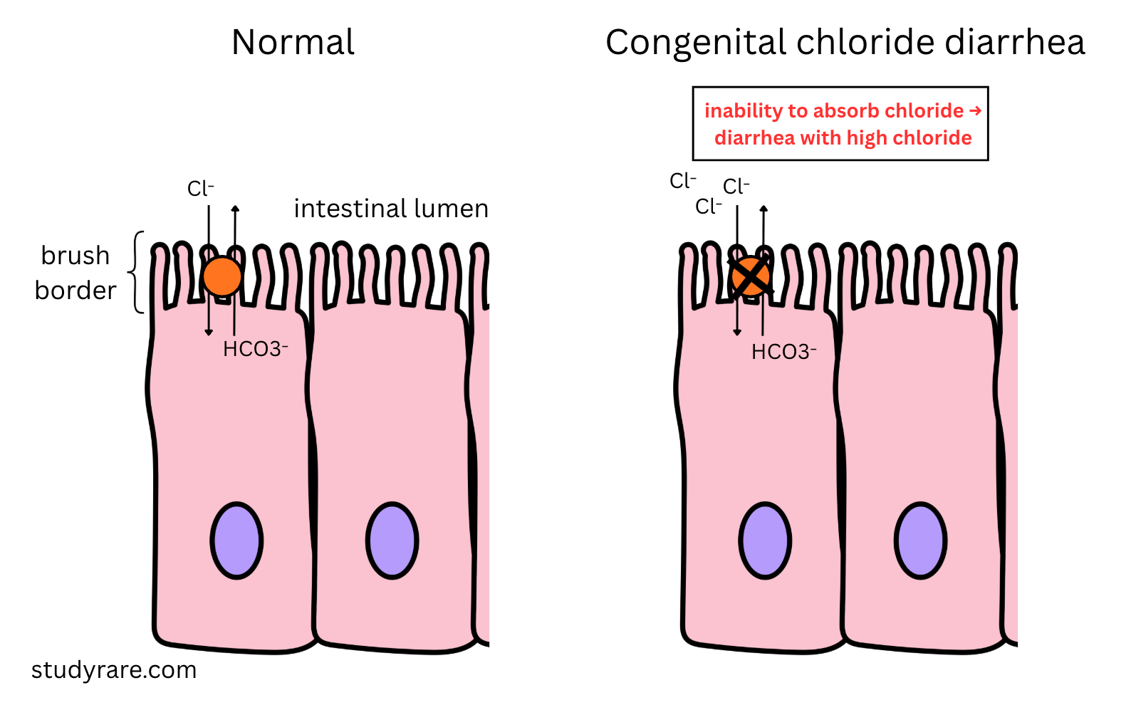
Patients with CCD present with a combination of features that include:
Watery, high-volume diarrhea from birth
Diarrhea that persists even when patient does not take nutrition by mouth
Severe dehydration and weight loss (this patient has lost 20% of his birth weight, which is significantly more than the expected 7-10% weight loss seen in healthy newborns during the first week of life)
Excessive stool Cl⁻ loss leading to metabolic alkalosis (more about this mechanism here)
Normal findings on small intestine biopsy (vs microvillus inclusion disease)
Prenatal ultrasound may show dilated bowel loops and polyhydramnios (shown and discussed further below).
Management of CCD
The management of CCD is with lifelong oral chloride supplementation in the form of sodium chloride and potassium chloride. In addition to electrolyte replacement, patients require adequate hydration and monitoring of serum electrolytes, growth, and development. In contrast to certain other causes of congenital diarrhea (discussed below), there are no dietary restrictions in CCD, as this is not a food intolerance. If CCD is untreated, it can be fatal in the first few weeks of life. However, with appropriate management, the prognosis is overall favorable and patients can achieve normal growth and development.
Monogenic causes of congenital diarrhea
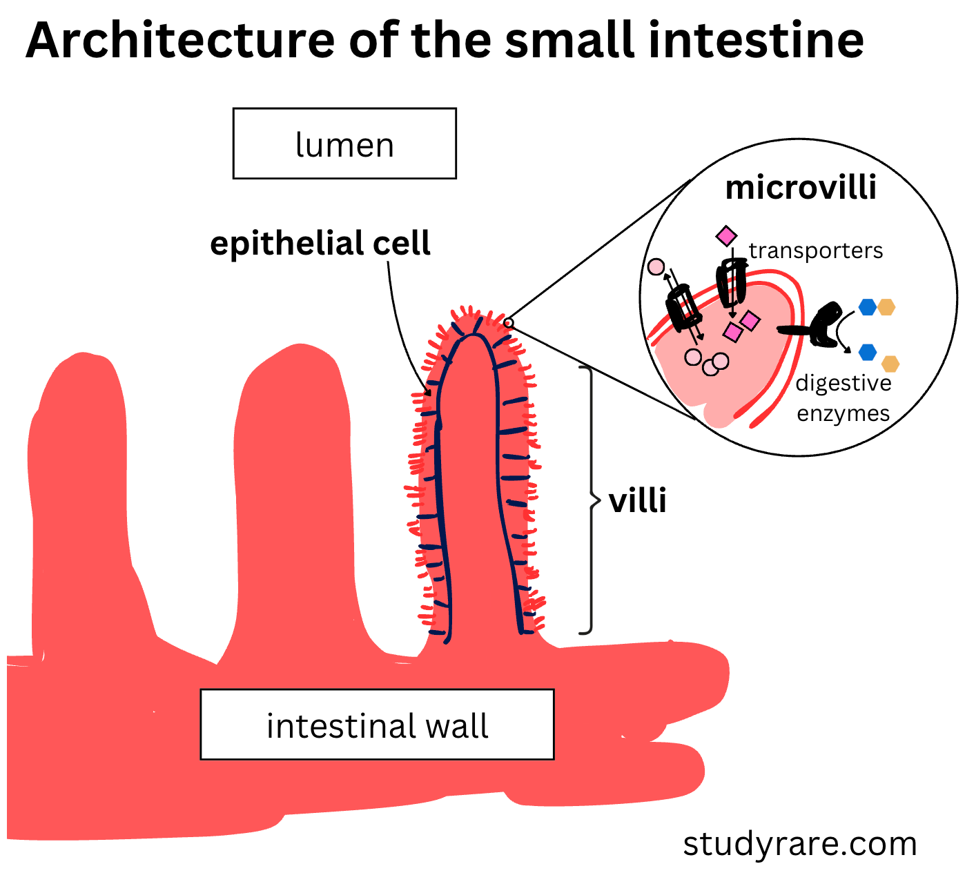
CCD is part of a family of congenital diarrheal disorders, which are inherited conditions that typically present in neonates and infants with severe diarrhea. These disorders often result from defects in the intestinal microvilli, which form the absorptive surface of the small intestine. The defects may affect the structure of the microvilli themselves (e.g., microvillus inclusion disease, which has a poor prognosis) or the function of enzymes and transporters found on the surface of the microvilli. The congenital diarrheal disorders that affect a single enzyme or transporter (rather than the entire structure of the microvilli) generally have a better prognosis and are in most cases treatable. The schematic below shows the architecture of intestinal villi and microvilli in more detail.
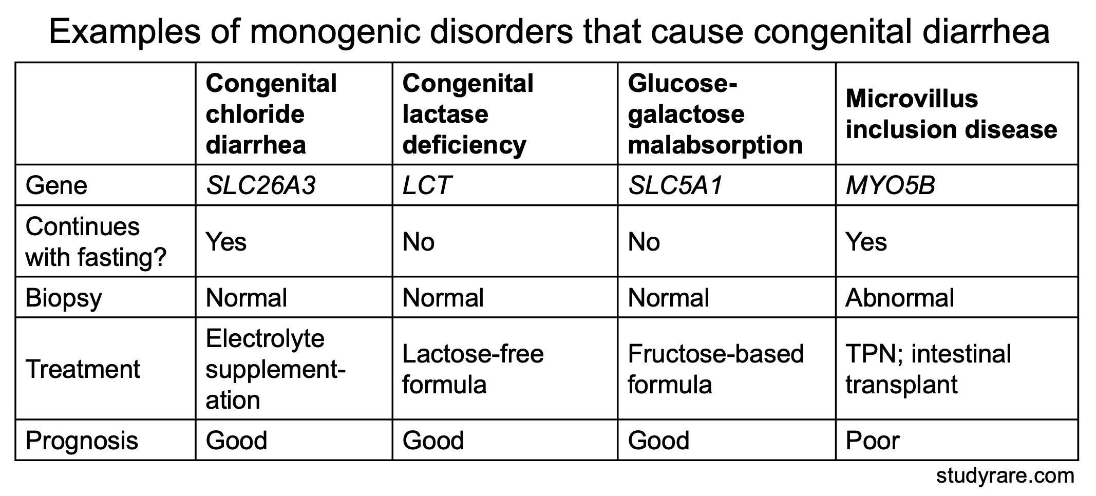
The table below compares CCD with other monogenic causes of congenital diarrhea. One important distinguishing feature of CCD is that the diarrhea persists despite stopping oral feeds and changing formulas.
Incorrect answer choices (Question 100)
(Choice A) Dietary disaccharides (disaccharide = two sugars linked together) such as lactose (the main sugar in breast milk), sucrose, and maltose must be broken down into monosaccharides (single sugars) before they can be absorbed by the intestines. If the disaccharides cannot be broken down due to a congenital enzyme deficiency, they remain in the intestinal lumen, attracting water and producing diarrhea. Typically, a dietary adjustment (e.g. removing the offending disaccharide) or stopping feeding will result in symptomatic improvement.
In the patient in question 100, the lack of improvement after switching to soy formula helps rule out congenital lactase deficiency, a disorder that presents in neonates after starting breast milk and that would be expected to improve with a lactose-free formula like soy. In addition, the lack of improvement of this patient after stopping oral feeds points away from congenital lactase deficiency.
(Choice C) Bile acids are an important component of bile, a substance that breaks down fats in the intestine. Patients with bile acid synthesis defects therefore have inadequate bile production and present with fat in the stool (steatorrhea), not watery diarrhea as was the case with our patient. Because bile is a breakdown product of bilirubin, patients who are unable to synthesize bile will also have hyperbilirubinemia and cholestatic jaundice (elevated conjugated bilirubin, hepatomegaly), and there was no mention of jaundice in this patient. Furthermore, the diarrhea in a bile acid synthesis defect would be expected to improve when dietary fat intake is eliminated, unlike in our patient.
(Choice D) The transport of dietary monosaccharides is disrupted in glucose-galactose malabsorption. This is a disorder caused by defects the sodium-glucose cotransporter (encoded by SLC5A1) that absorbs glucose and galactose from the intestinal lumen. Patients present in the neonatal period with severe watery diarrhea similar to CCD. However, unlike the patient in this question, patients improve after stopping oral feeds. Treatment is with a formula containing fructose, which uses a different transporter to enter the body.
Answer choices (Question 101)
Patients with CCD may have signs on prenatal ultrasound including dilated bowel loops (see image below) and polyhydramnios (Choice D). This is because in CCD, large volumes of fluid are secreted into the fetal intestinal lumen, which enlarges and produces a characteristic “honeycomb” pattern seen on ultrasound. The excess fluid in the intestinal lumen is then emptied into the amniotic cavity, causing polyhydramnios.
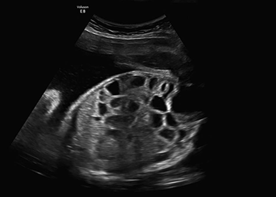
(Choice A) An absent stomach bubble is seen in esophageal atresia, not in CCD.
(Choice B) Increased nuchal translucency suggests chromosomal anomalies, especially aneuploidy (e.g., trisomy 21, Turner syndrome), and may be a sign of a lymphatic malformation. Increased nuchal translucency is not characteristic of congenital diarrheal syndromes.
(Choice C) Oligohydramnios is associated with renal agenesis or urinary tract obstruction. In contrast, CCD increases the fluid output into the amniotic space, causing polyhydramnios.
Learning objective
Congenital chloride diarrhea (CCD) is caused by the impaired absorption of chloride ions from the intestinal lumen into the body. Patients present with watery diarrhea in the neonatal period that persists despite stopping oral feeds and/or altering formula. Key diagnostic features include very high stool chloride (>90 mEq/L) and hypochloremic metabolic alkalosis. Prenatal ultrasound may show dilated bowel loops and polyhydramnios. Treatment requires lifelong electrolyte supplementation, and no dietary restriction is required.
2025 ABMGG General Exam Blueprint | V. Single gene inheritance → d) Single gene disorders → x) Other
2023 ABGC Exam Content Outline | Domain 1C. Genetic Conditions
Additional resources
The genetic architecture of congenital diarrhea and enteropathy (NEJM, 2025)
Honeycomb fetal abdomen: characteristic sign of congenital chloride diarrhea (2016)
Comments
Nothing yet. Say the first thing.
Sign in to join the conversation.