
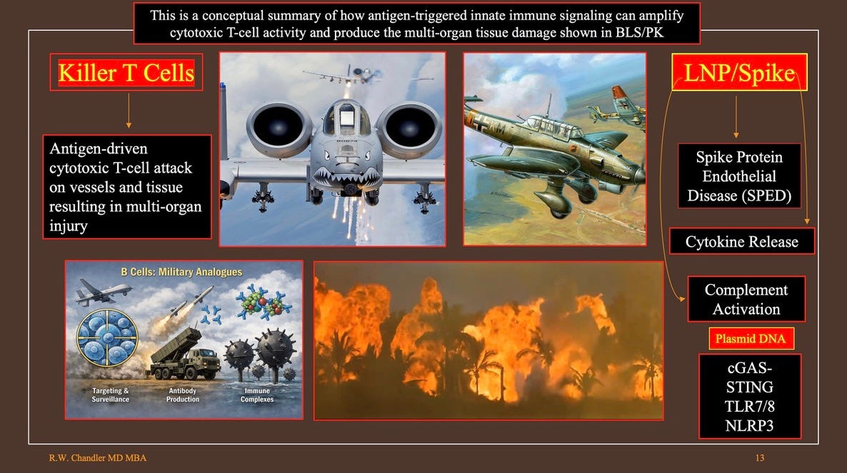
From the inventors of the Pfizer/BioNTech product, BNT162b2
The machinery of the transfected cell is utilized for in vivo translation of the message to the corresponding protein, which is the pharmacologically active product.1
Foreign proteins encoded by synthetic ribonucleic acid (RNA) have been associated with anaphylaxis, autoimmunity, clotting and bleeding, complement activation, cytokine storm, deposition of amyloid and unidentified foreign/non-local material (FM/NLM), endotheliitis, fibrin activation, lymphocytopenia, and lysis of elastin in arterial walls. That is but a partial list that does not include modification of the kinetics and morphology of blood clots creating resistance to thrombolytics and anti-coagulants, cryoprotein formation, and neoplasia from retained plasmid SV40 promoters and enhancers and suppressed p53 cancer suppressor. A suppressed suppressor = a promoter.
The story begins with where the injected material is distributed. The figure below is a distribution map of a lipid nano particle transport vehicle designed for human membrane penetration in stealth mode.
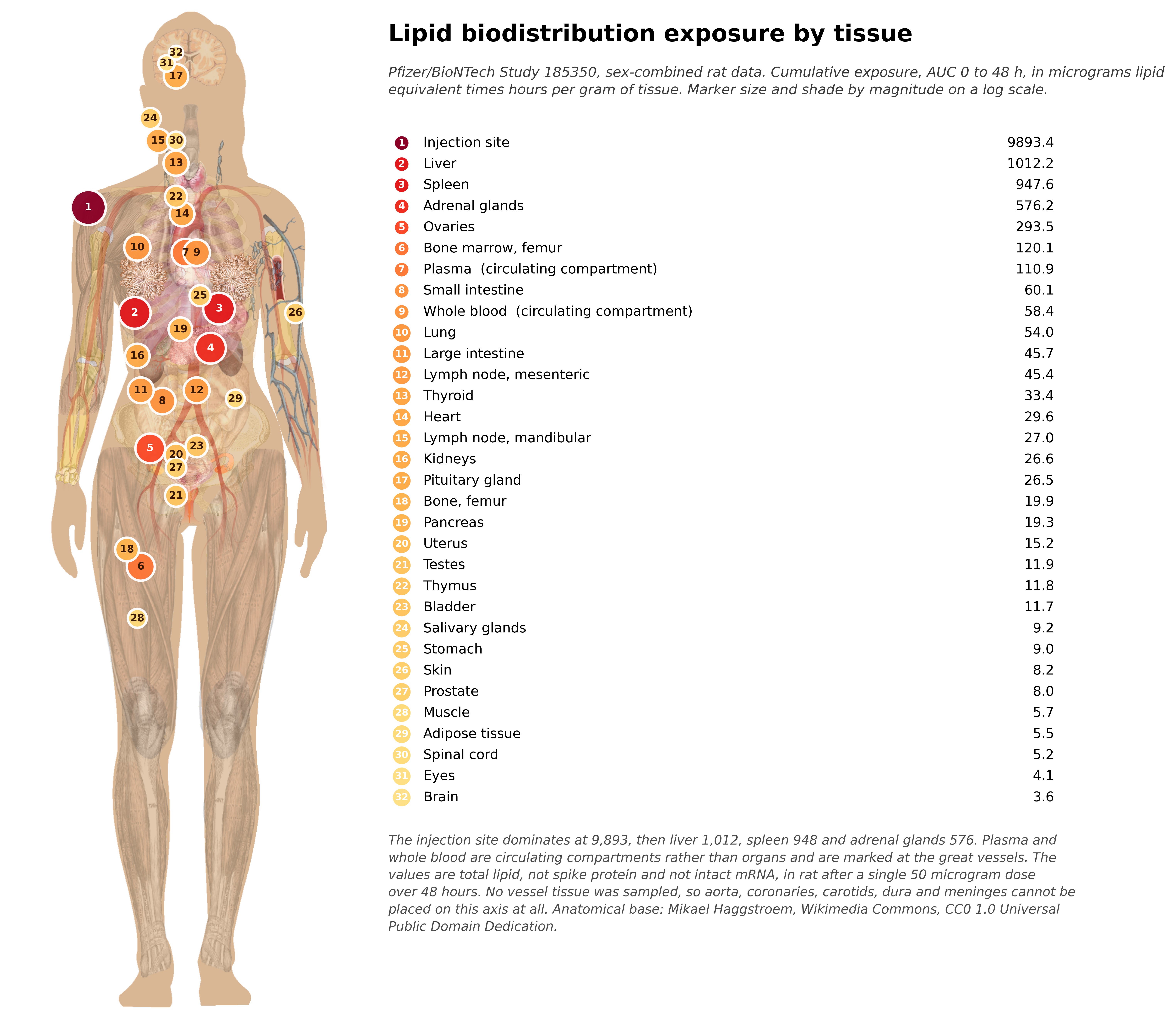
The Pfizer Documents (Pfizer BioNTech study #185350)2 present this biodistribution study of a lipid nano particle (LNP) delivery of genetic material encoded for Luciferase, a means to trace movement of the LNP package through the body. The study was done in rats using a LNP vehicle different than the product used in BNT162b2 which was either not tested or the results have been sequestered.
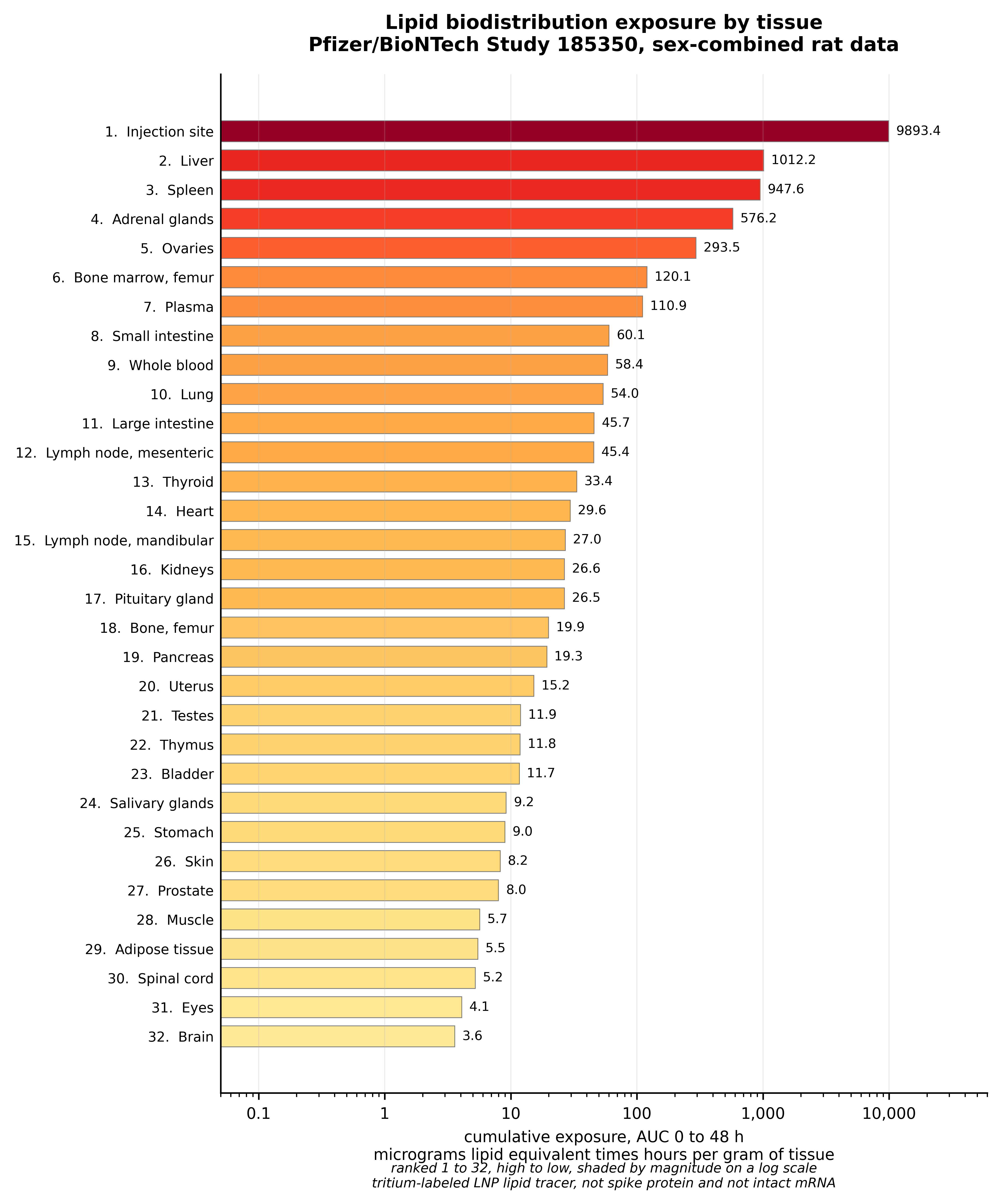
The study ended at 48 hours at which point LNP continued to concentrate in most organs. Below is the cumulative exposure by tissue/organ. The final concentrations were not determined.
mRNA and and the novel lipid vehicle was design to evade the body’s defense mechanisms then settle into tissues and begin transcribing synthetic genetic code into proteins. See https://www.nature.com/articles/nrd4278/figures/2
The duration of production and action, the amount of protein produced, the characterization of the proteins, tissue effects, toxicity, genotoxicity, carcinogenicity, and location of production and distribution were not studied. 3
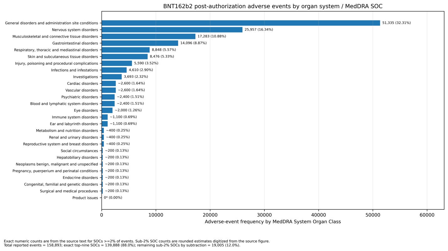
After an Emergency Use Authorization approval widespread injection of BNT162b2 Version 9 Process 2 began in the United Kingdom in Mid December and the United States in January 2021. By February 28, 2021 1223 deaths were reported to Pfizer along with an average of 3.8 Adverse Events for each of ~40,000 people reporting.4 Most cases had no reported outcome or followup. BNT162b2 Process 2 Version 9 never went through the 2 year RCT specified in the protocol.
German pathologist Arne Burkhardt was closing his pathology institute in Reutlingen, Germany in Spring of 2021 when requests from colleagues, friends and family members of persons recently deceased following Corona vaccination caused him to reverse course and begin a 5 year study of the histopathology and immunohistochemistry of organs and tissues from the deceased. Four other pathologists joined the effort culminating in publication May 30, 2026 of Corona-Vaccine Damage, Report of the “Pathologiekomferenz”5 by tredition online publishing.
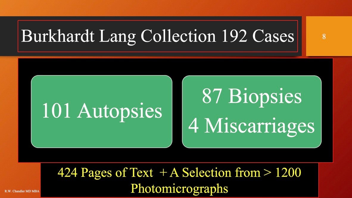
Each case had at least one autopsy by numerous pathologists working in institutions in Germany, Switzerland, and Austria. This body of work stands alone in size and detail. Three of Dr. Burkhardt’s lectures are featured on this substack.
Over the course of almost four years, I have compiled observations from this project in a database without an attempt to re-interpret the actual 1200+ photomicrographs from this work. Highly detailed findings were available for 86 of the 101 cases although in some cases specimens were limited due to spoilage and limited numbers of tissue blocks. The consultant pathologist has limited ability to obtain additional specimens (tissue block) given the referral nature of the case flow. Some cases could not be used due to lack of tissue blocks.
A series of reports is planned consisting of data analysis from this database. This report will summarize organ/tissue distribution of the vaccine related findings and death statistics.
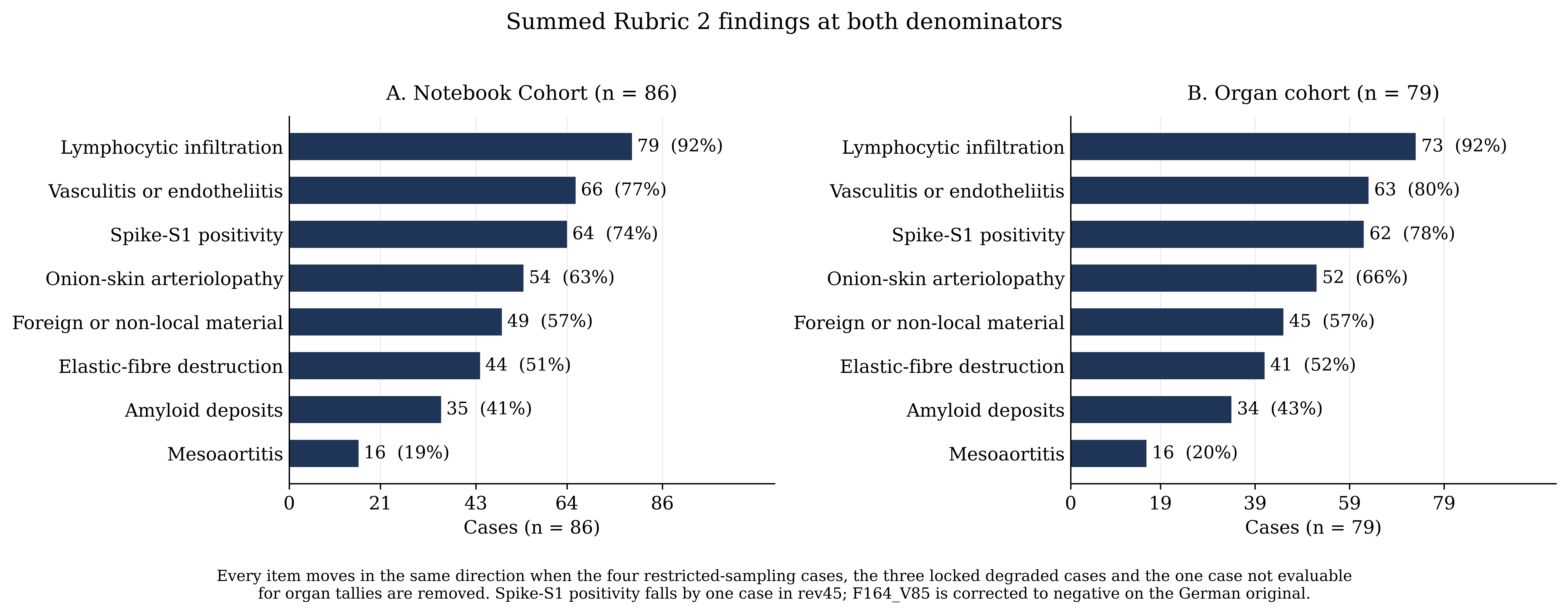
Tissue/organ level findings are available on 86 autopsy cases drawn from laboratory notebooks provided by Drs. Lang, Schwarz and Palmer.
The consultants identified 8 tissue levels findings they deemed characteristic of vaccine effects were aggregated as as Rubric 2, see below. Rubric 1 was applied to the full cohort n = 101 and results will be presented in subsequent reports. The 8 histological characteristics of vaccine damage are defined below.
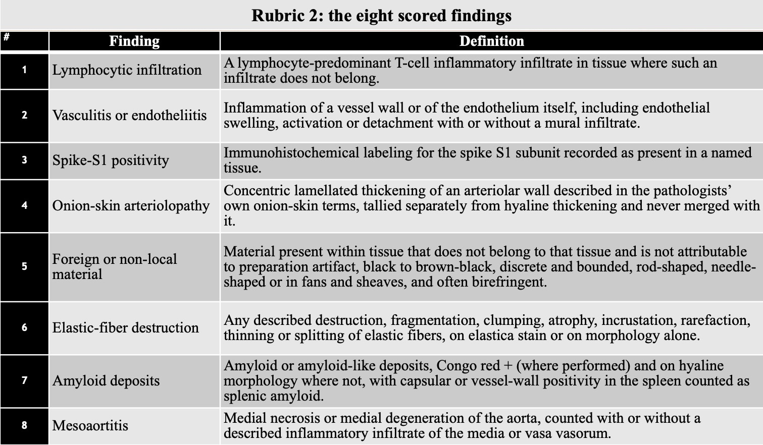
Briefly, lymphocytic infiltration is a sign of immunological activation and attack on tissues producing Spike protein so one might expect to see some overlap between lymphocytic infiltration and Spike protein locations. The primary target of this response is the circulatory system, specifically heart and muscular blood vessels although veins are also involved with clotting.
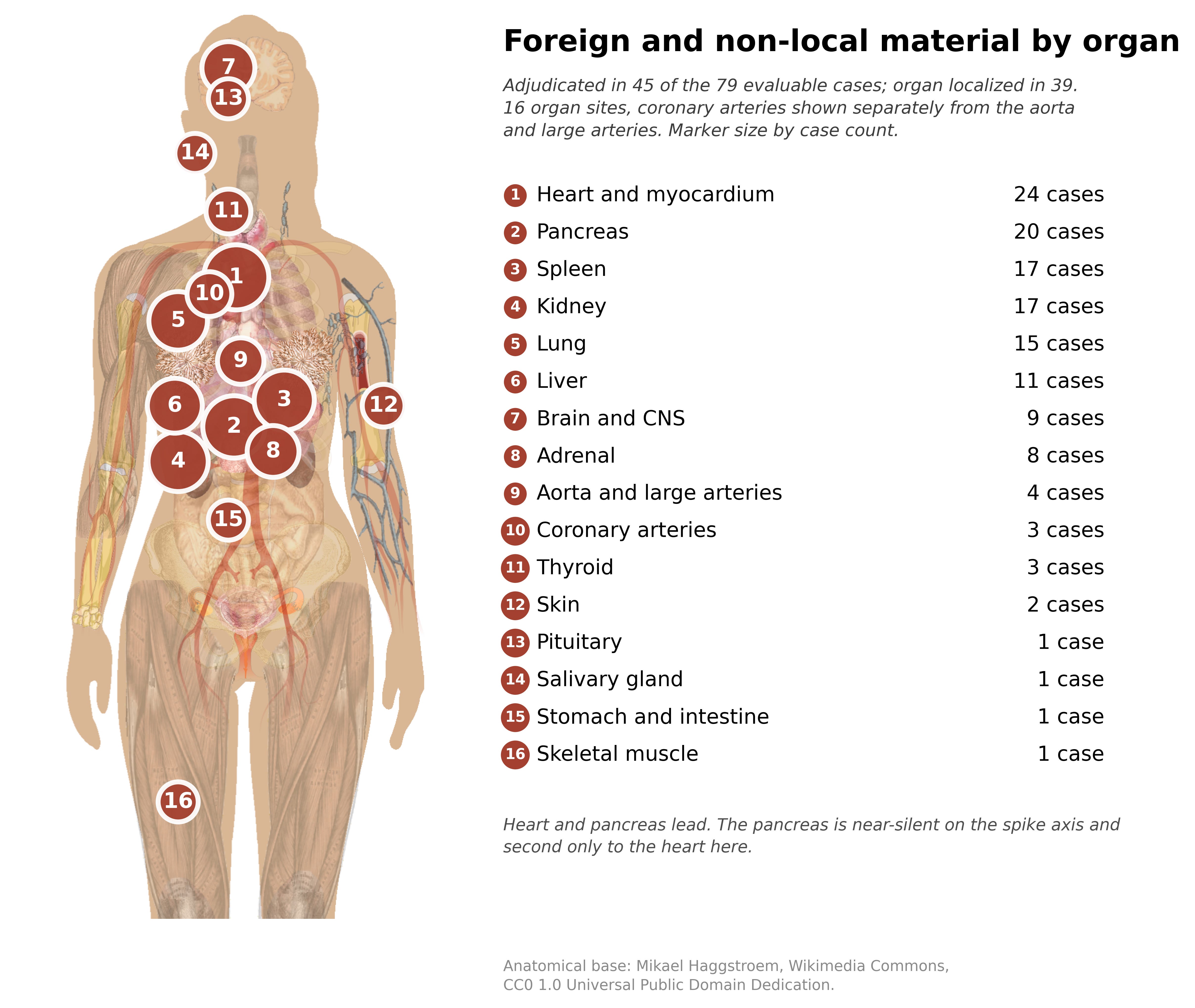
Three of the next four findings are deposition of material, both known and unknown: 1. in arterial walls = onion-skin arteriopathy, 2. in tissue seams and vessels = foreign or non-local material 3. arterial walls = amyloid. Remaining are destruction of elastin fibers in arterial walls and specific inflammation of the aorta = mesoaortitis. The figure below shows the distribution of these findings across the whole dataset on the left and on the right after removal of 7 cases of spoilage or limited tissue blocks leaving n = 79.
Widespread distribution of the LNP/modRNA (Pfizer and Moderna) as well as virus vectored (AstraZeneca and JNJ/Janssen) genetic code was associated with pathological findings in a median of eight organ systems. See below.
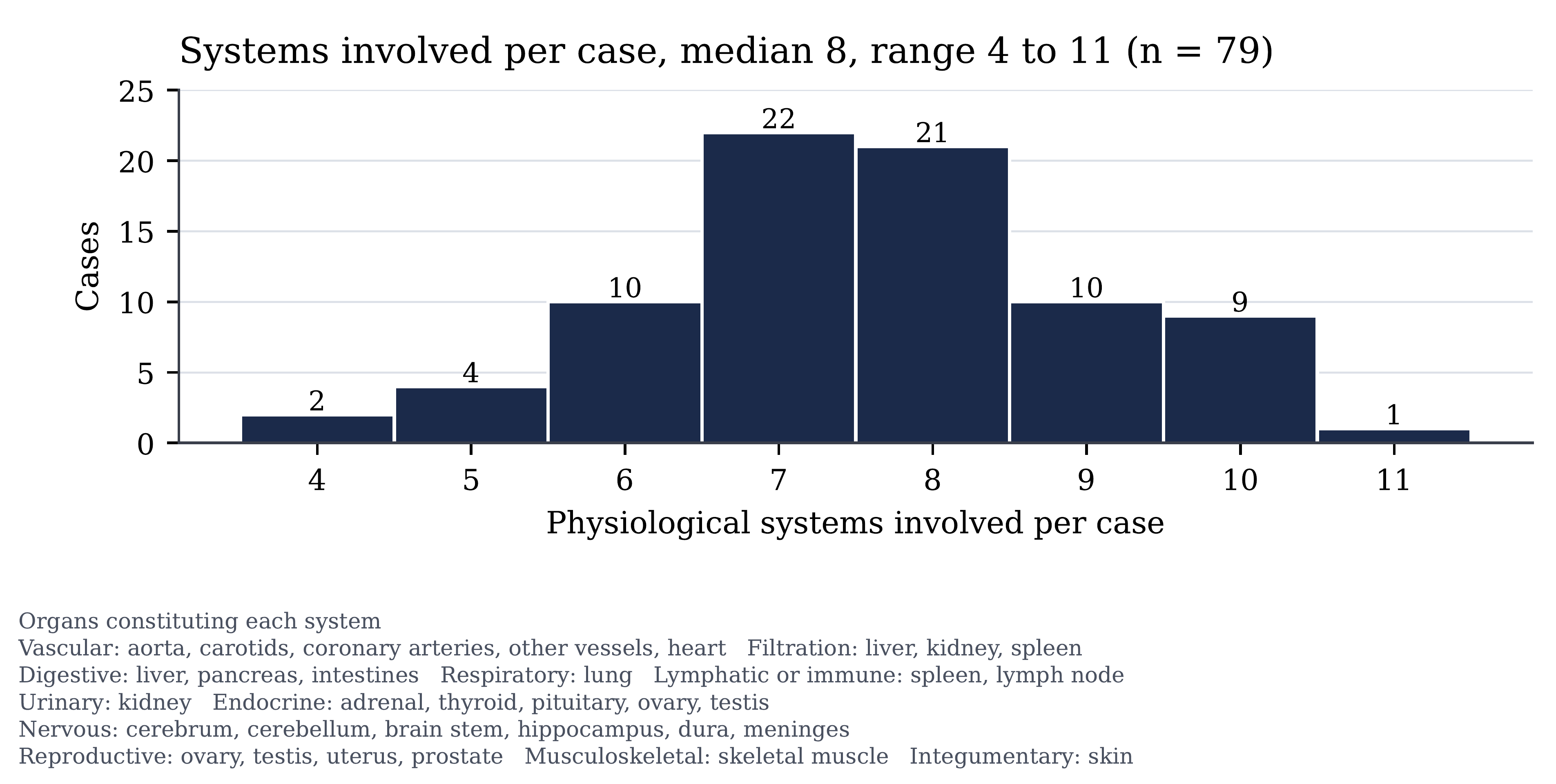
Remarkably, Case 1 had 11 systems involved. Dr. B was “gut hooked” by case 1 in fisherman speak. Time to re-open the lab!
The figure below shows where immunohistochemical stain for S1 spike protein was positive. Nucleocapsid stains were positive in four cases indicating SARS-CoV-2 protein.
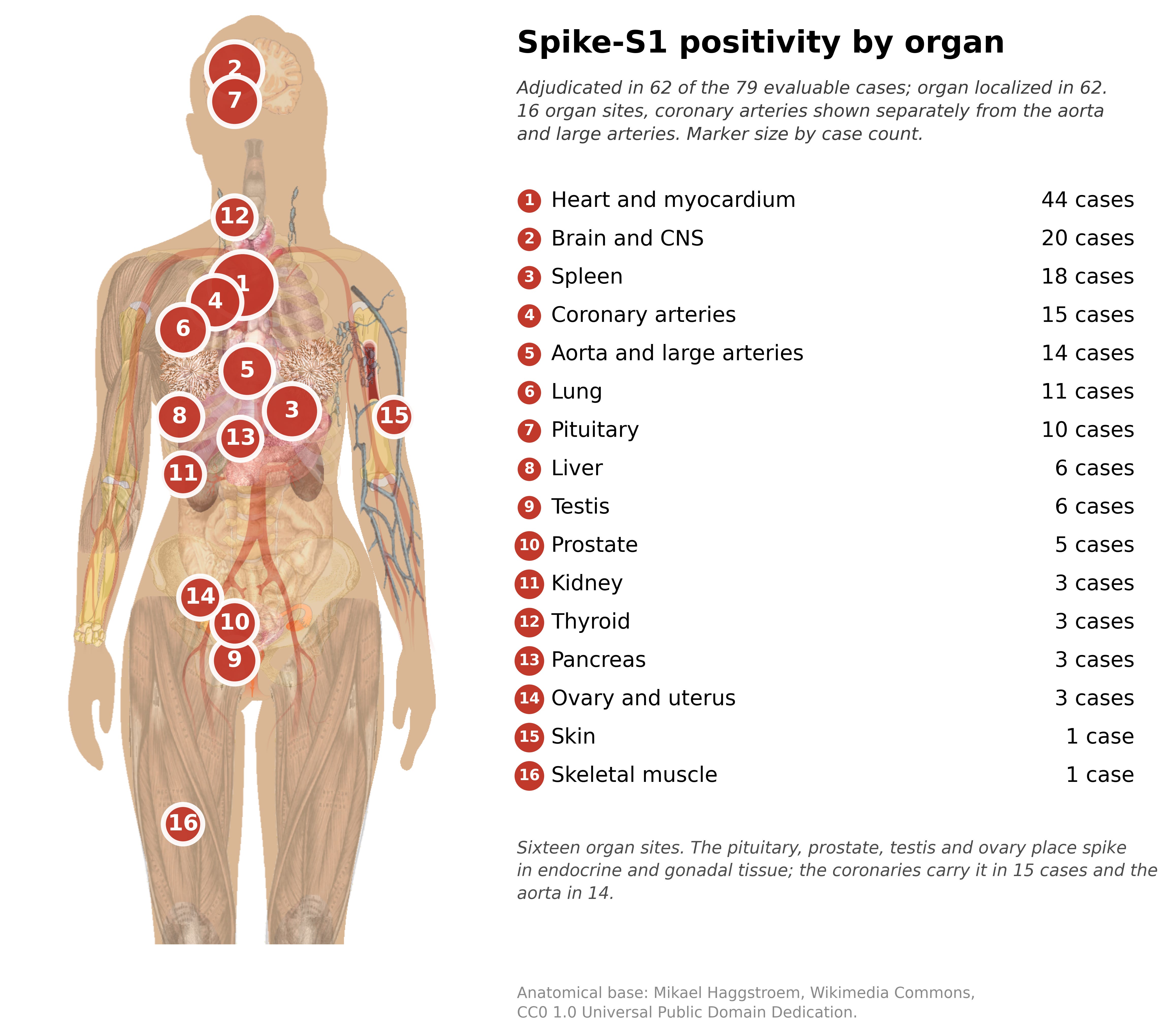
Dr. Burkhardt realized there was a sampling error factor in his work as a consultant and advised retaining the corpse to acquire additional tissue blocks until the examination completed.
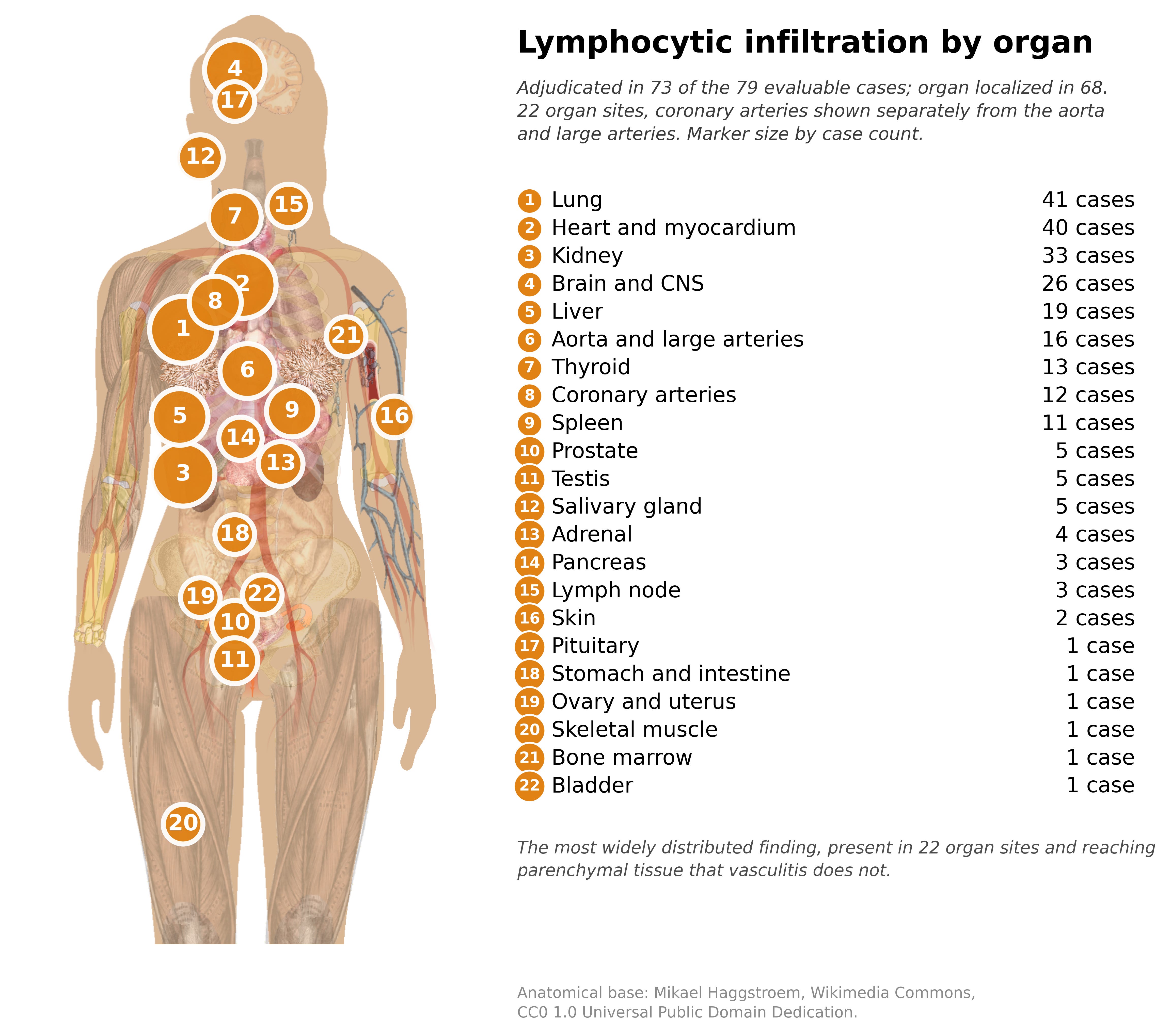
Following Spike protein production in the tissues comes the immunological response in the form of lymphocytes recruited by the non-self proteins located in these tissue and organs. T-cell lymphocytes dominate but B-cells are also present.
The figure below shows where lymphocytic infiltration was identified.
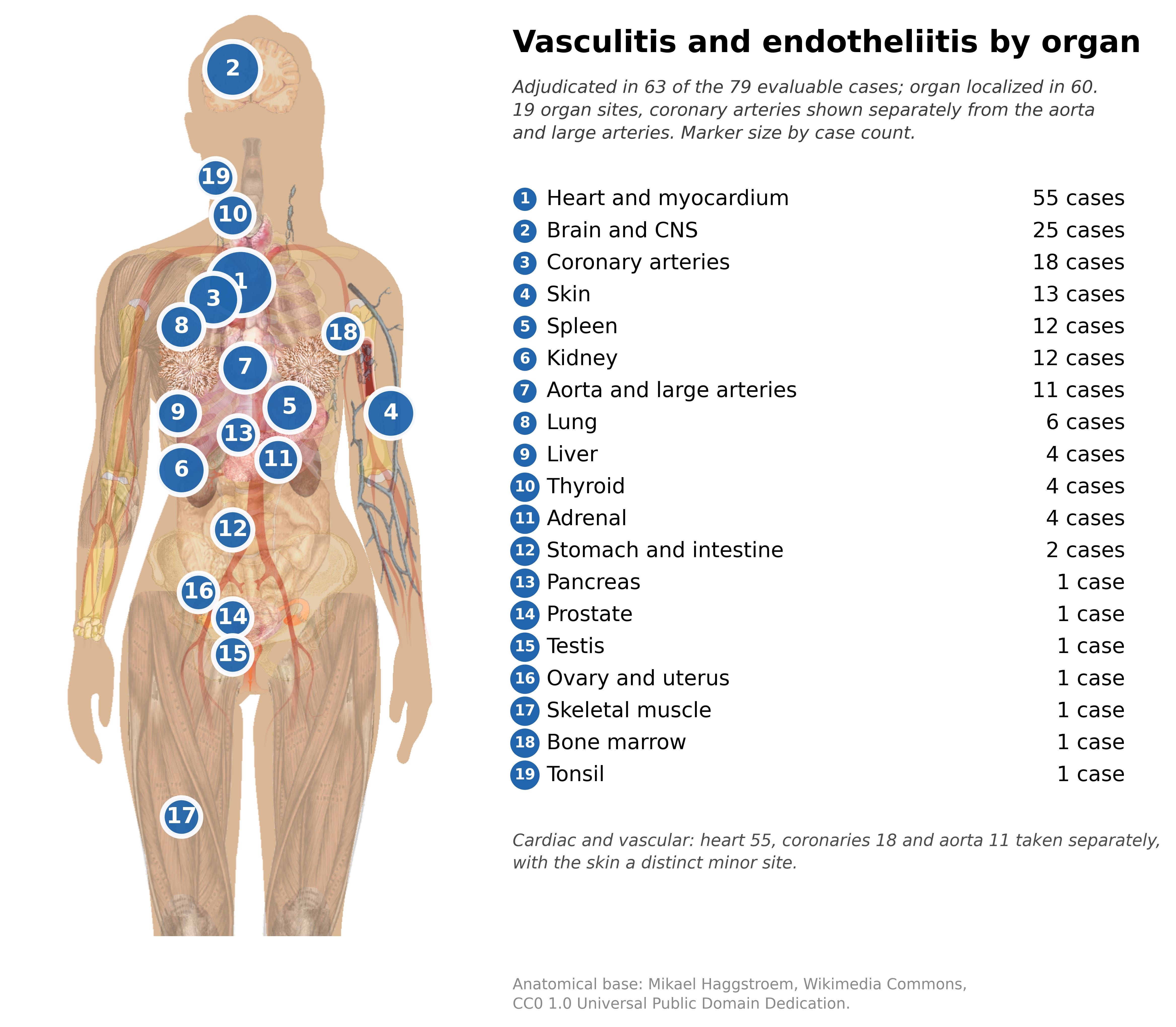
Damage to blood vessels was widely distributed. Below.
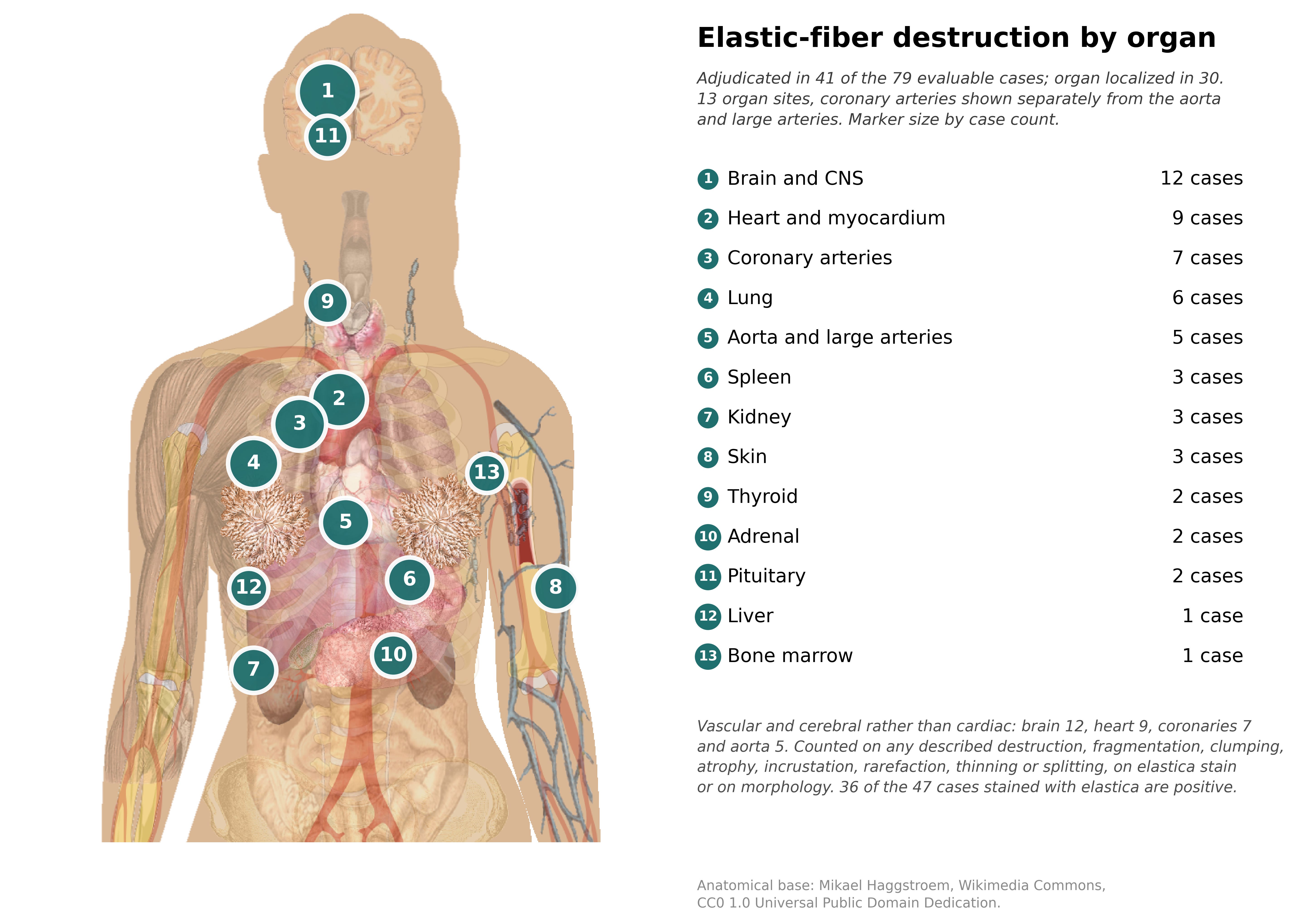
Destruction of the flexible fibers, elastin, in the muscular layer of arterial wall was a mechanism of mechanical failure of the vessels. Elastin once destroyed cannot be replaced except by scar tissue, collagen a rigid fiber. Below.
Case 10 below illustrates the combined effect of spike, lymphocytic infiltration, and tissue destruction in a 61 year old male 67 and 25 days after BNT162b2 (Pfizer). Cause of Death (COD) assigned by the pathologists in this case was Corona vaccination at a level of near certainty, level 3. COD level 0 = no clear connection with death, level 1 is vaccination as probable cause of death, level 2 = very probable and level 3 = near certainty.
Cause of Findings (COF) uses an inverse scale (developed by author based on rubric 2 findings) was also at a high degree of probability 1 = near certainty. Categories 2-4 are decreasing probability with level 4 = no clear relationship.
The mode of death (SADS = Sudden Adult Death Syndrome) was #2 or a witnessed death with unsuccessful resuscitation. The remaining levels — 1 being found dead, 3 being successfully resuscitated with later death after no recovery and 4 being chronic course.
This 61 year -old man died of a dissecting aneurysm in his ascending aorta that ruptured into the pericardial sac filling the sac with blood under pressure stopping the heart almost instantly. Below are some vaccine associated findings on histological examination.
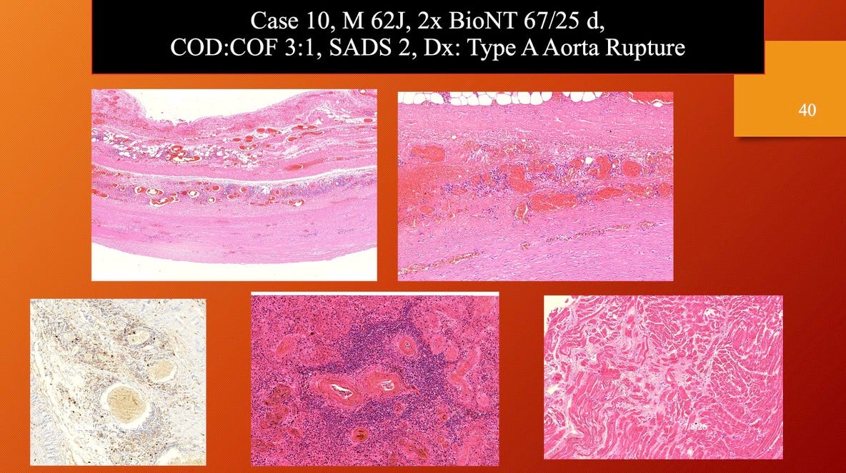
Top left shows dissection as a split in the middle layer of the aorta, top right shows the hemorrhage and fiber destruction in the arterial wall, bottom left illustrates spike S1 + stain, middle bottom shows lymphocytic infiltration around a thickened small artery (protein deposition), and bottom right shows scar tissue replacement of heart tissue. Once scar collagen forms there is no way to remove it. The presence of scar indicates a process that occurs over weeks to months.
Men dominate the sex statistic but the difference is under powered —does not reach statistical significance (p < 0.05). However, a higher percentage of fatalities in males is a familiar pattern from other datasets. A lower probability does not eliminate a causal connection particularly in an underpowered study sample.
Females have 3:1 more adverse events as seen in Pfizer document 5.3.6 and the observation extends across multiple datasets including the US Vaccine Adverse Event Reporting System (VAERS), Swedish, Danish, and multiple other data collections. During the early months of 2021 adverse events were as high as 4:1 F:M but then came down. Historically in VAERS going back decades, females reported 60-62% of adverse events across all vaccines.
Age is a different story with younger > older with a statistically significant p value of 0.016. A real age effect cannot be differentiated from sampling error in this dataset as sudden death of a young person may have motivated the referral.
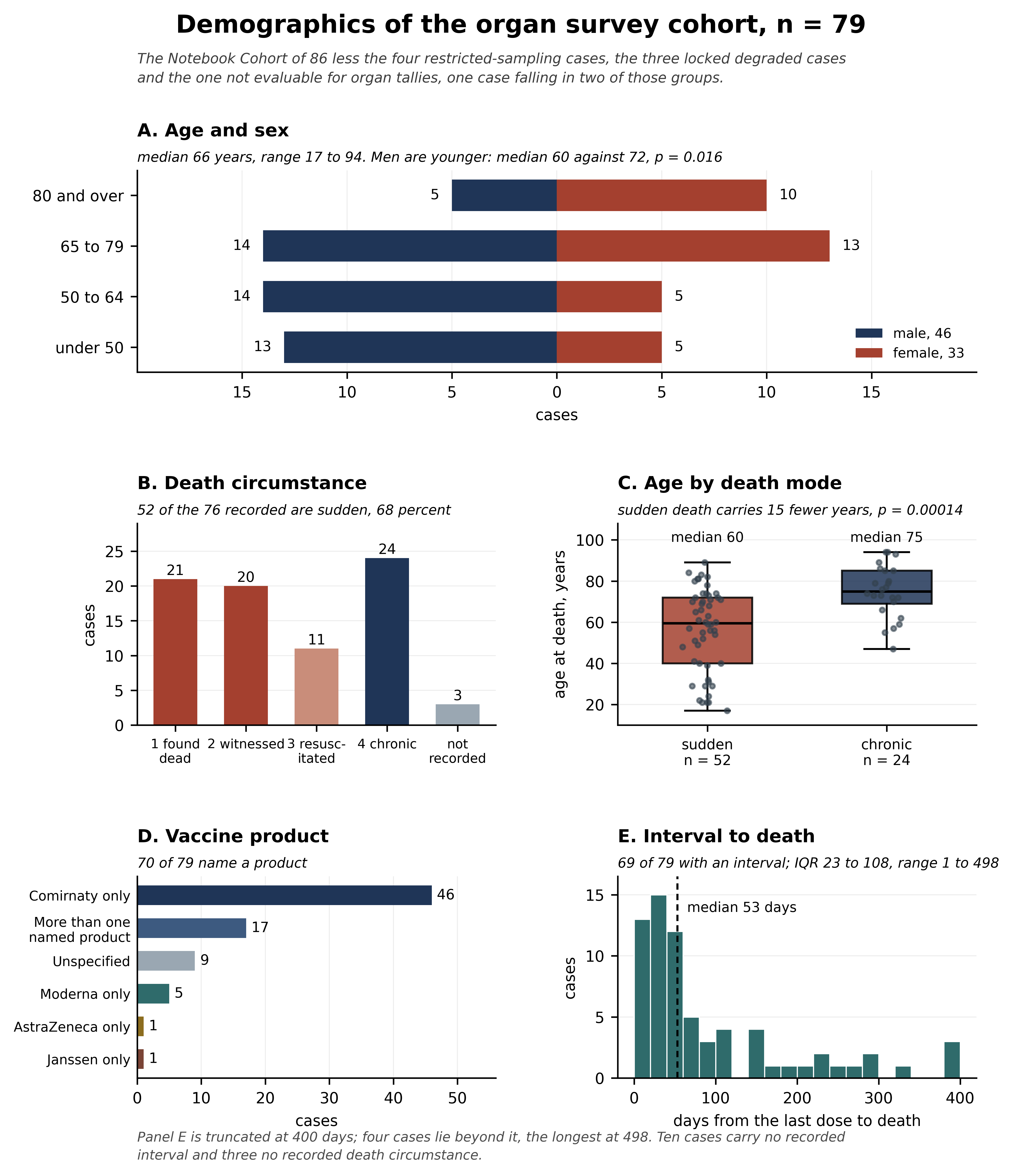
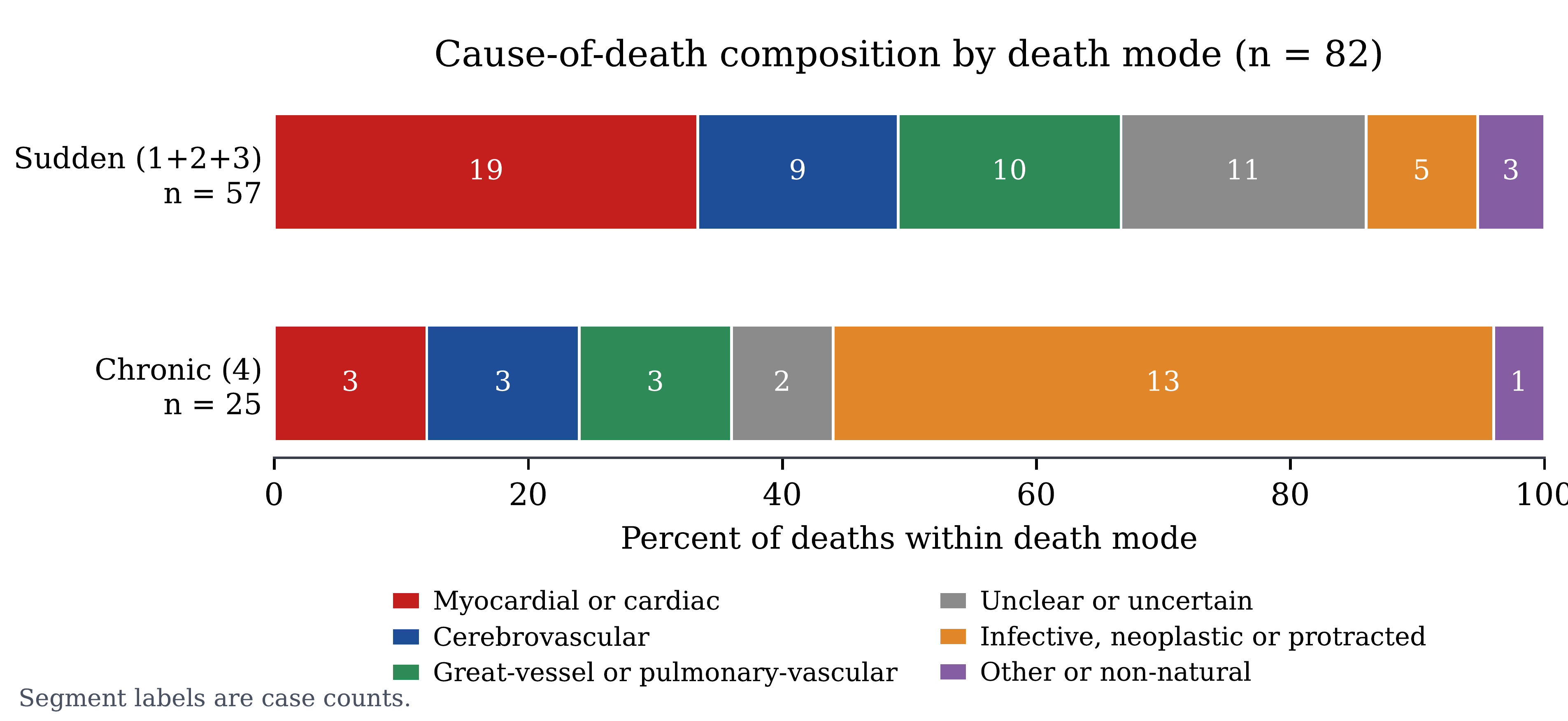
Pathological changes attributed to Corona vaccination are widely distributed and affect a median of 8 organ systems. Sudden death occurs in younger persons that are dominated by males but the sex difference did not reach statistical significance.
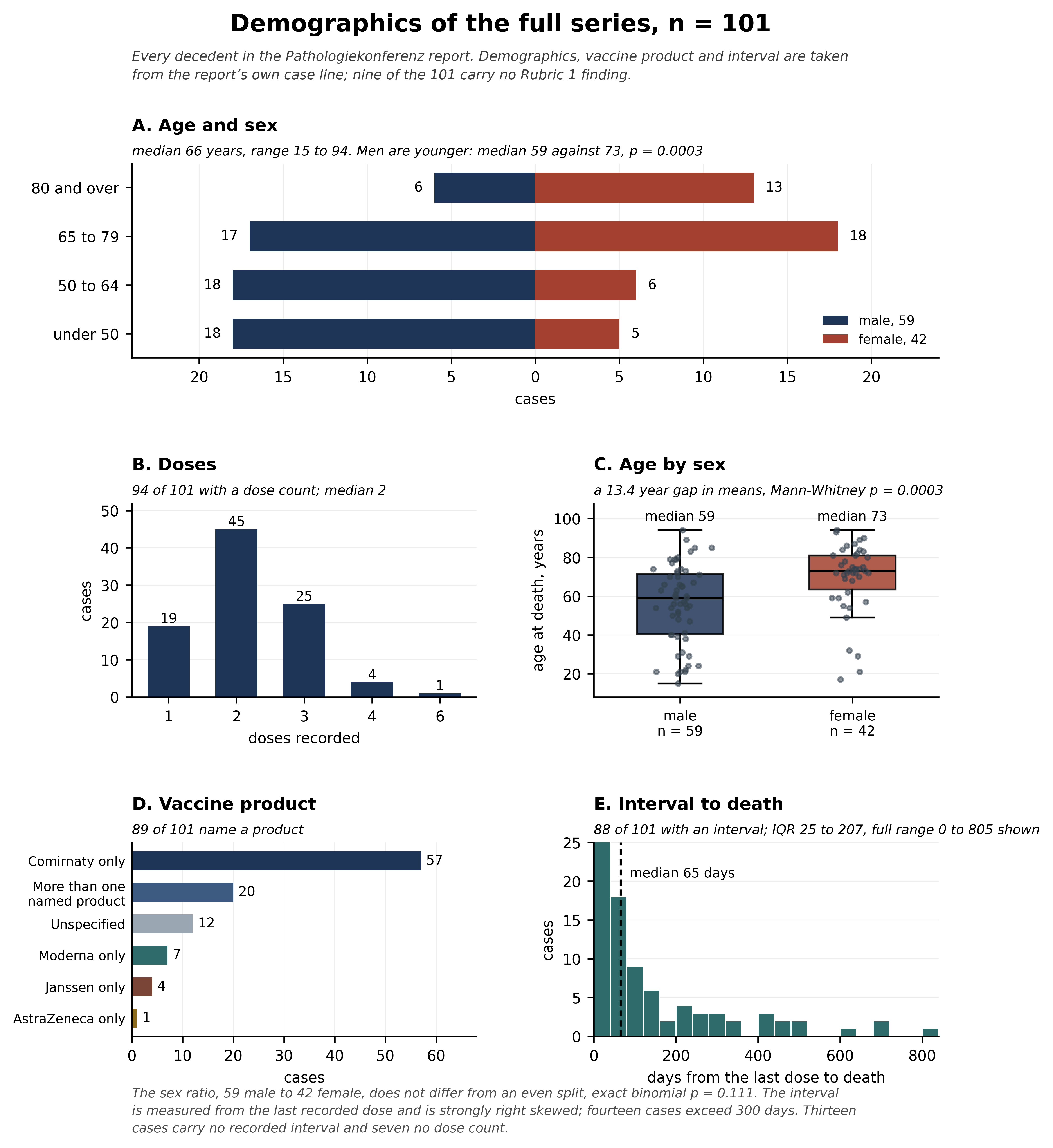
The cohort examined here was a subset of the full n = 101 and was selected for the highly granular data available for analysis.
The demographic data for the entire n = 101 cases is presented below. Overall, 73% of the 101 deaths were considered caused by the Corona vaccines at a level of probable to almost certain. Some causally unrelated cases had tissue evidence of vaccine injury (COF) but were deemed to not be the cause of death.
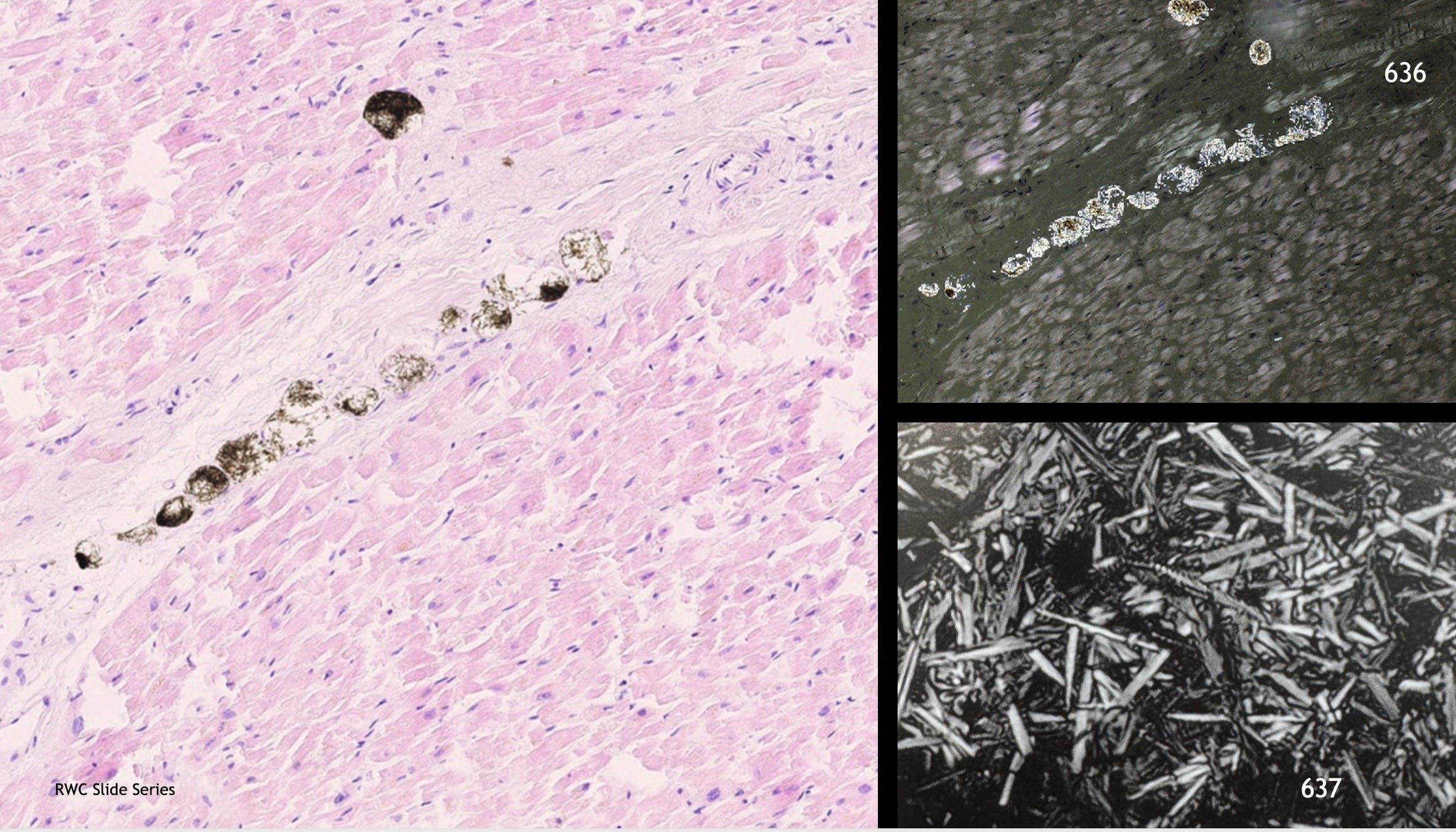
Blackish-brown birefringent (top right) crystalloid material possibly in lipid filled vacuoles (left, seen occupying seams in the myocardium) were present in about half of the autopsy cases. Heart, pancreas, spleen, kidney, lung and liver were the top 6 locations where this material was located. The bottom right laser microscopy image at 6000x magnification highlights the fine structure of this unidentified material. Some of these structures had a cork screw configuration at the end.
Sahin, U., Karikó, K. & Türeci, Ö. mRNA-based therapeutics — developing a new class of drugs. Nat Rev Drug Discov 13, 759–780 (2014). https://doi.org/10.1038/nrd4278
Pfizer BioNTech study #185350, https://archive.org/details/pfizer-study-185350-combined
https://phmpt.org/wp-content/uploads/2022/03/125742_S1_M2_24_nonclinical-overview.pdf
Pfizer Document 5.3.6, https://phmpt.org/wp-content/uploads/2022/04/reissue_5.3.6-postmarketing-experience.pdf
“The work, including its parts, is protected by copyright. The authors are responsible for the content. The authors expressly wish the book contents to be redistributed. They can be reached at: trediton GmbH, Department “Impressumservice,” Halenreie 40–44, 22359 Hamburg, Germany.” Front material in Pathologiekonferenz.
No posts
Comments
Nothing yet. Say the first thing.
Sign in to join the conversation.