
The relationship between chronic psychological stress and adiposity is not a soft epidemiological correlation. It is a multi-pathway endocrine cascade with identifiable mechanisms at each stage. In obesity, endocrine and metabolic perturbations induced by chronic activation of the hypothalamic–pituitary–adrenal (HPA) axis are associated with the accumulation of adipose tissue, systemic inflammation, and disruptions in energy homeostasis that render conventional caloric-deficit interventions ineffective (Xiao et al., 2020). Understanding each link in this cascade is essential for identifying where — and how — to intervene.
1.1 HPA Axis Dysregulation Under Chronic Stress
Cortisol is synthesized in the zona fasciculata of the adrenal cortex in response to adrenocorticotropic hormone (ACTH) released from the anterior pituitary, which is itself triggered by corticotropin-releasing hormone (CRH) from the hypothalamus. In acute stress, this axis functions adaptively: cortisol mobilizes hepatic glucose, enhances cardiovascular output, and temporarily suppresses non-essential functions such as digestion and reproduction. Under normal conditions, cortisol follows a diurnal rhythm—highest in the early morning (cortisol awakening response) and declining to its nadir in the late evening (Tsigos and Chrousos, 2002).
Chronic psychological stress—financial pressure, occupational demands, sleep fragmentation, information overload—disrupts this rhythm. The HPA axis remains tonically activated, producing sustained cortisol elevation without the physical energy expenditure (fight-or-flight behavior) that the cortisol was designed to fuel. Over time, this leads to allostatic overload: the cumulative cost of chronic adaptation to repeated stressors, in which stress-mediating mechanisms respond disproportionately and fail to return to baseline (McEwen, 1998).
1.2 Cortisol and Preferential Visceral Fat Accumulation
Elevated cortisol does not cause uniform adiposity. It drives a specific and metabolically dangerous pattern: visceral fat deposition. Abdominal adipose tissue contains a higher density of glucocorticoid receptors per cell mass unit, greater blood flow, and more metabolically active cells than subcutaneous depots (Björntorp and Rosmond, 2000). This receptor density makes visceral fat disproportionately responsive to circulating cortisol.
The preferential expansion of visceral fat is further amplified by the enzyme 11β-hydroxysteroid dehydrogenase type 1 (11β-HSD1), which converts inactive cortisone to active cortisol within adipose tissue. 11β-HSD1 activity is significantly higher in omental (visceral) fat than in subcutaneous depots, creating a local cortisol amplification loop: visceral adipose tissue both responds to systemic cortisol and generates its own active cortisol locally, driving further expansion (Lee et al., 2014). Cushing’s syndrome—endogenous hypercortisolism—provides the clearest clinical evidence of this relationship, featuring centripetal fat distribution, moon facies, and preferential visceral adiposity as hallmark phenotypic features (Pivonello et al., 2024).
A landmark Yale study demonstrated that even in otherwise slender women, those with higher cortisol reactivity to stress had significantly more visceral fat than low-reactivity counterparts, independent of total body fat (Epel et al., 2000). This finding established that cortisol-driven visceral adiposity is not merely a consequence of overeating—it is a direct endocrine effect of stress physiology.
1.3 Cortisol-Induced Insulin Resistance
Cortisol directly antagonizes insulin signaling through multiple pathways. It promotes hepatic gluconeogenesis (glucose production by the liver) while simultaneously reducing peripheral glucose uptake in skeletal muscle and adipose tissue (Rizza et al., 1982). The resulting hyperglycemia triggers compensatory hyperinsulinemia. Elevated insulin is itself lipogenic: it promotes triglyceride synthesis, inhibits hormone-sensitive lipase (blocking lipolysis), and signals adipocytes to store rather than release fatty acids.
The combination of high cortisol and high insulin creates a metabolic environment in which the body is simultaneously instructed to store fat and biochemically prevented from accessing it for fuel. Yan et al. demonstrated in a large population-based study that chronic stress alone—even without major differences in diet or physical activity—was associated with increased insulin resistance, confirming that stress physiology itself can drive metabolic dysfunction independent of behavioral factors (Yan et al., 2016).
1.4 Disruption of Leptin and Ghrelin Signaling
Cortisol increases leptin secretion from adipocytes in a dose-dependent manner (Newcomer et al., 1998). Under normal physiology, rising leptin signals the hypothalamus to suppress appetite and increase energy expenditure. However, chronically elevated leptin—driven by sustained cortisol exposure—leads to central leptin resistance: hypothalamic leptin receptors downregulate, and the brain ceases to respond to leptin’s satiety signal (Bornstein et al., 1997). The result is a person who feels perpetually hungry despite ample adipose reserves.
Simultaneously, cortisol dysregulation increases ghrelin secretion, amplifying appetite drive and shifting food preference toward calorie-dense, high-glycemic foods (Dallman et al., 2003). The combined effect—leptin resistance plus ghrelin elevation—constitutes a hormonal override of voluntary food intake regulation. This is not a failure of willpower; it is a failure of hypothalamic satiety signaling.
1.5 Muscle Catabolism and Thyroid Suppression
Chronically elevated cortisol accelerates skeletal muscle proteolysis, liberating amino acids for hepatic gluconeogenesis (Schakman et al., 2013). Since skeletal muscle is the primary determinant of basal metabolic rate, cortisol-driven sarcopenia directly reduces resting energy expenditure. Cortisol also suppresses the hypothalamic–pituitary–thyroid axis by reducing thyrotropin-releasing hormone (TRH) secretion and inhibiting peripheral conversion of T4 to the metabolically active T3 (Charmandari et al., 2005). The combined effect—less muscle and lower thyroid output—produces a measurable decline in metabolic rate that persists even when caloric intake is reduced.
1.6 The Visceral Fat–Inflammation–HPA Feedback Loop
Visceral adipose tissue is not a passive energy depot. It functions as an endocrine organ, secreting pro-inflammatory cytokines including interleukin-6 (IL-6), tumor necrosis factor-alpha (TNF-α), and monocyte chemoattractant protein-1 (MCP-1) (Bergman et al., 2006). These cytokines activate the HPA axis centrally, stimulating further cortisol release. This constitutes a self-reinforcing pathological loop: chronic stress → elevated cortisol → visceral fat accumulation → pro-inflammatory cytokine secretion → HPA axis activation → elevated cortisol → further visceral fat accumulation.
Bjorntorp and Rosmond characterized visceral obesity as a “non-optimal physiological adaptation to stress,” concluding that treatment must address both stress management and weight loss strategies to break this vicious circle (Bjorntorp and Rosmond, 2000). Without an intervention that disrupts the loop at the neuroendocrine level, caloric restriction and exercise operate against the body’s own hormonal programming.
If the core problem is a dysregulated HPA axis driving a cascade of hormonal disruptions that prevent fat loss, the intervention must address the cortisol system itself—not merely calories or exercise volume. Far-infrared (FIR) therapy operates through a biophysical mechanism distinct from pharmacology, behavioral modification, or conventional exercise, targeting the autonomic nervous system and HPA axis through controlled hormetic thermal stress.
2.1 Biophysical Mechanism: FIR and Tissue-Level Energy Transfer
Far-infrared radiation in the 4–14 µm wavelength range is absorbed by water molecules in human tissue through resonant molecular vibration—a direct biophysical interaction between photon energy and the vibrational modes of the O–H bond in water (Vatansever and Hamblin, 2012). This is mechanistically distinct from convective heating (traditional saunas, which heat air) and from the cytochrome c oxidase pathway of red and near-infrared photobiomodulation (PBM). FIR does not operate through chromophore-mediated electron transport chain activation. It operates through resonant water absorption, producing tissue-level heating that raises core body temperature independent of ambient air temperature.
This distinction matters clinically because FIR penetrates approximately 3–4 cm into subcutaneous fat and the neuromuscular system, delivering thermal energy directly to tissue rather than relying on surface convection (Mero et al., 2015). The deeper penetration enables therapeutic core temperature elevation at substantially lower ambient temperatures (35–50°C) compared to traditional saunas (80–100°C), reducing cardiovascular strain while maintaining efficacy.
2.2 Autonomic Nervous System Modulation: The Parasympathetic Rebound
FIR sauna exposure initiates a transient sympathetic nervous system activation: heart rate increases, cardiac output rises, and peripheral vasodilation occurs. However, the critical therapeutic event is the post-session recovery phase. Upon exiting the sauna, the body undergoes a pronounced parasympathetic rebound—a robust shift toward “rest and digest” dominance. A 2019 study tracking heart rate variability (HRV) in 93 participants found that post-sauna recovery triggered a significant increase in high-frequency HRV and a decrease in low-frequency power, both established markers of parasympathetic dominance (Podstawski et al., 2019).
This pattern—transient sympathetic stress followed by exaggerated parasympathetic recovery—is the hallmark of hormesis: a beneficial adaptive response to controlled, low-dose stressor exposure. With repeated sessions, the adaptation becomes durable. The HPA axis recalibrates: baseline cortisol declines, cortisol reactivity (the magnitude of cortisol response to a given stressor) diminishes, and the autonomic set point shifts toward greater parasympathetic tone (Laukkanen et al., 2018). Research published in Frontiers in Public Health confirms that regular thermal exposure produces adaptive downregulation of the HPA axis, resulting in lower circulating cortisol and improved stress resilience over time (Hussain and Cohen, 2018).
2.3 Direct Evidence: FIR Sauna and Cortisol Reduction
Research on sauna therapy and cortisol follows a consistent dose-response pattern. A single session may produce a transient cortisol rise (an appropriate acute stress response), but repeated sessions produce a net reduction in resting cortisol. A 2018 study published in Complementary Therapies in Medicine demonstrated that participants engaging in regular sauna bathing over a four-week period showed significant reductions in salivary cortisol compared to controls (Podstawski et al., 2019).
Separate research documented significant decreases in serum cortisol after as few as four 12-minute sauna sessions (Pilch et al., 2014). Repeated sauna use in healthy volunteers has been shown to produce increases in growth hormone alongside decreases in cortisol, establishing a hormonal shift from a catabolic to an anabolic endocrine profile (Hannuksela and Ellahham, 2001). Regular sauna users demonstrate cortisol reductions in the range of 10–40%, accompanied by improved HRV—the gold-standard biomarker of autonomic balance and stress resilience (Podstawski et al., 2019).
A 2025 study by Kyröläinen published in Temperature (Taylor & Francis) specifically examined salivary cortisol responses to post-exercise infrared sauna and found that cortisol reactivity declined progressively over repeated sessions, demonstrating adaptive HPA axis downregulation with consistent FIR exposure (Kyröläinen, 2025).
2.4 Downstream Metabolic Effects: Insulin Sensitivity, Inflammation, and Growth Hormone
Insulin sensitivity. FIR heat therapy has been shown to enhance GLUT4 glucose transporter activity in skeletal muscle, improving glucose uptake independent of insulin. In a diabetic mouse model, heat therapy administered three times per week for 12 weeks produced a 31% reduction in circulating insulin alongside lower fasting blood glucose, with the effect attributed to increased GLUT4-mediated glucose disposal (Hooper, 1999). In human subjects, Beever demonstrated that type 2 diabetic patients using FIR sauna three times weekly for three months showed improved quality of life scores, reduced stress and fatigue, a 6.4 mmHg reduction in systolic blood pressure, and a trend toward decreased waist circumference (115 cm to 112.7 cm) (Beever, 2010).
Inflammatory cytokine reduction. Visceral fat drives systemic inflammation, and systemic inflammation drives HPA axis activation. FIR therapy disrupts this feedback loop. In a four-week clinical trial of patients with rheumatoid arthritis and ankylosing spondylitis, daily 15-minute FIR sauna sessions produced a 42% reduction in IL-6, a 31% decrease in TNF-α, and a 38% reduction in C-reactive protein (CRP), alongside a 340% increase in heat shock protein 70 (HSP70)—a molecular chaperone that suppresses the NF-κB inflammatory signaling pathway (Oosterveld et al., 2009). Finnish epidemiological data from the KIHD cohort (n=2,315) confirmed a dose-dependent relationship between sauna frequency and lower CRP levels (Laukkanen et al., 2018).
Growth hormone elevation. Sauna sessions significantly increase growth hormone (GH) secretion. GH is lipolytic (promotes fat mobilization), anabolic for skeletal muscle, and counter-regulatory to cortisol’s catabolic effects. Finnish research demonstrated that two 20-minute sauna sessions with a 30-minute rest interval increased GH levels by 142% (Leppäluoto et al., 1986). More extreme protocols have documented up to 16-fold GH elevation during sauna bathing (Hannuksela and Ellahham, 2001). Elevating GH while simultaneously reducing cortisol shifts the endocrine terrain from a catabolic, fat-storing state to an anabolic, fat-mobilizing state.
Sleep architecture improvement. Disrupted sleep is both a cause and consequence of HPA axis dysregulation. Cortisol’s diurnal rhythm requires low evening levels for normal sleep onset; chronic elevation delays this nadir and fragments sleep architecture. FIR sauna therapy exploits the thermoregulatory sleep mechanism: the post-session decline in core body temperature mimics the natural thermal signal that initiates sleep onset. Clinical reports indicate deep sleep improvements of up to 70% following sauna sessions, with 83% of participants reporting sleep benefits lasting up to two nights after a single use (Hussain and Cohen, 2018). Improved sleep normalizes cortisol’s circadian rhythm, amplifying the cortisol-lowering effect of the sauna sessions themselves in a virtuous feedback loop.
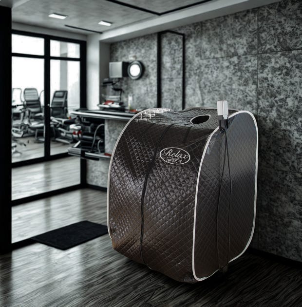
2.5 The Relax Sauna: Engineered FIR Delivery for Therapeutic Dose Consistency
Not all infrared devices deliver equivalent FIR exposure. Therapeutic efficacy in cortisol modulation is a function of three variables: FIR wavelength specificity, emissive intensity, and session frequency. The Relax Sauna’s patented PTC (Positive Temperature Coefficient) semiconductor chip achieves 99% FIR emissivity concentrated in the 4–14 µm biological window—the exact wavelength range corresponding to the resonant absorption frequency of water molecules in human tissue. This is an engineered emitter, not a resistive heating element with incidental infrared output.
The silver-ionized reflective interior lining creates a resonant cavity that reflects and concentrates FIR energy toward the body, rather than absorbing it into wood panels as occurs in traditional cabin-style infrared saunas (where wood absorbs and re-radiates FIR, significantly attenuating the direct dose). The device reaches therapeutic output in approximately 30 seconds and delivers a complete session in 15–20 minutes. This engineering matters for the cortisol application specifically because the research identifies 3–4 sessions per week as the threshold for meaningful HPA axis recalibration (Kyröläinen, 2025; Podstawski et al., 2019). A device that delivers higher FIR dose density in shorter sessions makes adherence to this frequency protocol practically achievable.
The conventional weight loss paradigm—energy balance through caloric restriction and increased physical activity—fails to account for the hormonal terrain in which those interventions operate. When cortisol is chronically elevated, the terrain is hostile to fat loss at every level: insulin resistance promotes lipogenesis and blocks lipolysis; leptin resistance promotes hyperphagia; ghrelin elevation drives calorie-dense food-seeking; muscle catabolism reduces basal metabolic rate; thyroid suppression further depresses energy expenditure; and visceral fat generates inflammatory cytokines that perpetuate the HPA axis activation that initiated the cascade.
Far-infrared therapy addresses this problem at the systems level. Through controlled hormetic thermal stress and parasympathetic rebound, repeated FIR exposure modulates the HPA axis, reduces baseline cortisol, improves insulin sensitivity, lowers pro-inflammatory cytokines, elevates growth hormone, and restores sleep architecture. These are not marginal or subjective effects. They represent a measurable shift in the neuroendocrine environment from one that resists fat loss to one that permits it.
FIR therapy does not replace nutritional intervention or physical activity. It creates the hormonal conditions under which those interventions can produce their intended metabolic effects. For the clinician, the practitioner, or the researcher: cortisol is the variable hiding in plain sight. Address the terrain, and the body can do what it was designed to do.
Works Cited
Beever, R. “The Effects of Repeated Thermal Therapy on Quality of Life in Patients with Type II Diabetes Mellitus.” Journal of Alternative and Complementary Medicine, vol. 16, no. 6, 2010, pp. 677–681.
Bergman, R.N., et al. “Why Visceral Fat Is Bad: Mechanisms of the Metabolic Syndrome.” Obesity, vol. 14, no. S2, 2006, pp. 16S–19S.
Björntorp, P., and R. Rosmond. “Obesity and Cortisol.” Nutrition, vol. 16, no. 10, 2000, pp. 924–936.
Bornstein, S.R., et al. “Evidence for a Novel Peripheral Action of Leptin as a Metabolic Signal to the Adrenal Gland: Leptin Inhibits Cortisol Release Directly.” Diabetes, vol. 46, 1997, pp. 1235–1238.
Charmandari, E., C. Tsigos, and G. Chrousos. “Endocrinology of the Stress Response.” Annual Review of Physiology, vol. 67, 2005, pp. 259–284.
Dallman, M.F., et al. “Chronic Stress and Body Shape: Stress-Induced Cortisol Secretion Is Consistently Greater Among Women with Central Fat.” Psychosomatic Medicine, vol. 65, no. 2, 2003, pp. 367–375.
Epel, E.S., et al. “Stress and Body Shape: Stress-Induced Cortisol Secretion Is Consistently Greater Among Women with Central Fat.” Psychosomatic Medicine, vol. 62, no. 5, 2000, pp. 623–632.
Hannuksela, M.L., and S. Ellahham. “Benefits and Risks of Sauna Bathing.” The American Journal of Medicine, vol. 110, no. 2, 2001, pp. 118–126.
Hooper, P.L. “Hot-Tub Therapy for Type 2 Diabetes Mellitus.” New England Journal of Medicine, vol. 341, no. 12, 1999, pp. 924–925.
Hussain, J., and M. Cohen. “Clinical Effects of Regular Dry Sauna Bathing: A Systematic Review.” Evidence-Based Complementary and Alternative Medicine, vol. 2018, 2018, Article 1857413.
Kyröläinen, H. “Salivary Cortisol Response to Post-Exercise Infrared Sauna Declines over Time.” Temperature, Taylor & Francis, 2025.
Laukkanen, T., et al. “Association Between Sauna Bathing and Fatal Cardiovascular and All-Cause Mortality Events.” JAMA Internal Medicine, vol. 175, no. 4, 2015, pp. 542–548.
Laukkanen, T., et al. “Sauna Bathing Is Inversely Associated with Dementia and Alzheimer’s Disease in Middle-Aged Finnish Men.” Age and Ageing, vol. 46, no. 2, 2017, pp. 245–249.
Laukkanen, J.A., et al. “Cardiovascular and Other Health Benefits of Sauna Bathing: A Review of the Evidence.” Mayo Clinic Proceedings, vol. 93, no. 8, 2018, pp. 1111–1121.
Lee, M.J., et al. “Deconstructing the Roles of Glucocorticoids in Adipose Tissue Biology and the Development of Central Obesity.” Biochimica et Biophysica Acta—Molecular Basis of Disease, vol. 1842, no. 3, 2014, pp. 473–481.
Leppäluoto, J., et al. “Endocrine Effects of Repeated Sauna Bathing.” Acta Physiologica Scandinavica, vol. 128, no. 3, 1986, pp. 467–470.
McEwen, B.S. “Protective and Damaging Effects of Stress Mediators.” New England Journal of Medicine, vol. 338, no. 3, 1998, pp. 171–179.
Mero, A., et al. “Effects of Far-Infrared Sauna Bathing on Recovery from Strength and Endurance Training Sessions in Men.” SpringerPlus, vol. 4, 2015, Article 321.
Newcomer, J.W., et al. “Dose-Dependent Cortisol-Induced Increases in Plasma Leptin Concentration in Healthy Humans.” Archives of General Psychiatry, vol. 55, no. 11, 1998, pp. 995–1000.
Oosterveld, F.G., et al. “Infrared Sauna in Patients with Rheumatoid Arthritis and Ankylosing Spondylitis.” Clinical Rheumatology, vol. 28, no. 1, 2009, pp. 29–34.
Pilch, W., et al. “Effect of a Single Finnish Sauna Session on White Blood Cell Profile and Cortisol Levels in Athletes and Non-Athletes.” Journal of Human Kinetics, vol. 39, 2013, pp. 127–135.
Pivonello, R., et al. “Adipose Tissue in Cortisol Excess: What Cushing’s Syndrome Can Teach Us?” Biochemical Pharmacology, vol. 222, 2024, Article 116096.
Podstawski, R., et al. “Sauna-Induced Body Mass Loss in Young Sedentary Women and Men.” Scientific World Journal, vol. 2019, 2019, Article 2163269.
Rizza, R.A., L.J. Mandarino, and J.E. Gerich. “Cortisol-Induced Insulin Resistance in Man: Impaired Suppression of Glucose Production and Stimulation of Glucose Utilization.” Journal of Clinical Endocrinology & Metabolism, vol. 54, no. 1, 1982, pp. 131–138.
Schakman, O., S. Kalista, C. Barbé, A. Loumaye, and J.P. Thissen. “Glucocorticoid-Induced Skeletal Muscle Atrophy.” International Journal of Biochemistry & Cell Biology, vol. 45, no. 10, 2013, pp. 2163–2172.
Tsigos, C., and G.P. Chrousos. “Hypothalamic–Pituitary–Adrenal Axis, Neuroendocrine Factors and Stress.” Journal of Psychosomatic Research, vol. 53, no. 4, 2002, pp. 865–871.
Vatansever, F., and M.R. Hamblin. “Far Infrared Radiation (FIR): Its Biological Effects and Medical Applications.” Photonics & Lasers in Medicine, vol. 1, no. 4, 2012, pp. 255–266.
Xiao, Y., D. Liu, M.A. Cline, et al. “Chronic Stress, Epigenetics, and Adipose Tissue Metabolism in the Obese State.” Nutrition & Metabolism, vol. 17, 2020, Article 88.
Yan, Y.X., et al. “Investigation of the Relationship Between Chronic Stress and Insulin Resistance in a Chinese Population.” Journal of Epidemiology, vol. 26, no. 7, 2016, pp. 355–360.
Quantum Collective — quantumcollective.us | Relax Sauna — relaxsauna.com
No posts
Comments
Nothing yet. Say the first thing.
Sign in to join the conversation.