
This is Preserving Hope’s first guest-post, but it needn’t be the last. We welcome submissions on anything relevant to the theory and practice of brain preservation. If you’d like to submit a piece, contact Ariel at ariel@brainpreservation.org
In his 23 October article, neuroscientist Ariel Zeleznikow-Johnston raises essential questions: Is terminal lucidity real? And if so, what are the significant implications for neuroscience, medicine, and biostasis?
“But if terminal lucidity is real, it means there are naturally occurring conditions under which even severely damaged brains can have their function restored. Instead of trying to prevent amyloid accumulation—an approach that has largely failed despite billions invested—we could study what’s happening during these episodes. If a dying brain can indeed spontaneously access “lost” memories, perhaps we could induce a living brain to do the same.”
Terminal lucidity describes a specific restoration of neurocognitive function—often seen in dementia, schizophrenia, or severe brain injury—marked by the return of orientation to person and/or place, and coherent thought shortly before death.
As Zeleznikow-Johnston correctly observes, the brains of patients with end-stage dementia are profoundly degraded. Macroscopically, there is pronounced cortical and hippocampal atrophy, widening of the sulci, ventricular enlargement, and an overall reduction in brain volume—changes most pronounced in Alzheimer’s disease, where the medial temporal lobes and association cortices appear markedly shrunken on CT and MRI. Microscopically, these brains exhibit extensive neuronal loss, gliosis, and synaptic loss, accompanied by the widespread deposition of extracellular β-amyloid plaques and intracellular neurofibrillary tangles composed of hyperphosphorylated tau, ultimately leading to the disintegration of brain architecture and severe disruption of neural connectivity. Superficially, this would seem incompatible with any resumption of normal cognition, much less the return of meaningful communication.
However, a closer look at this phenomenon—and at the pathophysiology of Alzheimer-type dementia, where it is most reported—may provide possible explanations.
I should note that I have observed terminal lucidity in long demented and aphasic patients on several occasions. Here, it is essential to consider several key factors that apply to the phenomenon as a whole. The first is that these patients had not undergone brain imaging by CT or MRI, nor had they been imaged and evaluated to determine which brain areas have been degraded, and are metabolically quiescent, and, perhaps more importantly, how connectivity and function may remain between brain areas in these patients.
It is crucial to define precisely what is meant by a “return of cognitive function.” In the cases I’ve observed, the return of improved mental function was limited and was prognostic of impending death. Patients engaged in limited conversation and recognition of loved ones, but dilatory back-and-forth autobiographical conversations did not occur.
Still, this seems astonishing given our perception of the disease burden and its implications for cognition and speech. Here, the words “our perception of the disease burden” are crucial. While “Big Pharma” and a large portion of the medical and neuroscience communities continue to support the β-amyloid hypothesis, recent work has increasingly focused on chronic inflammation and abnormal activation of brain microglia [2] [3] [4]. This view is supported by numerous clinical observations that there is a subset of patients with preserved cognitive function despite an extensive β-amyloid brain burden (and distribution), typically indistinguishable from that seen in severely demented patients[5] [6]. These patients also experience the gross neuroanatomical changes associated with Alzheimer’s Disease (AD) as listed above[7] [8] [9]. This suggests that β-amyloid may not be the root cause of the disease and that other mechanisms are likely at play.
Similarly, it is essential to consider the course and progression of AD in the overall patient population. Anyone who has cared for AD patients in a clinical or extended care facility (ECF) setting will be familiar with the significant variations in mental clarity and cognitive function that often occur diurnally (”sundowning”) and over the long-term course of the disease [10] [11]. Patients have good days and bad days; it is not uncommon for individuals who have been disoriented, delusional, and incoherent to experience periods—lasting hours or even a whole day—of remarkable lucidity and orientation[12] [13] [14]. These “lucid episodes” tend to decrease in frequency as the disease progresses, often ceasing altogether for intervals of months to years before death occurs. This waxing and waning of lucidity closely resembles the clinical course of motor and sensory function impairment in multiple sclerosis and other neuroinflammatory diseases, where fluctuating gradations of inflammation—the primary underlying mechanism—are present. Thus, exacerbations and improvements in the patients’ condition may not represent irreversible microstructural obliteration of brain structure.
It is also the case that some patients with end-stage dementia retain remarkable preservation of procedural memory, as evidenced by their ability to play musical instruments, such as the piano, when cued by being placed on the keys [15]. Interestingly, it has been observed that the music they play is most often that which has personal emotional gravitas, suggesting an interaction between different areas of the brain. The question that apparently is not asked or answered in these cases is: what is the neuroanatomical condition of patients who experience lucid intervals, and, just as importantly, what is the microscopic condition of the brains of these patients?
In many patients with early AD, and many more patients with mild cognitive impairment, the macroscopic and microvascular changes in the brain are extensive, or to put it more subjectively, shocking. There is often marked cerebral atrophy (a ~30% loss of brain mass) and extensive white matter changes indicative of microangiopathy. And yet, many such patients exhibit only mild symptoms, which caused them to seek medical attention and advanced imaging. The reality is that most patients with these conditions do not seek medical attention or are not evaluated until their activities of daily living are compromised. In any event, the degree of pathology that is compatible with rational speech, autobiographical memory, and social interaction is sometimes astounding.
Similarly, there are no robust studies of the histological or ultrastructural condition of the brains of patients as they proceed toward end-stage dementia. Neuroscientists are typically not clinicians, and this may create a blind spot in their understanding of the disease burden associated with irreversible loss of essential brain structure and, thus, function. The reality is that we know very little about the extent to which metabolic and functional linkages in the brain persist in advanced dementia, and we similarly— and perhaps crucially— do not know to what extent underlying neuroinflammation masks the preservation of what would otherwise be functional structure. Neurologists specializing in treating dementia increasingly see patients with “mild” or even no cognitive impairment, despite extensive pathological changes present in neuroimaging. The introduction of β-amyloid and tau radionuclide imaging has made this discrepancy increasingly apparent.
The question of how much brain structure is necessary for normal—or even above-average—cognitive function is perhaps best illustrated by adults with massive hydrocephalus, whose brains are reduced to a thin cortical mantle pressed against the skull (Figures 1 and 2).[16] [17]
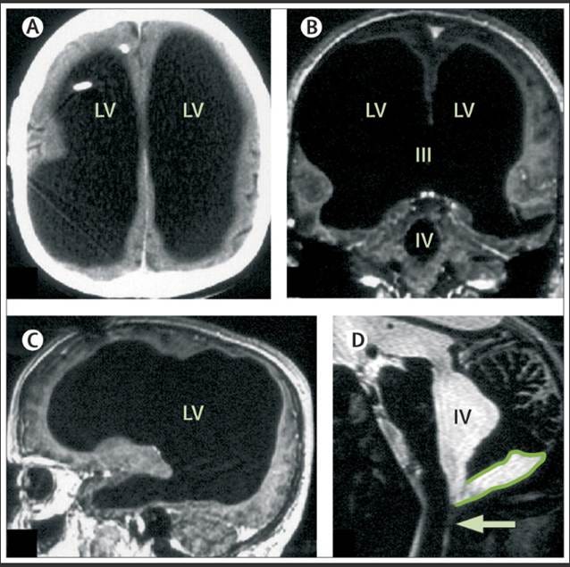
Figure 1: Massive ventricular enlargement in a patient with normal social functioning(A) CT; (B, C) T1-weighted MRI, with gadolinium contrast; (D) T2-weighted MRI. LV=lateral ventricle. III=third ventricle. IV=fourth ventricle. Arrow = Magendie’s foramen. The posterior fossa cyst is outlined in (D). This image is from Feuillet et al., see reference 20.
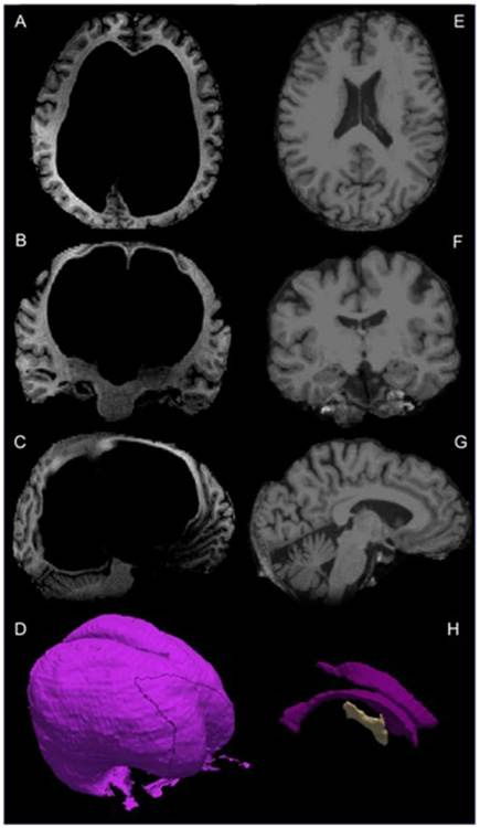
Figure 2: T1-weighted MRI images from Patient CS and an age- and sex-matched healthy control (HC1, age 60). Panels A–C show transverse, coronal, and sagittal slices of Patient CS’s brain, while panels E–G display the corresponding views for the control participant. Panel D depicts a three-dimensional reconstruction of Patient CS’s ventricular system. Panel H shows 3-D models of the left, right, and third ventricles generated from the Freesurfer average brain template derived from 35 individuals, as described by Dale et al. [18] This image is from Alders et al., see reference 19.
The typical brain structures in these patients have been massively compressed (and in some instances possibly obliterated) and cannot be evaluated by routine clinical neuroanatomical criteria. Volumetric analyses sometimes show only 10–20% of normal brain volume in individuals with intact social and intellectual function [19] [20]. Neuroscientists attempting to conduct micro- and macro-connectomic studies (postmortem) in such cases would be flummoxed and would be forced to develop refined or alternative modalities [21]. Indeed, in the mid-1980s, when I showed similar images of a functionally normal patient to a neurologist and neuroscientist, [22] he flatly stated that the patient must be demented and refused to believe that anything even approaching normal cognition was possible in such a case. Since that time, this phenomenon has been validated repeatedly by other clinicians [23] [24]. This phenomenon has also been observed in rats [25].
Also of possible relevance is the transient restoration of consciousness and communication observed in a small subset of patients with chronic disorders of consciousness (DOC)—such as minimally conscious or vegetative states—after administration of the Z-drug zolpidem1 [26] [27] [28] [29]. In these patients—especially those with severe hypoxic-ischemic injury—the brain appears catastrophically damaged on CT or MRI. Yet, responders typically retain the structural integrity of the anterior forebrain—especially the thalamus, striatum, and cortical mantle—despite diffuse injury [30]. Functional imaging in responders demonstrates profound baseline hypometabolism in fronto-parietal and thalamo-striatal systems, followed by marked focal increases in cerebral perfusion and metabolism—again, particularly in the frontal lobes, thalamus, and striatum—during the zolpidem-induced interval of improved function [31]. In contrast, non-responders tend to exhibit extensive cortical necrosis, disruption of white matter pathways, or brainstem injury that precludes re-engagement of the anterior forebrain network [32]. The point is that patients with apparently devastating global brain injury—who historically never recovered—may nonetheless retain the physical brain structures encoding personal identity.
Neuroscientists who claim that brains undergoing 30–50% dehydration during conventional cryoprotective perfusion have lost essential synaptic or macroscopic neural connectivity (and, by extension, their personal identity) should exercise caution before drawing such conclusions. While criticism of such preservation methods—relative to aldehyde-stabilized cryopreservation—is warranted, definitive conclusions should await direct ultrastructural evaluation of rehydrated tissue, or dehydrated tissue subjected to expansion microscopy by transmission electron microscopy (TEM) or focused-ion-beam scanning electron microscopy (FIB-SEM) to confirm or refute neuronal and connectome preservation. It is worth remembering that patients often make complete or nearly complete recoveries from severe traumatic brain injuries that would likely present a dismal to hopeless picture if they were evaluated by TEM or FIB-SEM immediately after the injury and before neuronal repair and rewiring restore function. Thus, it would seem prudent for biostasis neuroscientists to establish a reference gallery of what constitutes truly irreparable damage under natural conditions of injury and recovery.
A perhaps related phenomenon which has had a direct adverse effect on biostasis patients is the “terminal rally,” the “last rally, or the “last surge” which is different from terminal lucidity. In the terminal rally, acutely dying patients briefly recover from a short period of obtundation or unconsciousness and/or state that they are “feeling much improved” within hours or minutes before dying [33] [34]. I have observed this half a dozen times in long-obtunded or terminally ill patients dying from cancer, renal failure, or other chronic diseases. Patients who have been acutely unresponsive (days to a week or two), confused, or immobilized from fatigue may suddenly become lucid, recognize loved ones, speak coherently, and behave appropriately.
The hazard this poses for patients presenting for biostasis is that it is often mistaken for a sign that the patient’s condition has markedly improved and stabilized. This is especially problematic in patients who do not have an explicit terminal diagnosis, but whose condition is grave due to their dire global health situation. For example, patient A-1001, cryopreserved in 1976, was a long-bed-bound 79-year-old male with a long history of Type II diabetes, systemic atherosclerosis, left cerebral infarct (1967), and severe bacterial pneumonia (the proximate cause of medicolegal death (MLD)). After days of being hypoxic and obtunded or unconscious, the patient unexpectedly rallied, sat up in bed, and requested and ate a full dinner. This was so reassuring that most of the standby, stabilization, and transport (SST) team were allowed to take a much-needed break and go to dinner at a nearby restaurant, during which the patient experienced MLD [35]. Dr. James Beford, the first man to be cryopreserved, also experienced much improved well-being shortly before dying, and this also resulted in his being unattended when he experienced MLD [36].
The cause of the terminal rally is unknown. Several speculative mechanisms have been proposed, including alterations in neurotransmitter balance, particularly catecholamine and serotonin levels, and the terminal release of endorphins, which may briefly restore consciousness. Another putative mechanism is that reduced cerebral inhibition from hypoxia or alterations in regional perfusion may transiently disinhibit partially functional neural circuits, producing a short-lived return of coherent function. Except possibly for increased endorphin release, none of these explanations seems credible to the author. In addition to the hazards the terminal rally presents to effective biostasis, it is notorious for causing emotional distress to family members who experience false hope that the patient is recovering, leading them to leave the bedside, resulting in their not being present for their loved one’s death, or worse, causing the loved one to die alone [37] [38].
In summary, it is not unreasonable to postulate, and even prognosticate, that in most patients with end-stage dementia, particularly in extensive multi-infarct dementia, personal identity has been destroyed. Conversely, we should not be surprised when a few patients demonstrate remarkable conservation of brain function and cognition even in the presence of extensive clinical and neuroanatomical damage. The brain is a highly complex and deeply redundant organ. Consider the early to mid-20th-century work of Karl Lashley, who progressively removed portions of the cerebral cortex from rats in an attempt to locate the anatomical site of memory and find the “engram.” After training rats to run mazes, he systematically removed increasing portions of their cerebral cortex to see whether these memories could be erased. Lashley found that no discrete cortical area appeared essential for maze learning; instead, performance declined roughly in proportion to the total amount of cortex removed.[39] From this, he formulated two influential principles: mass action—the idea that the cortex functions as a whole in complex learning—and equipotentiality—the idea that remaining cortical regions can substitute for those destroyed.[40] Thus, terminal lucidity may be a related phenomenon requiring no paradigm shift in the neurobiology of memory and personal identity.
[1] Zeleznikow-Johnston, A. (2024, October 23). Is terminal lucidity real? Or are we just desperate to say one last goodbye? Preserving Hope. https://preservinghope.substack.com/p/is-terminal-lucidity-real
[2] Herrup, K. (2015). The case for rejecting the amyloid cascade hypothesis. Nature Neuroscience, 18(6), 794–799. https://doi.org/10.1038/nn.4017
[3] Heneka, M. T., Carson, M. J., El Khoury, J., Landreth, G. E., Brosseron, F., Feinstein, D. L., Kummer, M. P. (2015). Neuroinflammation in Alzheimer’s disease. The Lancet Neurology, 14(4), 388–405. https://doi.org/10.1016/S1474-4422(15)70016-5
[4] Malpetti, M., Kievit, R. A., Passamonti, L., Jones, P. S., Tsvetanov, K. A., Rittman, T., Rowe, J. B. (2020). Microglial activation and tau burden predict cognitive decline in Alzheimer’s disease. Brain, 143(5), 1588–1602. doi 10.1093/brain/awaa088
[5] Aizenstein, H. J., Nebes, R. D., Saxton, J. A., Price, J. C., Mathis, C. A., Tsopelas, N. D., Ziolko, S. K., James, J. A., Snitz, B. E., Houck, P. R., Bi, W., Cohen, A. D., Lopresti, B. J., DeKosky, S. T., Halligan, E. M., Klunk, W. E., & Becker, J. T. (2008). Frequent amyloid deposition without significant cognitive impairment among the elderly. Archives of Neurology, 65(11), 1509–1517. https://doi.org/10.1001/archneur.65.11.1509
[6] Bennett, D. A., Schneider, J. A., Arvanitakis, Z., Kelly, J. F., Aggarwal, N. T., Shah, R. C., Wilson, R. S., Cochran, E. J., & Wilson, R. S. (2006). Neuropathology of older persons without cognitive impairment from two community-based studies. Neurology, 66(12), 1837–1844. doi: 10.3233/JAD-2009-1227
[7] Price, J. L., & Morris, J. C. (1999). Tangles and plaques in nondemented aging and “preclinical” Alzheimer’s disease. Annals of Neurology, 45(3), 358–368. doi: 10.1002/1531-8249(199903)45:3<358::aid-ana12>3.0.co;2-x.
[8] Villemagne, V. L., Burnham, S., Bourgeat, P., Brown, B., Ellis, K. A., Salvado, O., Szoeke, C., Macaulay, S. L., Martins, R., Maruff, P., Ames, D., Rowe, C. C., & Masters, C. L. (2013). Amyloid-β deposition, neurodegeneration, and cognitive decline in preclinical Alzheimer’s disease. The Lancet Neurology, 12(4), 357–367. doi: 10.1016/S1474-4422(13)70044-9
[9] Villain, N., Chetelat, G., Grassiot, B., Bourgeat, P., Jones, G., Ellis, K. A., Ames, D., Martins, R. N., Eustache, F., Masters, C. L., Rowe, C. C., & Villemagne, V. L. (2012). Regional dynamics of amyloid-β deposition in healthy elderly, mild cognitive impairment, and Alzheimer’s disease. Brain, 135(7), 2126–2139. https://doi.org/10.1093/brain/aws125
[10] Volicer, L., Harper, D. G., Manning, B. C., Goldstein, R., & Satlin, A. (2001). Sundowning and circadian rhythms in Alzheimer’s disease. The American Journal of Psychiatry, 158(5), 704-711. doi: 10.1176/appi.ajp.158.5.704
[11] Lee, D. T. F., Woo, J., Mackenzie, A. E., & Cheng, T. S. (2010). Sundown syndrome in persons with dementia: An update. Dementia and Geriatric Cognitive Disorders, 30(4), 202-208. doi: 10.4306/pi.2011.8.4.275
[12] Normann, H. K., Asplund, K., & Norberg, A. (1989). Episodes of lucidity in people with severe dementia as narrated by formal carers. Journal of Advanced Nursing, 28(7), 1295–1300. DOI: 10.1046/j.1365-2648.1998.00845
[13] Gilmore-Bykovskyi, A., Block, L., Benson, C., & colleagues. (2023). “It’s you”: Caregiver and clinician perspectives on lucidity in people living with dementia. The Gerontologist, 63(1), 13–27. doi: 10.1093/geront/gnac095
[14] Ross, J. P., & Post, S. G. (2024). Lucidity in the deeply forgetful: A scoping review. Journal of Alzheimer’s Disease. Advance online publication. doi: 10.3233/JAD-230094
[15] Crystal, H. A., Grober, E., & Masur, D. (1989). Preservation of musical memory in Alzheimer’s disease. Journal of Neurology, Neurosurgery & Psychiatry, 52(12), 1415–1416. https://doi.org/10.1136/jnnp.52.12.1415
[16] Feuillet, L., Dufour, H., & Pelletier, J. (2007). Brain of a white-collar worker. The Lancet, 370(9583), 262.
[17] Alders, G. L., Smits, M., & van der Flier, W. M. (2018). Volumetric MRI analysis of a case of severe ventriculomegaly. Frontiers in Human Neuroscience, 12, 361. https://doi.org/10.1016/S0140-6736(07)61127-1
[18] Dale, A. M., Fischl, B., and Sereno, M. I. (1999). Cortical surface-based analysis: I. Segmentation and surface reconstruction. Neuroimage 9, 179–194. doi: 10.1006/nimg.1998.0395
[19] Alders, G. L., Smits, M., & van der Flier, W. M. (2018). Volumetric MRI analysis of a case of severe ventriculomegaly. Frontiers in Human Neuroscience, 12, 361. https://doi.org/10.1016/S0140-6736(07)61127-1
[20] Lewin, R. (1980). Is your brain really necessary? Science, 210(4475), 1232–1234. https://doi.org/10.1126/science.7434023
[21] Bigler, E. D. (1988). The neuropsychology of hydrocephalus. Archives of Clinical Neuropsychology, 3(1), 81–100. doi.org/
[22] Lewin, R. (1980). Is your brain really necessary? Science, 210(4475), 1232–1234. https://doi.org/10.1126/science.7434023
[23] Nahm M, Rousseau D, Greyson B. Discrepancy Between Cerebral Structure and Cognitive Functioning: A Review. The Journal of nervous and mental disease. 2017;205:967–972. doi: 10.1097/NMD.0000000000000752
[24] Masdeu, J. C., Pascual, B., Bressi, F., Casale, M., Prieto, E., Arbizu, J., & Fernández-Seara, M. A. (2009). Ventricular wall granulations and draining of cerebrospinal fluid in chronic giant hydrocephalus. Archives of Neurology, 66(2), 262–267. https://doi.org/10.1001/archneurol.2008.547
[25] Ferris, C. F., Cai, X., Qiao, J., Switzer, B., Baun, J., Morrison, T., Iriah, S., Madularu, D., Sinkevicius, K. W., & Kulkarni, P. (2019). Life without a brain: Neuroradiological and behavioral evidence of neuroplasticity necessary to sustain brain function in the face of severe hydrocephalus. Scientific Reports, 9(1), 16479. https://doi.org/10.1038/s41598-019-53042-3
[26] Shames, J. L., & Ring, H. (2008). Transient reversal of anoxic brain injury–related minimally conscious state after zolpidem administration: A case report. Archives of Physical Medicine and Rehabilitation, 89(2), 386–388. https://doi.org/10.1016/j.apmr.2007.08.137
[27] Kim, C., Kwon, B. S., Nam, K. Y., Park, J. W., & Lee, H. J. (2016). Zolpidem-induced arousal by paradoxical GABAergic stimulation: A case report with F-18 flumazenil positron emission tomography and single photon emission computed tomography study. Annals of Rehabilitation Medicine, 40(1), 177–181. https://doi.org/10.5535/arm.2016.40.1.177
[28] Sutton, J. A., & Clauss, R. P. (2017). A review of the evidence of zolpidem efficacy in neurological disability after brain damage due to stroke, trauma and hypoxia: A justification of further clinical trials. Brain Injury, 31(8), 1019–1027. https://doi.org/10.1080/02699052.2017.1300836
[29] Cohen, S. I., & Duong, T. T. (2008). Increased arousal in a patient with anoxic brain injury after administration of zolpidem. American Journal of Physical Medicine & Rehabilitation, 87(3), 229–231. https://doi.org/10.1097/PHM.0b013e318161971
[30] Williams, S. T., Conte, M. M., Goldfine, A. M., Noirhomme, Q., Gosseries, O., Thonnard, M., Beattie, B., Hersh, J., Katz, D. I., Victor, J. D., Laureys, S., & Schiff, N. D. (2013). Common resting brain dynamics indicate a possible mechanism underlying zolpidem response in severe brain injury. eLife, 2, e01157. https://doi.org/10.7554/eLife.01157
[31] Brefel-Courbon C, Payoux P, Ory F, Sommet A, Slaoui T, Raboyeau G, Lemesle B, Puel M, Montastruc JL, Demonet JF, Cardebat D. Clinical and imaging evidence of zolpidem effect in hypoxic encephalopathy. Ann Neurol. 2007 Jul;62(1):102-5. doi: 10.1002/ana.21110. PMID: 17357126.
[32] Hermoye, L., Lemaire, J.-J., & Laureys, S. (2013). Neuroimaging of patients with disorders of consciousness: From bench to bedside? Future Neurology, 8(6), 601–603. https://doi.org/10.2217/fnl.13.53
[33] Fenwick, P., Lovelace, H., & Brayne, S. (2010). Comfort for the dying: Five-year retrospective and one-year prospective studies of end of life experiences. Archives of Gerontology and Geriatrics, 51(2), 173–179. https://doi.org/10.1016/j.archger.2009.10.004
[34] Brayne, S., Lovelace, H., & Fenwick, P. (2008). End-of-life experiences and the dying process in a Gloucestershire nursing home as reported by nurses and care assistants. American Journal of Hospice and Palliative Medicine, 25(3), 195–206. doi: 10.1177/1049909108315302
[35] Darwin, M. G. (n.d.). Cryopreservation patient A-1001, case report: Frederick Rockwell Chamberlain, Jr. [Unpublished manuscript].
[36] Nelson, R. F. (1973). We froze the first man. Chilton Book Company.
[37] Crossroads Hospice & Palliative Care. (2022, September 7). End-of-life rally: How to make the most of it. Hospice Education, Caregiving Tips. https://www.crossroadshospice.com/hospice-palliative-care-blog/2022/september/07/end-of-life-rally-how-to-make-the-most-of-it/
[38] Karlawish, J., Peterson, A., Kleid, M., Harkins, K., Largent, E. A., Stites, S. D., Coykendall, C., & Clapp, J. T. (2024). Caregiver accounts of lucid episodes in persons with advanced dementia. The Gerontologist, 64(6), gnad170. https://doi.org/10.1093/geront/gnad170
[39] Lashley, K. S. (1950). In search of the engram. Symposia of the Society for Experimental Biology, 4, 454–482.
[40] Lashley, K. S. (1929). Brain mechanisms and intelligence: A quantitative study of injuries to the brain. Chicago, IL: The University of Chicago Press.
It is important to emphasize that these patients do not regain baseline function and remain significantly impaired in both neurocognitive and motor domains.
No posts
Comments
Nothing yet. Say the first thing.
Sign in to join the conversation.