
Living cells are in a constant state of chemical activity.
They consume fuel, transfer electrons, maintain ion gradients, repair damage, respond to stress, and neutralize reactive molecules. Most of the energy moving through these processes becomes biological work or heat. A minute fraction can emerge as light.
This phenomenon is called ultraweak photon emission, or UPE. The photons are often called biophotons, although UPE is the more precise term. The particles themselves are ordinary photons. What is unusual is the extremely low rate at which they are detected.
Reported surface emissions commonly range from a few to several thousand photons per square centimeter per second, depending on the organism, physiological state, spectral window, detector, and experimental conditions. Measured wavelengths can extend from the near-ultraviolet through visible light and into the near-infrared. These levels are far below human visual sensitivity. Detecting them requires light-tight chambers, low-noise photomultiplier tubes or cooled cameras, long exposures, careful calibration, and strict control of background light. [1]
UPE is not the same process that makes a firefly glow. Bioluminescent organisms possess specialized chemical systems that produce visible light, often for functions such as signaling, hunting, or defense. UPE is also distinct from fluorescence, which depends on prior excitation by an external light source, and from the thermal infrared radiation associated with body heat.
Another important distinction is delayed luminescence, the faint afterglow that can persist after a biological sample has been illuminated. UPE is generated spontaneously by ongoing chemistry. Delayed luminescence follows exposure to external light and must be carefully controlled in experiments. [9]
UPE is a form of endogenous, ultraweak chemiluminescence produced primarily during oxidative metabolic and stress reactions.
Where the Photons Come From
Oxygen makes efficient energy metabolism possible, but oxygen chemistry also generates reactive oxygen species. These include superoxide, hydrogen peroxide, hydroxyl radicals, and singlet oxygen.
Reactive oxygen species are not simply biological poisons. At controlled levels, they participate in cellular signaling, immune defense, adaptation, and regulation. When their production overwhelms antioxidant and repair systems, however, they contribute to oxidative stress and can damage lipids, proteins, pigments, and nucleic acids.
Some oxidative reactions produce electronically excited molecular species. These species contain more energy than they do in their stable ground states. When they relax, part of the excess energy can be released as a photon.
The basic sequence is
Metabolism or stress
→ reactive oxygen chemistry
→ oxidation of biomolecules
→ electronically excited products
→ photon emission
Known emitting species include triplet-excited carbonyls, excited pigments, and singlet oxygen. Because these products release energy in different spectral regions, wavelength distribution may carry more useful information than total brightness alone. [1][2]
UPE does not report every biochemical event occurring inside an organism. It preferentially reflects reactions capable of producing excited molecular states, followed by the fraction of emitted photons that escape the tissue and reach a detector.
The signal is therefore an optical consequence of chemistry occurring within a biological system.
A Faint Record of Physiological Activity
Because UPE is linked to oxidative metabolism, its intensity and distribution can change when physiology changes.
In a 2009 study, researchers placed five healthy volunteers in a light-tight room and imaged their upper bodies with a cryogenically cooled charge-coupled device camera. Photon emission was weakest during the morning, increased through the afternoon, peaked in the late afternoon, and then declined.
Its spatial pattern differed from the participants’ thermal images, and photon intensity did not significantly correlate with oral temperature. This supported the conclusion that UPE was not simply tracking body temperature. The study was small and did not establish a clinical test, but it demonstrated that human UPE can be spatially imaged and that the measured signal varies over time. [3]
Plants provide an especially useful experimental system because injury can be introduced at a known location and the resulting response can be followed directly.
When researchers mechanically wounded Arabidopsis thaliana leaves, photon emission increased around the damaged tissue. The spectral pattern changed as well, consistent with wound-induced oxidative chemistry and the formation of excited molecular products. [4]
The emission provided a measurable trace of the plant’s biochemical response while that response was unfolding.
A scar preserves evidence that an event occurred. UPE may reveal part of the chemistry taking place during the event itself.
What Recent Imaging Added
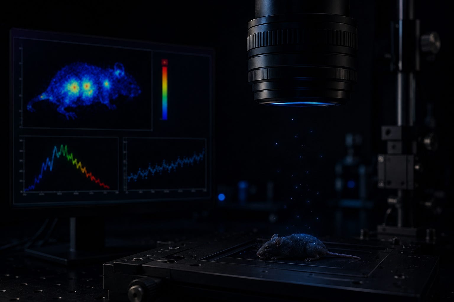
In 2025, researchers used highly sensitive EMCCD and CCD cameras to compare ultraweak emission from living mice with measurements taken from the same animals after death. They reported significant optical contrast between the two conditions.
The same investigation examined plants exposed to increased temperature, physical injury, and several chemical treatments. Heat and damage increased the detected emission, while chemical exposure altered its characteristics. The work demonstrated that modern sensors can produce spatial images from extremely low photon counts. [5]
The result does not create a universal optical definition of life or death. It came from particular organisms, detectors, procedures, temperatures, and observation periods. It also does not mean every living tissue produces one characteristic brightness or that postmortem emission follows a universal timetable.
Its importance is more practical: living and postmortem conditions produced detectably different optical patterns under the study’s experimental setup, while stress altered the signal generated by plants.
Sensitive instruments are beginning to make extremely faint biological chemistry visible.
Information Is Not Automatically Communication
A photon can carry information about the reaction that produced it.
Its wavelength, timing, intensity, and location may correlate with oxidation, metabolism, injury, temperature, inflammation, or another physiological condition. Researchers can potentially use those correlations even when the organism did not evolve the emission for signaling.
That does not establish that cells use UPE as a communication channel.
Smoke contains information about combustion, but a fire does not necessarily produce smoke to send a message. In the same way, UPE may be scientifically informative while remaining a chemical byproduct.
Establishing a genuine optical signaling mechanism would require more than detecting photons or finding correlations between nearby biological samples. Researchers would need to identify
a defined emitter
a biological receiver
sensitivity to particular wavelengths or intensities
a reproducible physiological response
loss of that response when the optical path is blocked
restoration through wavelength- and dose-matched artificial light
exclusion of chemical, electrical, thermal, acoustic, and mechanical alternatives
independent replication
Photonic communication remains a legitimate research question. It is not a general conclusion established by the existence of UPE. Current reviews describe possible biological roles and suggestive experiments, but persistent problems remain with confounding factors, signal intensity, receptor mechanisms, and experimental control. [9]
UPE also should not be assumed to form a coherent quantum field. A critical review of photon-count statistics concluded that biological UPE itself is experimentally well established, but found no reliable evidence that it possesses the claimed coherence or other nonclassical optical properties. [6]
From Faint Emission to Biological Measurement
The strongest near-term opportunity may not be decoding a cellular language. It may be developing a new measurement channel.
Researchers have explored UPE in connection with
plant injury and environmental stress
oxidative reactions in cells and tissues
changes in skin and whole-body physiology
seed condition and germination potential
responses to drugs or experimental treatments
food quality and deterioration
A 2024 study, for example, used a platform to collect both UPE and delayed-luminescence data, with ultraviolet excitation used for the delayed-luminescence component. Researchers combined those optical measurements with germination data from five legume species stored under different conditions.
The resulting models showed species-dependent relationships between photon measurements and seed quality. Some species produced substantially stronger classification performance than others, illustrating both the promise and the difficulty of translating these signals into a general-purpose measurement. [8]
The appeal is straightforward. Because UPE is produced endogenously, some applications might not require fluorescent dyes, radioactive tracers, genetic reporters, or destructive sampling.
The central obstacle is specificity.
Why Brightness Alone Is Not Enough
A rise in photon emission could reflect
increased metabolic activity
oxidative stress
inflammation or tissue injury
heat exposure
chemical treatment
residual light-induced afterglow
At the same time, similar internal chemistry can produce different measurements at the surface. Photons may be absorbed or scattered differently depending on tissue depth, pigmentation, geometry, oxygen availability, and the wavelengths involved.
This creates two separate problems
Different biological processes can produce similar optical signals.
Similar biological processes can produce different detected signals.
A useful instrument must therefore do more than count photons. It must connect an optical pattern to a validated biological state.
That will probably require combining several dimensions
intensity + wavelength + location + timing + physiological context + independent biochemical evidence
The goal is not a universal brightness score. It is a calibrated relationship between a measured optical pattern and a specific, independently verified process.
A faint signal can reveal that something changed. It cannot automatically identify what changed or why.
The Distance Between Detection and Diagnosis
Human UPE has been investigated for decades, but measurement is not the same as clinical validation.
A systematic review published in 2014 searched records through October 2011 and included 56 human studies. Only one was categorized as a randomized controlled trial. Twenty-seven were controlled clinical studies, while twenty-eight were observational or descriptive. The investigators also noted that no established quality-assessment framework fit this body of research particularly well. [7]
That review is now dated and does not include later imaging work. Its value is historical: it shows that the phenomenon had already produced a substantial literature while evidence for dependable diagnostic use remained limited.
For UPE to become a reliable biological or medical tool, studies will need
calibrated and independently characterized detectors
standardized dark-adaptation procedures
explicit control of delayed luminescence
predefined analysis methods
blinded comparisons
adequately powered samples
repeated measurements
external biochemical reference standards
independent laboratory replication
direct estimates of diagnostic sensitivity and specificity
Better cameras can reveal weaker signals. They cannot, by themselves, determine what those signals mean.
A New Window Into Living Systems
The importance of ultraweak photon emission lies in what it may reveal about ordinary biological chemistry.
Metabolic and oxidative reactions can create electronically excited molecules. Some of those molecules release photons as they return to lower-energy states. The collective output is invisible to human eyes but measurable with sufficiently sensitive instruments.
That output can vary with metabolism, time, injury, temperature, oxidative stress, and chemical exposure. It has also been observed to change after death under specific experimental conditions. The pattern recorded by an instrument is influenced both by the biology producing the light and by the physical conditions governing whether that light reaches the detector. [1][5]
The scientific question is no longer whether living systems emit extremely faint photons. They do.
The question is whether researchers can separate biologically meaningful patterns from noise, confounding factors, tissue optics, and measurement artifacts. If they can, UPE may become a valuable, noninvasive way to observe selected aspects of physiological change as they occur.
The chemistry is already producing a signal.
The work ahead is learning how to interpret it accurately.
Sources
1. Cifra, M., and Pospíšil, P. “Ultra-Weak Photon Emission from Biological Samples: Definition, Mechanisms, Properties, Detection and Applications.” Journal of Photochemistry and Photobiology B: Biology 139 (2014): 2–10. DOI: 10.1016/j.jphotobiol.2014.02.009.
2. Pospíšil, P., Prasad, A., and Rác, M. “Role of Reactive Oxygen Species in Ultra-Weak Photon Emission in Biological Systems.” Journal of Photochemistry and Photobiology B: Biology 139 (2014): 11–23. DOI: 10.1016/j.jphotobiol.2014.02.008.
3. Kobayashi, M., Kikuchi, D., and Okamura, H. “Imaging of Ultraweak Spontaneous Photon Emission from Human Body Displaying Diurnal Rhythm.” PLOS ONE 4, no. 7 (2009): e6256. DOI: 10.1371/journal.pone.0006256.
4. Prasad, A., Gouripeddi, P., Devireddy, H. R. N., Ovsii, A., Rachakonda, D. P., van Wijk, R., and Pospíšil, P. “Spectral Distribution of Ultra-Weak Photon Emission as a Response to Wounding in Plants: An In Vivo Study.” Biology 9, no. 6 (2020): 139. DOI: 10.3390/biology9060139.
5. Salari, V., Seshan, V., Frankle, L., England, D., Simon, C., and Oblak, D. “Imaging Ultraweak Photon Emission from Living and Dead Mice and from Plants under Stress.” The Journal of Physical Chemistry Letters 16, no. 17 (2025): 4354–4362. DOI: 10.1021/acs.jpclett.4c03546.
6. Cifra, M., Brouder, C., Nerudová, M., and Kučera, O. “Biophotons, Coherence and Photocount Statistics: A Critical Review.” Journal of Luminescence 164 (2015): 38–51. DOI: 10.1016/j.jlumin.2015.03.020.
7. Ives, J. A., van Wijk, E. P. A., Bat, N., et al. “Ultraweak Photon Emission as a Non-Invasive Health Assessment: A Systematic Review.” PLOS ONE 9, no. 2 (2014): e87401. DOI: 10.1371/journal.pone.0087401.
8. Griffo, A., Sehmisch, S., Laager, F., et al. “Non-Invasive Methods to Assess Seed Quality Based on Ultra-Weak Photon Emission and Delayed Luminescence.” Scientific Reports 14 (2024): 26838. DOI: 10.1038/s41598-024-74207-9.
9. Mould, R. R., Mackenzie, A. M., Kalampouka, I., et al. “Ultra Weak Photon Emission: A Brief Review.” Frontiers in Physiology 15 (2024): 1348915. DOI: 10.3389/fphys.2024.1348915.
No posts
Comments
Nothing yet. Say the first thing.
Sign in to join the conversation.