
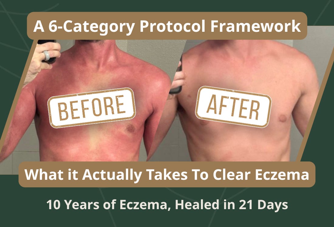
If you missed Part 1: Ethan came to us with full-body eczema severe enough to cost him work and basic daily function. His dermatologist had him on steroid creams. His GI doctors had told him everything looked fine. Neither had run a stool test.
We ran one. Here’s what it showed:
Active H. pylori infection.
Pancreatic elastase below normal, meaning his digestive capacity was compromised at the source.
Fecal calprotectin elevated - active intestinal inflammation, the kind that reflects real immune activation, not just a “sensitive gut.”
Secretory IgA elevated - his mucosal immune system was in a state of ongoing immune engagement.
Akkermansia muciniphila: undetectable. This is an important species but it was absent.
Dysbiotic bacterial overgrowth pattern across multiple species: Bacillus, Pseudomonas, Staphylococcus, Streptococcus, Prevotella.
His skin was the external manifestation, but his gut was the source.
Here’s the thing about a case like Ethan’s: running the test is not the protocol.
What you do with the data is the protocol.
These findings don’t all resolve the same way. H. pylori requires targeted antimicrobial support. Akkermansia depletion isn’t a “take a probiotic” situation, it requires specific reseeding and a mucosal terrain capable of supporting its return. Elevated calprotectin tells you the gut lining is under active immune assault and can’t begin repairing until that assault is addressed. Low elastase tells you every meal Ethan ate was arriving in his small intestine only partially broken down, creating a constant antigenic load.
None of this is visible from the outside. None of it shows up in standard bloodwork. None of it is on the radar of a practitioner who doesn’t test for it.
More than 50% of the global population carries H. pylori - most never know it [1]. The connection between reduced gut microbial diversity and eczema severity is documented in peer-reviewed literature [12]. Ethan’s case isn’t an anomaly. It’s a pattern I’ve seen across thousands of patients.
Below, I’m sharing the mockup protocol categories I built for this case: what each category addresses, why this specific patient needed it, and what class of intervention we used. This isn’t a copy-paste protocol. It’s an education in what real root-cause work looks like. If you recognize yourself in Ethan’s story, that recognition matters. I’ll show you what to do about it at the bottom.
Comments
Nothing yet. Say the first thing.
Sign in to join the conversation.