
I am thrilled to share today’s article with you. It is the most inspiring observation I have made since I began pursuing cancer treatment through repurposed drugs. It has now been six years since Dr. Harvey Risch replied to my email within five minutes on a Saturday afternoon in 2020.
Today’s hypothesis is the most promising refinement to the cancer protocols since my collaboration with Dr. Paul Marik began two years ago. For regular readers, this piece provides the evidence and PubMed support behind my last two articles. For newcomers, it offers a brief summary of the framework.
Last summer, Dr. Paul Marik and I published a brochure on the ‘10 Deadliest Cancers,’ presenting an evidence-based repurposed-drug protocol for addressing them.
The ‘10 Deadliest Cancers’ framework emerged from a Substack post I wrote on April 20, 2025, in which I used a power-plant metaphor to explain the Warburg effect and the roles of HIF-1 alpha, lactate, c-Myc, and hexokinase 2 in these ten lethal cancers.
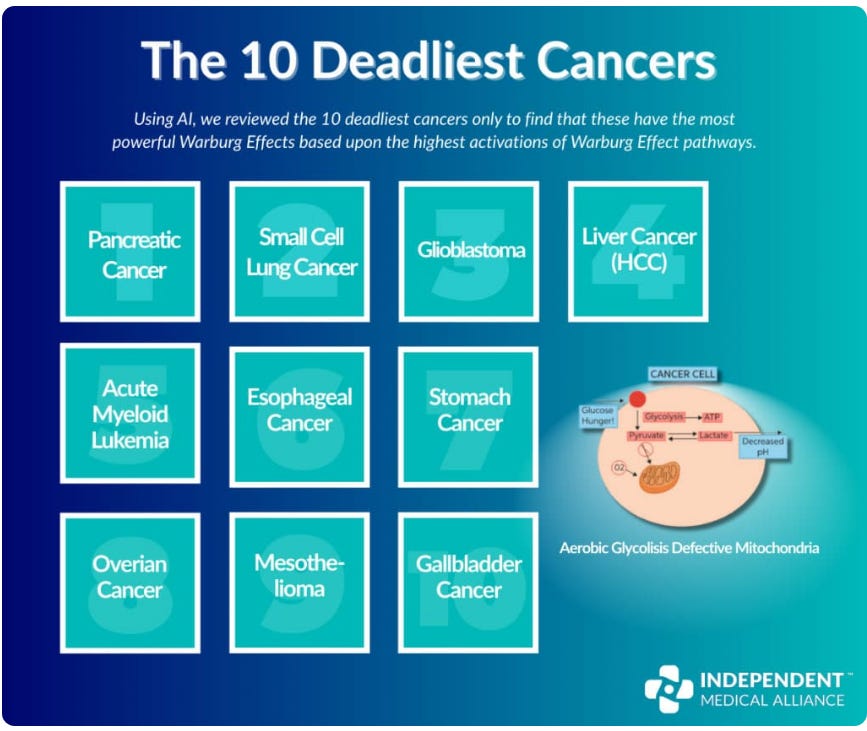
Our work on cancer stem cell and metabolic pathways led Dr. Marik and me to conclude that blocking the molecular drivers of aerobic glycolysis could suppress cancer. Here is a quick AI summary:
HIF-1 alpha, lactate, and Hexokinase 2 act as a highly synchronized, self-sustaining feedback loop that drives and locks cancer cells into the Warburg effect. Together, they ensure that the cell continuously prioritizes rapid glucose consumption and lactate production, even when oxygen is fully available.
From that point, we derived a set of Warburg-blocking agents that now form the backbone of many of our protocols, published on the IMA website as the “10 Deadliest Cancers” monograph.
However, when I recently reviewed this list of deadly cancers I was struck by another common denominator of these 10 deadly cancers and it was beyond the Warburg Effect.
It has to do with the desmoplastic shield: a fibrous, scar-like enclosure that many solid tumors form, creating a hypoxic core where immune cells fail to protect us and can instead be co-opted by the tumor.
The most extreme example is that of Pancreatic Cancer where the pressures inside this enclosed cancer-friendly stronghold are so high that blood vessels collapse, and no therapeutics, whether traditional chemotherapy or repurposed drugs can enter.
It is the tumor’s walled-off garrison, allowing the cancer to operate with impunity. Inside are high levels of lactic acid and Tregs, MDSCs, and special T cells expressing PD-L1, all our own immune cells that have been turned against us and now fight for the cancer’s survival.
All of this is documented in Dr. Marik's comprehensive, highly referenced Cancer Care Volume 2—well-established science within our field.
But what was new to me was that virtually all solid lethal cancers have a desmoplastic shield and the deadliest, in general, have the most developed. Furthermore, the strength of this shield correlates directly with the degree of hypoxia and immune evasion.
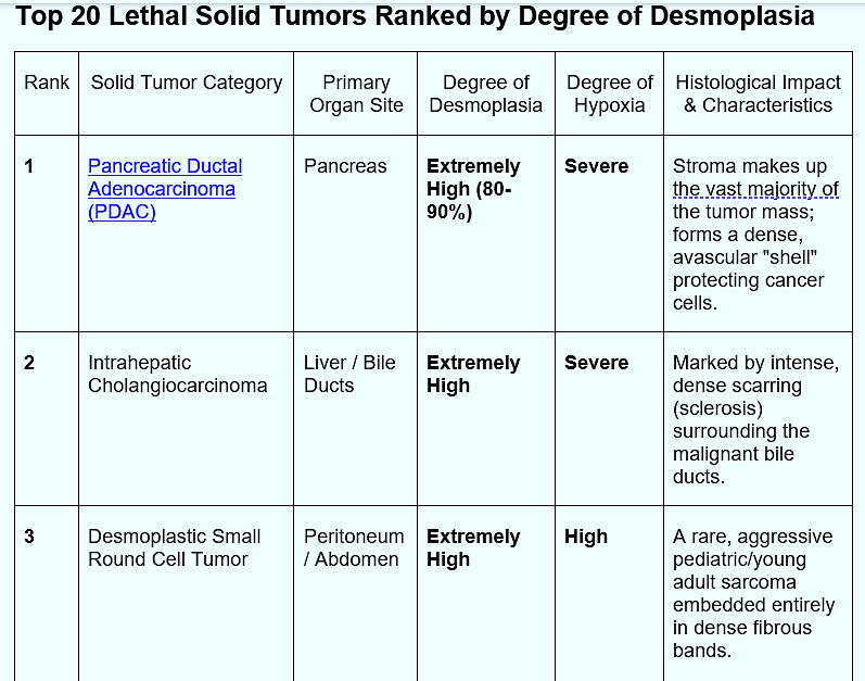
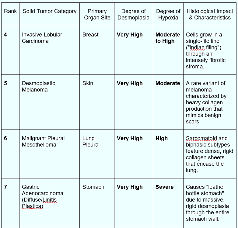
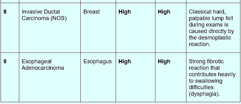
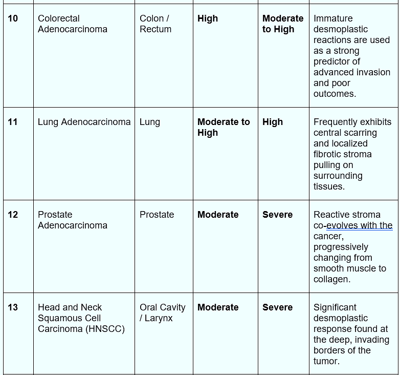
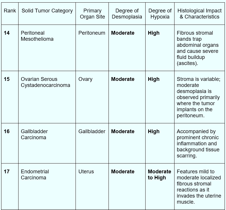
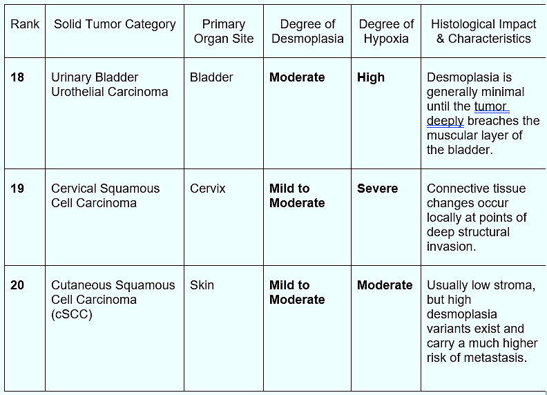
Desmoplasia and hypoxia are closely linked biological phenomena. A dense, high-pressure fibrous matrix crushes intratumoral blood vessels. This mechanical compression shuts off oxygen delivery, induces profound cellular hypoxia, and stabilizes HIF-1α.
The principle that “structure follows function” — historically stated as “form follows function” — originated in 19th-century architecture before being adopted by modern biology.
While scientists routinely identify biological structures, engineers and architects often have a deeper framework for understanding why those structures exist, because their disciplines require them to build systems from the ground up rather than merely observe pre-existing ones.
The phrase was originally coined as "form ever follows function" by the American architect Louis Sullivan in his 1896 essay, The Tall Office Building Artistically Considered.
Sullivan’s Architectural Intent: Sullivan, considered the "father of skyscrapers," argued that the outer shape, height, and layout of a building should be dictated purely by its internal purpose and utility, rather than arbitrary decorative precedents.
Later, evolutionary biologists and physiologists adapted this maxim. In biology, structure reflects natural selection: organisms that developed structural mutations serving essential functions survived, making structure a historical record of function.
The questions I kept returning to were:
Why does a physical shield exist in nearly all lethal solid tumors, correlating with both immune evasion and hypoxia?
Why do less deadly cancers have weaker shields?
Does 50% shield destruction correspond to a 50% improvement in immune function and survival?
The shield must serve a greater purpose than supporting Warburg metabolism alone. If so, then we are clearly missing something in our repurposed cancer protocols. My hunch is that the shield serves cancer’s immune evasion.
If this hypothesis is correct, corroborating evidence should exist. More importantly, it suggests that we may be able to reverse immune evasion and harness the host immune system to overcome cancer through repurposed drugs.
I believe the answer may also help explain the riddle of Ben Williams and how he survived glioblastoma. For readers who might argue that glioblastomas do not have a desmoplastic shield, I have two answers.
First, you may not have read my last Substack.
Second, Glioblastomas contain the very agent that builds the shield elsewhere: the Cancer-Associated Fibroblast, known in gliomas as the Glioma-Associated Fibroblast (GAF). While GAFs don't build a rigid, physical wall in brain tissue, they still erect a potent chemical and immunological shield around the tumor's hypoxic core.
Cancer-Associated Fibroblasts (CAFs) are the key to the architectural mystery of the tumor shield and its function. Not surprisingly, they have been the subject of intense research by engineers.
Dr. Meenal Datta, senior author of the study and assistant professor of aerospace and mechanical engineering at Notre Dame, said:
“A decade ago, we didn’t even know perivascular fibroblasts existed within the brain, and not just in the lining of the skull…My lab’s expertise is examining tumors from an engineering and systems-based approach and looking at the novel mechanical features in rare cancers that may have been understudied or overlooked.”
Maksym Zarodniuk, a PhD student in bioengineering and first author of the study, performed an analysis involving bulk and single-cell RNA sequencing of patient tumors. The data showed clear clinical differences between patients with more perivascular fibroblasts in their brain tumors than those with less.
Brain cancer patients with more perivascular fibroblasts in their tumors were more likely to experience poor responses to immunotherapy and have poor outcomes.
The research group found that the fibroblasts supported the creation of an immunosuppressive tumor microenvironment which facilitated immune evasion. The team felt that these perivascular fibroblasts might promote cancer stem cell growth and contribute to therapeutic resistance. Thus, the fibroblasts are believed to be a major source of metastases and relapse.
The situation is remarkably similar in blood cancers like acute myeloid leukemia. In a 2023 study, Ding and colleagues noted that CAFs constitute the largest proportion of stromal cells in the hematological tumor microenvironment. The team reported that CAF-mediated remodeling of the TME contributes to drug resistance, relapse, and tumor progression.
In another study by Gu and colleagues on the role of CAFs in acute leukemia, the researchers found that CAFs play a role in:
Reprogramming the malignant extracellular matrix.
Promoting tumor angiogenesis.
Fueling cancer cells.
Supporting cancer cell proliferation.
Promoting metastasis and invasion.
Deregulating metabolism.
Driving epigenetic reprogramming.
Promoting stemness.
Resisting cell death.
Shaping the TME.
Conferring therapeutic resistance.
In short, Dr. Gu and colleagues concluded:
The team went further and stated:
CAFs are well-equipped to assume the role of Master Organizer in the cancer by interacting with cancer cells and other stromal cells and immune cells in the TME and producing cancer specific ECM and secretome. Therefore, targeting CAFs to destroy cancer might be a potent therapeutic protocol for improving and perfecting cancer therapy.
Beyond the physical barrier and immune exclusion created by the desmoplastic shield, the cancer-associated fibroblasts that build it perform an equally insidious metabolic function known as the Reverse Warburg effect.
First described by Pavlides, Lisanti, and colleagues in 2009, this process involves tumor-derived signals — TGF-β, reactive oxygen species, and HIF-1α stabilization — that force CAFs into aerobic glycolysis.
The activated fibroblasts then export large quantities of lactate, pyruvate, ketone bodies, and other high-energy metabolites through monocarboxylate transporter 4 (MCT4). Adjacent cancer cells, frequently expressing MCT1, take up these substrates and feed them into their own mitochondrial oxidative phosphorylation machinery, generating ATP while sparing glucose for biosynthetic pathways needed for rapid proliferation.
This metabolic symbiosis turns the stromal fortress into a self-sustaining nutrient factory. The same fibroblasts that elevate interstitial pressure, collapse vessels, and create profound hypoxia are simultaneously supplying the energy and carbon skeletons that allow the cancer cells trapped inside the shield to thrive.
In highly desmoplastic tumors such as pancreatic cancer, this dual structural and metabolic support helps explain both therapy resistance and the remarkable ability of the tumor to maintain growth despite limited vascular delivery of drugs or nutrients. Interrupting the Reverse Warburg coupling—whether by targeting CAF activation, MCT-mediated lactate shuttling, or the upstream TGF-β signals that drive it—therefore represents a logical extension of any strategy aimed at dismantling the desmoplastic shield.
By now, everyone knows the story of Dr. Gregory Riggins' mice: after treating a pinworm outbreak in his brain-tumor-implanted research mice with Fenbendazole (a veterinary dewormer), their tumors unexpectedly stopped growing. This serendipitous discovery opened the door to cancer research using Mebendazole, its human-grade equivalent.
By way of review, these are the “ten deadliest cancers” against which we can now direct Mebendazole.
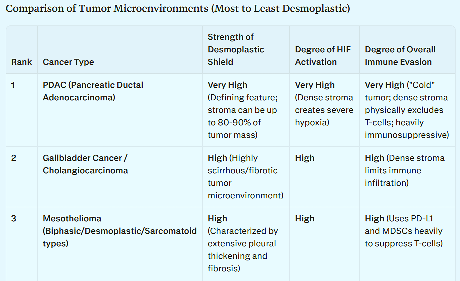
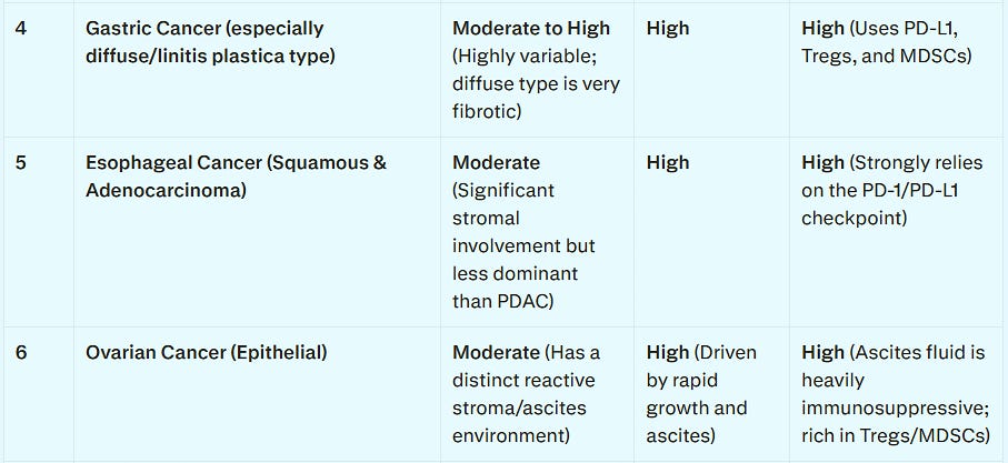
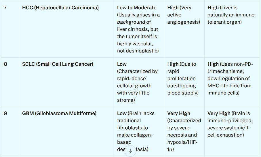

Mebendazole (MBZ) suppresses desmoplasia, cancer-associated fibroblasts (CAFs), and hypoxia-inducible factors (HIF) by altering structural scaffolds and transcriptional pathways within the tumor microenvironment (TME).
Key Mechanisms
Suppression of Desmoplasia / Stromal De-bulking: MBZ significantly decreases the volume of trichrome-positive fibrotic connective tissue and dense extracellular matrix (ECM) deposits. By disrupting microtubule-dependent collagen deposition, it lowers interstitial fluid pressure and opens physical passageways for cytotoxic CD8+ T-cells to infiltrate the tumor core.
Deactivation of CAFs: MBZ directly targets and suppresses α-smooth muscle actin (α-SMA)-positive activated pancreatic stellate cells and CAFs. It accomplishes this by disrupting the ciliary and cytosolic β-tubulin dynamics necessary for CAF structural maintenance, signaling, and motility.
Inhibition of HIF: Under hypoxic solid-tumor conditions, MBZ suppresses the protein induction and accumulation of HIF-1α, HIF-2α, and HIF-1β. This reduces downstream targets responsible for angiogenesis and metabolic reprogramming.
Myeloid Reprogramming (M2 → M1): MBZ acts as a potent myeloid-reprogramming agent. It directly stimulates CD14+ monocyte lineages via TLR8/NLRP3 inflammasome pathways and drives a functional phenotypic switch in Tumor-Associated Macrophages (TAMs) from the pro-tumor M2 state to the anti-tumor M1 state.
Pro-inflammatory Cytokine Influx & Immune Recruitment: The M1 shift triggers robust local release of TNF-α and IFN-γ, recruiting natural killer (NK) cells and naïve T-lymphocytes into the tumor core.
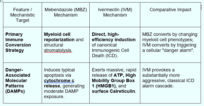
Microtubule-Mediated Immunogenic Cell Death (ICD): By disrupting β-tubulin polymerization, MBZ induces G2/M arrest and apoptosis. This damage-associated cascade initiates mild ICD, forcing exposure of surface “eat-me” signals such as calreticulin.
While both drugs help overcome immune evasion, Ivermectin (IVM) is widely considered a much more potent, direct driver of canonical Immunogenic Cell Death (ICD). It acts as a specialized biochemical alarm system, whereas MBZ converts the tumor microenvironment primarily through physical debulking and myeloid system remodeling.
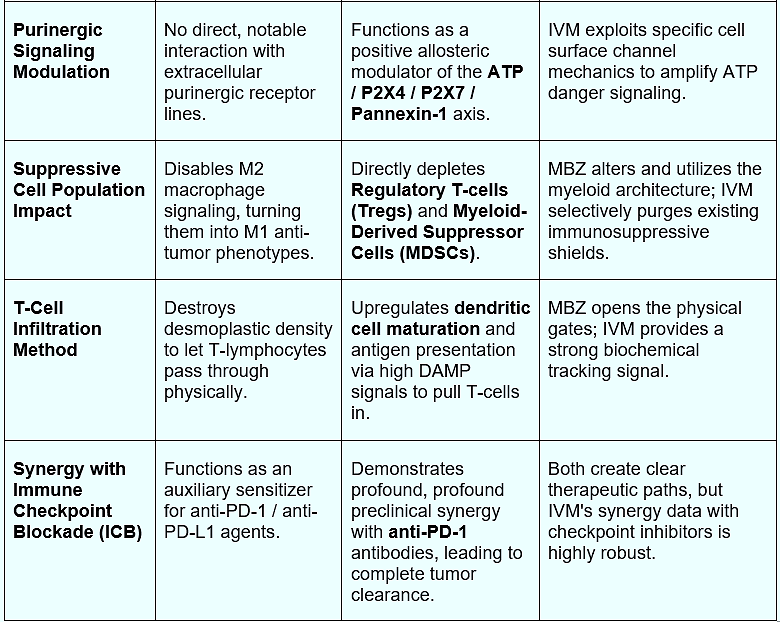
Transforming Growth Factor-Beta (TGF-β) is recognized as the “master regulator” of cancer-associated fibroblasts (CAFs) because it simultaneously controls their activation, spatial heterogeneity, structural remodeling, and immunosuppressive functions within the tumor microenvironment (TME).
While several growth factors and inflammatory cytokines can influence stroma, no other signaling molecule orchestrates the lifecycle and multi-functional programming of CAFs as comprehensively as TGF-β. For more mechanistic detail, see Appendix I.
A 2026 study by Hulscher et al. reported an 84.4% Clinical Benefit Ratio in 197 cancer patients using Ivermectin and Mebendazole—with nearly half achieving regression or no evidence of disease. Response rates this high from two non-toxic, generic drugs point to a deeper mechanistic synergy.
I believe that synergy lies in their shared assault on the desmoplastic shield. This fibrotic structure, purposefully constructed by Cancer-Associated Fibroblasts (CAFs) under the command of TGF-β, is what makes solid tumors lethal. Ivermectin disarms it chemically, blocking TGF-β receptors and Smad signaling to silence the CAF activation command.
Mebendazole disarms it physically, depolymerizing the microtubules CAFs need to build and deposit collagen. Together, they don’t just attack the cancer cell—they deconstruct the stronghold protecting it.
Building on this synergy, here are four repurposed five-agent cocktails designed to maximize TGF-β suppression and dismantle the desmoplastic shield from multiple angles.
Comments
Nothing yet. Say the first thing.
Sign in to join the conversation.